IL3 Antibodies
Background
IL3 is a hematopoietic growth factor secreted by activated T cells and other immune cells. Its structure belongs to the four-helix bundle cytokine family and regulates the proliferation and differentiation of various hematopoietic progenitor cells by specifically binding to cell surface receptors. This cytokine plays a core role in promoting the development of myeloid cells (such as granulocytes and macrophages), and is also involved in regulating immune responses and inflammatory processes. IL3 was independently discovered by multiple research teams in the 1980s. Its gene is located on human chromosome 5q31.2, a region closely related to myelodysplastic disorders and allergic diseases. As a typical representative of pleiotropic cytokines, IL3 has become an important model for studying hematopoietic regulation, immune signal transduction and targeted therapy, providing a key theoretical basis for understanding the role of cytokine networks in physiological and pathological processes.
Structure of IL3
The molecular weight of the protein encoded by the IL3 gene is approximately 15-17 kDa, and its precise molecular weight varies by species.
| Species | Human | Mouse | Rat |
| Molecular Weight (kDa) | 15.2 | 16.1 | 15.8 |
| Primary Structural Differences | Has the characteristic of the N - glycosylation sites | The C-terminal extends by three amino acids | The 56th position is serine rather than proline |
This protein is composed of 133 amino acids and presents a typical four-helical bundle topological structure. Its primary structure consists of four highly conserved α -helices (named A, B, C, and D respectively), which are connected by flexible ring regions to form a spatial conformation. The receptor binding interface formed by helix A and D contains the crucial glutamate-asparagine domain. The disulfide bond formed by cysteine at position 16 and 84 is essential for maintaining structural stability. There is a species-specific amino acid variation in the helical D region at the C-terminal, which directly affects its binding affinity with the receptor.
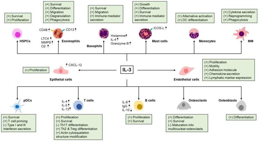 Fig. 1 Effect of IL-3 on immune and non-immune cells.1
Key structural properties of IL3:
- Typical four-helix bundle topological configuration (A-D helix)
- Hydrophobic core spiral relative spatial orientation
- Glutamic acid at position 56 and arginine at position 112 form a salt bridge stable structure
- C-terminal helix D contains receptor binding specific determinates
- Disulfide bonds (Cys16-Cys84) ensure structural integrity
- Glycosylation modification in the AB loop region affects the half-life of proteins
Functions of IL3
The main function of the IL3 gene is to regulate the differentiation of hematopoietic cells and immune responses. In addition, it is also involved in pathological processes such as inflammatory responses, the occurrence of allergic diseases, and the regulation of the tumor microenvironment.
| Function | Description |
| Hematopoietic regulation | Promote the proliferation and differentiation of pluripotent hematopoietic progenitor cells and maintain the development of myeloid cell lineages such as granulocytes and macrophages. |
| Immune regulation | Activate dendritic cell function, enhance T cell response, and participate in the polarization of Th2-type immune response. |
| Inflammation mediation | Under the stimulation of allergens, basophils and mast cells are induced to release inflammatory mediators such as histamine. |
| Tumor-related | In some parts of the abnormally high expression in leukemia, promote tumor cell survival by autocrine loop. |
| Tissue repair | By mobilizing macrophages to participate in the repair and remodeling process after tissue damage. |
The signal transduction of IL3 follows the typical pattern of the JAK-STAT pathway and shares the receptor β subunit with GMA-CSF and IL-5. However, its specificity is determined by the α chain. This characteristic makes it have both specificity and functional overlap in the cytokine network.
Applications of IL3 and IL3 Antibody in Literature
1. Hirsch, Hans H., et al. "Interleukin-3 mRNA Stabilization by a trans-Acting Mechanism in Autocrine Tumors Lacking Interleukin-3 Gene Rearrangements." Journal of Biological Chemistry 270.35 (1995): 20629-20635. https://doi.org/10.1074/jbc.270.35.20629
The article indicates that in PB-3c tumors transformed from v-Ha-ras, class I tumors significantly enhance the stability of IL3 mRNA (t>3h) through the trans-action mechanism and promote IL3 secretion. Class II tumors are characterized by rapid degradation of IL3 mRNA (t<0.5h). c-myc and IL6 transcripts are unstable in both types of tumors.
2. Tajer, Parisa, et al. "IL3 has a detrimental effect on hematopoietic stem cell self-renewal in transplantation settings." International Journal of Molecular Sciences 23.21 (2022): 12736. https://doi.org/10.3390/ijms232112736
Research shows that when expanding human hematopoietic stem cells using cytokine combinations, the addition of IL3 will reduce their implantation and regeneration ability in the transplantation model, which is not conducive to the self-renewal of stem cells. Therefore, it is recommended to exclude IL3 in the gene therapy protocols.
3. Podolska, Malgorzata J., et al. "IL-3: key orchestrator of inflammation." Frontiers in immunology 15 (2024): 1411047. https://doi.org/10.3389/fimmu.2024.1411047
Studies show that interleukin-3 (IL-3) is not only a hematopoietic factor but also a key regulator of inflammatory responses. It acts on various immune and non-immune cells through the specific receptor CD123, either exacerbating pathological inflammation or promoting its resolution in different diseases, thus becoming a potential clinical therapeutic target.
4. Luo, Xiong-jian, et al. "The interleukin 3 gene (IL3) contributes to human brain volume variation by regulating proliferation and survival of neural progenitors." PloS one 7.11 (2012): e50375. https://doi.org/10.1371/journal.pone.0050375
Research has found that variations in the promoter region of the interleukin-3 (IL-3) gene (rs31480) regulate the proliferation and survival of neural progenitor cells by influencing its expression level, thereby leading to differences in brain volume among different populations and genders, revealing a new function of IL-3 in brain development.
5. Gündogdu, Mehtap S., et al. "The haematopoietic GTPase RhoH modulates IL3 signalling through regulation of STAT activity and IL3 receptor expression." Molecular cancer 9.1 (2010): 225. https://doi.org/10.1186/1476-4598-9-225
Studies have shown that RhoH protein is a key negative regulatory factor of IL-3 signaling. High expression of RhoH inhibits cell proliferation by activating STAT1 and up-regulating p21/p27. Low expression of RhoH increases the expression of IL-3 receptor (CD123) and enhances STAT5 activity, thereby promoting cell growth. This reveals a new mechanism of RhoH in leukemia.
Creative Biolabs: IL3 Antibodies for Research
Creative Biolabs specializes in the production of high-quality IL3 antibodies for research and industrial applications. Our portfolio includes monoclonal antibodies tailored for ELISA, Flow Cytometry, Western blot, immunohistochemistry, and other diagnostic methodologies.
- Custom IL3 Antibody Development: Tailor-made solutions to meet specific research requirements.
- Bulk Production: Large-scale antibody manufacturing for industry partners.
- Technical Support: Expert consultation for protocol optimization and troubleshooting.
- Aliquoting Services: Conveniently sized aliquots for long-term storage and consistent experimental outcomes.
For more details on our IL3 antibodies, custom preparations, or technical support, contact us at email.
Reference
- Podolska, Malgorzata J., et al. "IL-3: key orchestrator of inflammation." Frontiers in immunology 15 (2024): 1411047. https://doi.org/10.3389/fimmu.2024.1411047
Anti-IL3 antibodies
 Loading...


Hot products 
-
Mouse Anti-CCDC6 Recombinant Antibody (CBXC-0106) (CBMAB-C5397-CQ)

-
Mouse Anti-GLP1R Recombinant Antibody (4F3) (CBMAB-G0521-LY)

-
Mouse Anti-ADAM29 Recombinant Antibody (V2-179787) (CBMAB-A1149-YC)

-
Mouse Anti-GFAP Recombinant Antibody (20) (CBMAB-G2914-LY)

-
Mouse Anti-AAV9 Recombinant Antibody (V2-634029) (CBMAB-AP023LY)

-
Mouse Anti-FAS2 Monoclonal Antibody (1D4) (CBMAB-0071-CN)

-
Rabbit Anti-AKT2 (Phosphorylated S474) Recombinant Antibody (V2-556130) (PTM-CBMAB-0605LY)

-
Mouse Anti-CCDC25 Recombinant Antibody (CBLC132-LY) (CBMAB-C9786-LY)

-
Mouse Anti-DMPK Recombinant Antibody (CBYCD-324) (CBMAB-D1200-YC)

-
Mouse Anti-AMIGO2 Recombinant Antibody (CBYY-C0756) (CBMAB-C2192-YY)

-
Mouse Anti-CD24 Recombinant Antibody (ALB9) (CBMAB-0176CQ)

-
Mouse Anti-CCL18 Recombinant Antibody (64507) (CBMAB-C7910-LY)

-
Mouse Anti-ENO1 Recombinant Antibody (8G8) (CBMAB-E1329-FY)

-
Mouse Anti-CTNND1 Recombinant Antibody (CBFYC-2414) (CBMAB-C2487-FY)

-
Mouse Anti-dsRNA Recombinant Antibody (2) (CBMAB-D1807-YC)

-
Mouse Anti-ENPP1 Recombinant Antibody (CBFYE-0159) (CBMAB-E0375-FY)

-
Mouse Anti-ARG1 Recombinant Antibody (CBYCL-103) (CBMAB-L0004-YC)

-
Mouse Anti-DISP2 Monoclonal Antibody (F66A4B1) (CBMAB-1112CQ)

-
Mouse Anti-ADV Recombinant Antibody (V2-503423) (CBMAB-V208-1364-FY)

-
Mouse Anti-ALOX5 Recombinant Antibody (33) (CBMAB-1890CQ)

- AActivation
- AGAgonist
- APApoptosis
- BBlocking
- BABioassay
- BIBioimaging
- CImmunohistochemistry-Frozen Sections
- CIChromatin Immunoprecipitation
- CTCytotoxicity
- CSCostimulation
- DDepletion
- DBDot Blot
- EELISA
- ECELISA(Cap)
- EDELISA(Det)
- ESELISpot
- EMElectron Microscopy
- FFlow Cytometry
- FNFunction Assay
- GSGel Supershift
- IInhibition
- IAEnzyme Immunoassay
- ICImmunocytochemistry
- IDImmunodiffusion
- IEImmunoelectrophoresis
- IFImmunofluorescence
- IGImmunochromatography
- IHImmunohistochemistry
- IMImmunomicroscopy
- IOImmunoassay
- IPImmunoprecipitation
- ISIntracellular Staining for Flow Cytometry
- LALuminex Assay
- LFLateral Flow Immunoassay
- MMicroarray
- MCMass Cytometry/CyTOF
- MDMeDIP
- MSElectrophoretic Mobility Shift Assay
- NNeutralization
- PImmunohistologyp-Paraffin Sections
- PAPeptide Array
- PEPeptide ELISA
- PLProximity Ligation Assay
- RRadioimmunoassay
- SStimulation
- SESandwich ELISA
- SHIn situ hybridization
- TCTissue Culture
- WBWestern Blot
