RAI2 Antibodies
Background
RAI2 is a nuclear protein that was initially found to be induced to express by retinoic acid. It plays a tumor-suppressing role in various solid tumors. This gene interacts with the transcriptional co-repressor CtBP through its encoded protein, regulating the expression of Wnt signaling pathway and cell cycle-related genes. Studies have shown that RAI2 is often downregulated or silenced due to high methylation in the promoter region in colorectal cancer, breast cancer, and gastric cancer. Its low expression is closely related to poor patient prognosis, changes in immune infiltration, and chemotherapy resistance. As a tumor suppressor, RAI2 inhibits tumor cell proliferation, migration, and invasion by various mechanisms such as inhibiting the AKT signaling pathway, regulating stem cell characteristics, and inducing cell apoptosis. It is expected to become a new molecular target for cancer diagnosis and treatment.
Structure of RAI2
RAI2 is a nuclear protein with a molecular weight of approximately 45 kDa. Its sequence and structure vary among different species. This protein contains multiple functional domains and mainly interacts with the transcriptional co-repressor CtBP through its C-terminal region.
| Species | Human | Mouse | Rat | Simian |
| Molecular Weight (kDa) | 45.2 | 45.1 | 45.0 | 45.2 |
| CtBP binding domain | Located at the C-terminal | Highly conserved sequence | Highly conserved sequence | Highly conserved sequence |
The RAI2 protein contains approximately 420 amino acids. Its secondary structure is mainly composed of α-helices and random coils, enabling it to mediate multivalent interactions with CtBP through repetitive SLiM motifs. This binding can induce the aggregation of CtBP to form intranuclear filaments, thereby relieving its transcriptional repression of downstream target genes. The nuclear localization signal of RAI2 enables it to be mainly located in the cell nucleus. By regulating the expression of Wnt signaling pathway and cell cycle-related genes, it plays a role in inhibiting the proliferation, migration, and invasion of tumor cells.
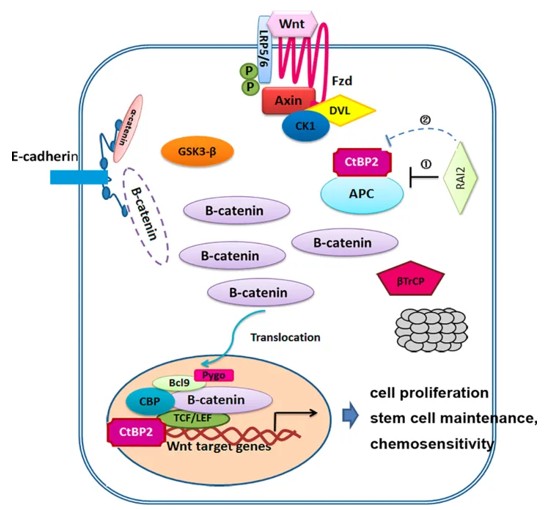 Fig. 1 Mechanisms model for the regulation of RAI2 in Wnt/β-catenin signaling.1
The core structural features of RAI2:
- Contains repetitive SLiM motifs, mediating multivalent binding with CtBP
- C-terminal CtBP binding domain, regulating transcriptional repression function
- Nuclear localization signal, ensuring its localization in the cell nucleus
- Predominantly unstructured coiled-coil secondary structure, providing conformational flexibility
- Forms intranuclear long filaments by inducing CtBP aggregation, relieving the inhibition of target genes
Functions of RAI2
The main function of RAI2 is to exert tumor suppression in various solid tumors. However, it is also involved in regulating various biological processes such as cell differentiation, immune infiltration, and chemotherapy sensitivity.
| Function | Description |
| Wnt signal inhibition | RAI2 inhibits the activity of the Wnt/β-catenin pathway by interacting with CtBP, reducing the expression of downstream target genes such as c-Myc and CyclinD1. |
| Cell cycle regulation | RAI2 blocks the cell cycle progression by down-regulating cycle-related genes such as CCNA2 and CDC20, thereby inhibiting tumor cell proliferation. |
| Stem cell characteristic inhibition | RAI2 reduces the ability of tumor cells to form spheres and inhibits the expression of stemness markers such as LGR5, weakening the self-renewal ability of tumor stem cells. |
| Chemotherapy sensitization | RAI2 expression restoration can enhance the sensitivity of colorectal cancer and gastric cancer cells to chemotherapy drugs such as oxaliplatin and fluorouracil. |
| Immune infiltration regulation | RAI2 expression level is related to the density of tumor-infiltrating lymphocytes and the abundance of PD-L1 expression, and may participate in the remodeling of the tumor immune microenvironment. |
The tumor suppressive function of RAI2 is mainly achieved through its interaction with CtBP: when RAI2 is highly expressed, it induces the aggregation of CtBP to form nuclear filaments, relieving the transcriptional inhibition of target genes by CtBP, thereby exerting the role of inhibiting tumor progression. In colorectal cancer, breast cancer, and gastric cancer, RAI2 is often silenced due to high methylation of the promoter region, and its low expression is significantly associated with poor patient prognosis.
Applications of RAI2 and RAI2 Antibody in Literature
1. Zhang, Weitao, et al. "Retinoic acid-induced 2 (RAI2) is a novel antagonist of Wnt/β-catenin signaling pathway and potential biomarker of chemosensitivity in colorectal cancer." Frontiers in Oncology 12 (2022): 805290. https://doi.org/10.3389/fonc.2022.805290
The article indicates that low expression of RAI2 is an independent poor prognostic marker for colorectal cancer. RAI2 inhibits the Wnt signal by binding to CtBP2, reduces the nuclear entry of β-catenin, thereby suppressing stem cell characteristics and increasing chemotherapy sensitivity.
2. Lou, Xiaoli, et al. "RAI2 acts as a tumor suppressor with functional significance in gastric cancer." Aging (Albany NY) 15.21 (2023): 11831. https://doi.org/10.18632/aging.205135
The article indicates that RAI2 is expressed at a low level in gastric cancer, and is associated with high immune infiltration and PD-L1. It inhibits the proliferation, migration and invasion of cancer cells by regulating multiple pathways, and is a potential tumor suppressor target.
3. Goradia, Nishit, et al. "Master corepressor inactivation through multivalent SLiM-induced polymerization mediated by the oncogene suppressor RAI2." Nature Communications 15.1 (2024): 5241. https://doi.org/10.1038/s41467-024-49488-3
The article indicates that RAI2 induces the aggregation of transcriptional repressor factor CtBP by repeating the SLiM motif, forming long intranuclear filaments and thereby inhibiting their function. The expression of RAI2 is significantly reduced in the treatment of resistant prostate cancer, revealing a new mechanism by which its inactivation drives disease progression.
4. Jiao, Ying, et al. "Comprehensive analysis of the expression and prognosis for RAI2: A promising biomarker in breast cancer." Frontiers in Oncology 13 (2023): 1134149. https://doi.org/10.3389/fonc.2023.1134149
The article indicates that RAI2 is expressed at a low level in breast cancer. Patients with high expression have a better prognosis and more immune infiltration. It exerts its tumor-suppressing effect by regulating pathways such as the cell cycle, and genes related to the cell cycle such as CCNA2 are the key downstream targets.
5. Yan, Wenji, et al. "Retinoic acid-induced 2 (RAI2) is a novel tumor suppressor, and promoter region methylation of RAI2 is a poor prognostic marker in colorectal cancer." Clinical epigenetics 10.1 (2018): 69. https://doi.org/10.1186/s13148-018-0501-4
The article indicates that RAI2 is inactivated in colorectal cancer due to high promoter methylation, and methylation is an independent adverse prognostic marker. RAI2 exerts a tumor-suppressive effect by inhibiting the AKT pathway, including inhibiting proliferation, migration, invasion and promoting apoptosis.
Creative Biolabs: RAI2 Antibodies for Research
Creative Biolabs specializes in the production of high-quality RAI2 antibodies for research and industrial applications. Our portfolio includes monoclonal and polyclonal antibodies tailored for ELISA, Flow Cytometry, Western blot, immunohistochemistry, and other diagnostic methodologies.
- Custom RAI2 Antibody Development: Tailor-made solutions to meet specific research requirements.
- Bulk Production: Large-scale antibody manufacturing for industry partners.
- Technical Support: Expert consultation for protocol optimization and troubleshooting.
- Aliquoting Services: Conveniently sized aliquots for long-term storage and consistent experimental outcomes.
For more details on our RAI2 antibodies, custom preparations, or technical support, contact us at email.
Reference
- Zhang, Weitao, et al. "Retinoic acid-induced 2 (RAI2) is a novel antagonist of Wnt/β-catenin signaling pathway and potential biomarker of chemosensitivity in colorectal cancer." Frontiers in Oncology 12 (2022): 805290. Distributed under Open Access license CC BY 4.0, without modification. https://doi.org/10.3389/fonc.2022.805290
Anti-RAI2 antibodies
 Loading...


Hot products 
-
Mouse Anti-CA9 Recombinant Antibody (CBXC-2079) (CBMAB-C0131-CQ)

-
Mouse Anti-CD63 Recombinant Antibody (CBXC-1200) (CBMAB-C1467-CQ)

-
Mouse Anti-4-Hydroxynonenal Recombinant Antibody (V2-502280) (CBMAB-C1055-CN)

-
Mouse Anti-GFAP Recombinant Antibody (5) (CBMAB-G0346-LY)

-
Mouse Anti-CSPG4 Recombinant Antibody (CBFYM-1050) (CBMAB-M1203-FY)

-
Mouse Anti-CAPZB Recombinant Antibody (CBYY-C0944) (CBMAB-C2381-YY)

-
Mouse Anti-BSN Recombinant Antibody (219E1) (CBMAB-1228-CN)

-
Mouse Anti-CECR2 Recombinant Antibody (CBWJC-2465) (CBMAB-C3533WJ)

-
Mouse Anti-ALPL Antibody (B4-78) (CBMAB-1009CQ)

-
Mouse Anti-ACTN4 Recombinant Antibody (V2-6075) (CBMAB-0020CQ)

-
Mouse Anti-BRCA2 Recombinant Antibody (CBYY-0790) (CBMAB-0793-YY)

-
Mouse Anti-APC Recombinant Antibody (CBYC-A661) (CBMAB-A3036-YC)

-
Rabbit Anti-AP2M1 (Phosphorylated T156) Recombinant Antibody (D4F3) (PTM-CBMAB-0610LY)

-
Mouse Anti-CCN2 Recombinant Antibody (CBFYC-2383) (CBMAB-C2456-FY)

-
Human Anti-SARS-CoV-2 S1 Monoclonal Antibody (CBFYR-0120) (CBMAB-R0120-FY)

-
Rat Anti-AChR Recombinant Antibody (V2-12500) (CBMAB-0990-CN)

-
Mouse Anti-BBS2 Recombinant Antibody (CBYY-0253) (CBMAB-0254-YY)

-
Mouse Anti-14-3-3 Pan Recombinant Antibody (V2-9272) (CBMAB-1181-LY)

-
Rat Anti-C5AR1 Recombinant Antibody (8D6) (CBMAB-C9139-LY)

-
Mouse Anti-F11R Recombinant Antibody (402) (CBMAB-0026-WJ)

- AActivation
- AGAgonist
- APApoptosis
- BBlocking
- BABioassay
- BIBioimaging
- CImmunohistochemistry-Frozen Sections
- CIChromatin Immunoprecipitation
- CTCytotoxicity
- CSCostimulation
- DDepletion
- DBDot Blot
- EELISA
- ECELISA(Cap)
- EDELISA(Det)
- ESELISpot
- EMElectron Microscopy
- FFlow Cytometry
- FNFunction Assay
- GSGel Supershift
- IInhibition
- IAEnzyme Immunoassay
- ICImmunocytochemistry
- IDImmunodiffusion
- IEImmunoelectrophoresis
- IFImmunofluorescence
- IGImmunochromatography
- IHImmunohistochemistry
- IMImmunomicroscopy
- IOImmunoassay
- IPImmunoprecipitation
- ISIntracellular Staining for Flow Cytometry
- LALuminex Assay
- LFLateral Flow Immunoassay
- MMicroarray
- MCMass Cytometry/CyTOF
- MDMeDIP
- MSElectrophoretic Mobility Shift Assay
- NNeutralization
- PImmunohistologyp-Paraffin Sections
- PAPeptide Array
- PEPeptide ELISA
- PLProximity Ligation Assay
- RRadioimmunoassay
- SStimulation
- SESandwich ELISA
- SHIn situ hybridization
- TCTissue Culture
- WBWestern Blot
