
SLPI Antibodies
Background
The SLPI gene encodes secretory leukocyte protease inhibitor, which is mainly secreted by mucosal epithelial cells and belongs to the serine protease inhibitor family. This protein protects tissues from excessive inflammatory damage by inhibiting the activity of proteins such as neutrophil elastase. It also plays a key role in immune regulation and wound repair. This gene was first identified in 1986 and its expression is precisely regulated by inflammatory signals. It is particularly important in barrier tissues such as the respiratory tract and reproductive tract. Its unique dual-function domain - the N-terminal has antibacterial activity, and the C-terminal is responsible for protease inhibition - provides a classic model for studying innate immunity and protease regulation, and is of great value for research in fields such as infectious diseases, chronic lung diseases, and tumor microenvironments.
Structure of SLPI
The protein encoded by the SLPI gene has a molecular weight of approximately 12-14 kDa. The difference in this range mainly results from the varying degrees of glycosylation modification. This protein consists of 107 amino acid residues and its primary structure contains two clearly defined functional domains: the N-terminal domain has broad-spectrum antibacterial activity, while the C-terminal domain is responsible for serine protease inhibition function. At the secondary structure level, the SLPI protein is rich in β-sheet and α-helix, which together form a compact and stable three-dimensional spherical structure. The key amino acids at the active center of the protein - especially the leucine-methionine residues located at the reaction center - are precisely positioned through a characteristic reverse parallel β-sheet structure, which is the structural basis for its specific inhibition of targets such as elastase. This unique dual-domain design enables SLPI to simultaneously participate in innate immune defense and regulation of protease activity.
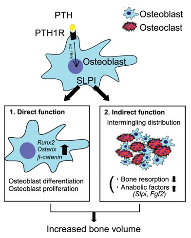 Fig. 1 Model of SLPI functions in bone metabolism.1
Key structural properties of SLPI:
- Compact globular structure composed of two independent functional domains
- The core area is rich in reverse parallel β -folded configurations
- Active centers rely on conservative disulfide bond network stable space conformation
- Pairs of leucine-methionine residues constitute recognition sites for specific binding of serine proteases
Functions of SLPI
The protein encoded by the SLPI gene mainly functions to inhibit serine proteases such as tissue protease G and elastase to regulate inflammatory responses. Additionally, it is involved in various physiological and pathological processes, including innate immune responses and tissue repair.
| Function | Description |
| Proteinase Inhibition | Specifically inhibits the serine proteases released by neutrophils, protecting tissues from excessive proteolysis damage. |
| Anti-inflammatory regulation | By inhibiting protease activity, it reduces the release of pro-inflammatory cytokines and may directly affect the functions of immune cells such as macrophages. |
| Antibacterial Activity | The N-terminal domain of it possesses broad-spectrum antibacterial capability and can directly damage the bacterial cell membrane. |
| Tissue Repair | By regulating the balance of proteases and cell migration, it promotes epithelial regeneration and wound healing. |
| Immune Homeostasis Maintenance | Forming a protective barrier on the mucosal surface, balancing the defense response and preventing excessive inflammation from causing self-damage. |
The inhibitory mechanism of this protein exhibits highly efficient specificity. Its inhibition constant (Ki) is at the nanomolar level, which is much lower than that of many other protease inhibitors. This enables it to rapidly neutralize a large amount of proteases in the local microenvironment and act as a crucial "molecular sentinel".
Applications of SLPI and SLPI Antibody in Literature
1. Morimoto, Akito, et al. "SLPI is a critical mediator that controls PTH-induced bone formation." Nature Communications 12.1 (2021): 2136. https://doi.org/10.1038/s41467-021-22402-x
The research has found that the serine protease inhibitor SLPI is a key mediator for parathyroid hormone (PTH) to promote bone formation. PTH induces high expression of SLPI in osteoblasts, which can enhance osteogenic differentiation and inhibit osteoclast function by increasing the contact between bone cells, thereby coordinating the balance of bone metabolism.
2. Kwiecinska, Patrycja, et al. "SLPI controls neutrophil migration abilities and impacts neutrophil skin infiltration in experimental psoriasis." Cellular and Molecular Life Sciences 82.1 (2025): 74. https://doi.org/10.1007/s00018-025-05606-y
The study found that the absence of the SLPI gene significantly affects the skin infiltration of neutrophils in the psoriasis mouse model. The lack of SLPI leads to abnormal interactions between neutrophils and the vascular wall, reduced efficiency of transvascular migration, and changes in their distribution and movement patterns within the skin tissue.
3. Brown, Ryan, et al. "SLPI deficiency alters airway protease activity and induces cell recruitment in a model of muco-obstructive lung disease." Frontiers in Immunology 15 (2024): 1433642. https://doi.org/10.3389/fimmu.2024.1433642
The research has found that SLPI is an important innate immune protein in the airway, with the ability to resist proteases and inhibit the anti-inflammatory effect mediated by NF-κB. In mucous obstructive lung diseases such as chronic obstructive pulmonary disease and cystic fibrosis, the level of SLPI often decreases due to inflammation or degradation by neutrophil elastase, which may exacerbate airway immune damage. The study explored its pathological role through the ENaC-Tg and SLPI knockout mouse models.
4. Nugteren, Sandrine, and Janneke N. Samsom. "Secretory Leukocyte Protease Inhibitor (SLPI) in mucosal tissues: Protects against inflammation, but promotes cancer." Cytokine & Growth Factor Reviews 59 (2021): 22-35. https://doi.org/10.1016/j.cytogfr.2021.01.005
The research has found that SLPI is a multifunctional protein expressed at the mucosal barrier, which can inhibit proteases, kill microorganisms and block the NF-κB-mediated inflammatory response, thereby maintaining tissue homeostasis. However, its overexpression in cancer cells may promote tumor metastasis.
5. Sun, Jie, et al. "SLPI suppresses hepatocellular carcinoma progression via endoplasmic reticulum stress induced apoptosis." International Journal of Biological Sciences 18.1 (2022): 140. https://doi.org/10.7150/ijbs.65676
The study found that SLPI is significantly under-expressed in hepatocellular carcinoma (HCC), and its low level indicates a poorer prognosis for the patients. Functionally, SLPI enhances tumor cell apoptosis mediated by endoplasmic reticulum stress by activating the MAPK signaling pathway, thereby inhibiting the proliferation and invasion of HCC and exerting a potential tumor suppressor effect.
Creative Biolabs: SLPI Antibodies for Research
Creative Biolabs specializes in the production of high-quality SLPI antibodies for research and industrial applications. Our portfolio includes monoclonal antibodies tailored for ELISA, Flow Cytometry, Western blot, immunohistochemistry, and other diagnostic methodologies.
- Custom SLPI Antibody Development: Tailor-made solutions to meet specific research requirements.
- Bulk Production: Large-scale antibody manufacturing for industry partners.
- Technical Support: Expert consultation for protocol optimization and troubleshooting.
- Aliquoting Services: Conveniently sized aliquots for long-term storage and consistent experimental outcomes.
For more details on our SLPI antibodies, custom preparations, or technical support, contact us at email.
Reference
- Morimoto, Akito, et al. "SLPI is a critical mediator that controls PTH-induced bone formation." Nature Communications 12.1 (2021): 2136. Distributed under Open Access license CC BY 4.0, without modification. https://doi.org/10.1038/s41467-021-22402-x
Anti-SLPI antibodies
 Loading...
 Compare




Hot products 
-
Mouse Anti-C5B-9 Recombinant Antibody (CBFYA-0216) (CBMAB-X0304-FY)

-
Rabbit Anti-AKT3 Recombinant Antibody (V2-12567) (CBMAB-1057-CN)

-
Rabbit Anti-ABL1 (Phosphorylated Y245) Recombinant Antibody (V2-505716) (PTM-CBMAB-0465LY)

-
Armenian hamster Anti-CD40 Recombinant Antibody (HM40-3) (CBMAB-C10365-LY)

-
Mouse Anti-BCL6 Recombinant Antibody (CBYY-0435) (CBMAB-0437-YY)

-
Mouse Anti-DES Monoclonal Antibody (440) (CBMAB-AP1857LY)

-
Mouse Anti-CIITA Recombinant Antibody (CBLC160-LY) (CBMAB-C10987-LY)

-
Mouse Anti-CD164 Recombinant Antibody (CBFYC-0077) (CBMAB-C0086-FY)

-
Mouse Anti-14-3-3 Pan Recombinant Antibody (V2-9272) (CBMAB-1181-LY)

-
Mouse Anti-AQP2 Recombinant Antibody (G-3) (CBMAB-A3359-YC)

-
Mouse Anti-AFM Recombinant Antibody (V2-634159) (CBMAB-AP185LY)

-
Mouse Anti-FOXL1 Recombinant Antibody (CBXF-0845) (CBMAB-F0462-CQ)

-
Mouse Anti-ADGRL2 Recombinant Antibody (V2-58519) (CBMAB-L0166-YJ)

-
Mouse Anti-AGO2 Recombinant Antibody (V2-634169) (CBMAB-AP203LY)

-
Mouse Anti-CD24 Recombinant Antibody (2Q1282) (CBMAB-C1624-CN)

-
Mouse Anti-CD19 Recombinant Antibody (CBXC-1224) (CBMAB-C1491-CQ)

-
Mouse Anti-AMOT Recombinant Antibody (CBYC-A564) (CBMAB-A2552-YC)

-
Rat Anti-4-1BB Recombinant Antibody (V2-1558) (CBMAB-0953-LY)

-
Mouse Anti-CTNND1 Recombinant Antibody (CBFYC-2414) (CBMAB-C2487-FY)

-
Mouse Anti-BCL6 Recombinant Antibody (CBYY-0442) (CBMAB-0445-YY)

- AActivation
- AGAgonist
- APApoptosis
- BBlocking
- BABioassay
- BIBioimaging
- CImmunohistochemistry-Frozen Sections
- CIChromatin Immunoprecipitation
- CTCytotoxicity
- CSCostimulation
- DDepletion
- DBDot Blot
- EELISA
- ECELISA(Cap)
- EDELISA(Det)
- ESELISpot
- EMElectron Microscopy
- FFlow Cytometry
- FNFunction Assay
- GSGel Supershift
- IInhibition
- IAEnzyme Immunoassay
- ICImmunocytochemistry
- IDImmunodiffusion
- IEImmunoelectrophoresis
- IFImmunofluorescence
- IGImmunochromatography
- IHImmunohistochemistry
- IMImmunomicroscopy
- IOImmunoassay
- IPImmunoprecipitation
- ISIntracellular Staining for Flow Cytometry
- LALuminex Assay
- LFLateral Flow Immunoassay
- MMicroarray
- MCMass Cytometry/CyTOF
- MDMeDIP
- MSElectrophoretic Mobility Shift Assay
- NNeutralization
- PImmunohistologyp-Paraffin Sections
- PAPeptide Array
- PEPeptide ELISA
- PLProximity Ligation Assay
- RRadioimmunoassay
- SStimulation
- SESandwich ELISA
- SHIn situ hybridization
- TCTissue Culture
- WBWestern Blot
