TBCA Antibodies
Background
The TBCA protein encoded by the TBCA gene, as a key component of the molecular chaperone complex, mainly participates in the correct folding of β-tubulin and the assembly process of tubulin heterodimers. This protein plays a significant role in maintaining genomic stability by maintaining the conformational stability of tubulin and ensuring the normal formation of the spindle during cell division. Research has found that mutations in the TBCA gene can disrupt the microtubule network structure, leading to abnormal neuronal development, which is closely related to the occurrence of various neurodegenerative diseases. As a core regulatory factor of the eukaryotic microtubule assembly system, its unique folding mechanism provides an important model for the study of protein conformational diseases. In recent years, the structure of the TBCA-tubulin complex resolved by cryo-electron microscopy technology has further deepened people's understanding of the folding pathway mediated by molecular chaperone.
Structure of TBCA
TBCA is a microtubulin folding chaperone with a molecular weight of approximately 12.8 kDa, and its precise molecular weight varies slightly among different eukaryotes.
| Species | Human | Mouse | Yeast | Arabidopsis thaliana | Zebrafish |
| Molecular Weight (kDa) | 12.8 | 12.7 | 13.2 | 12.9 | 12.6 |
| Primary Structural Differences | Highly conservative β -folded core | 90% homology to humans | The N-end extension area | Special acidic C-terminal | Retain all functional residues |
This protein is composed of 109 amino acids and forms a typical α+β mixed folded conformation. The core of its three-dimensional structure consists of four anti-parallel β -folds, with three sets of α -helices distributed on both sides, jointly forming hydrophobic binding pockets. This pocket directly recognizes the intermediate domain of β -tubulin through phenylalanine at position 37 and leucine at position 73. It is particularly worth noting the acidic ring region formed at positions 85-92, where aspartic acid at position 87 and glutamic acid at position 91 stabilize the GTP binding site of tubulin through a hydrogen bond network, ensuring that β -tubulin can correctly complete the folding process.
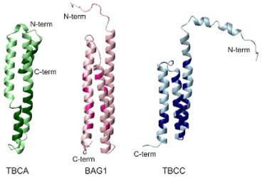 Fig. 1 Comparison of the interacting face of TBCA, BAG1 and TBCC N-terminal domain.1
Key structural properties of TBCA:
- Typical α+β mixed fold conformation
- The hydrophobic core is composed of four anti-parallel β -folds
- Conserved acidic amino acid residues are involved in β -tubulin recognition
- The C-terminal flexible region regulates tubulin dimer assembly
Functions of TBCA
The core function of the protein encoded by the TBCA gene is to mediate the folding and assembly of β -tubulin. Its main functional system includes:
| Function | Description |
| Tubulin folding | As a molecular chaperone, it directly binds to the unfolded β -tubulin, preventing its aggregation and promoting the acquisition of the correct three-dimensional conformation. |
| Dimer assembly | The β -tubulin that assists in the folding process forms a functional heterodimer with α -tubulin. |
| Maintenance of microtubule networks | By regulating the supply quality of tubulin monomers, the normal dynamics of spindle microtubules during cell division can be guaranteed. |
| Stress protection | Stabilize the tubulin pool under conditions such as oxidative stress to prevent fibrotic aggregation caused by misfolding. |
| Neurodevelopmental support | In particular, it ensures a continuous supply of tubulin in neurons to support axon transport and synaptic plasticity. |
This protein forms a specific recognition interface with the GTP binding domain of β -tubulin through its C-terminal acidic region. This interaction does not rely on ATP hydrolysis but achieves the folding catalysis of substrate proteins through conformational selection mechanisms, providing structurally correct functional units for microtubule assembly.
Applications of TBCA and TBCA Antibody in Literature
1. Nolasco, Sofia, et al. "Colchicine blocks tubulin heterodimer recycling by tubulin cofactors TBCA, TBCB, and TBCE." Frontiers in cell and developmental biology 9 (2021): 656273. https://doi.org/10.3389/fcell.2021.656273
Research has found that colicine, which is used to treat pericarditis, can inhibit the decomposition of tubulin dimer by TBCE/TBCB, leading to an increase in free TBCA. This, in turn, interferes with tubulin recovery by affecting the TBCA system, which may be its new anti-inflammatory mechanism.
2. Hillary, Robert F., et al. "Genome‐and epigenome‐wide studies of plasma protein biomarkers for Alzheimer's disease implicate TBCA and TREM2 in disease risk." Alzheimer's & Dementia: Diagnosis, Assessment & Disease Monitoring 14.1 (2022): e12280. https://doi.org/10.1002/dad2.12280
Research has found that the higher the levels of TBCA and TREM2 proteins in the blood, the lower the risk of Alzheimer's disease (AD). The research, through genomic methods, has revealed the genetic and epigenetic factors that regulate the levels of these AD-related proteins.
3. Danquah, Bright D., et al. "Mass Spectrometric analysis of antibody—Epitope peptide complex dissociation: Theoretical concept and practical procedure of binding strength characterization." Molecules 25.20 (2020): 4776. https://doi.org/10.1371/journal.pone.0042536
Research has found that the Tbca16 gene in mice negatively regulates the level of Tbca13 mRNA during testicular maturation through its sense and antisense transcripts. This post-transcriptional regulatory mechanism may play a key role in spermatogenesis that requires microtubule recombination.
4. Dai J, Gao J, Jiahui Gao, and Hongchao Dong. "Prognostic relevance and validation of ARPC1A in the progression of low-grade glioma." Aging (Albany NY) 16.14 (2024): 11162. https://doi.org/10.18632/aging.205952
Research has found that the set composed of mitochondrial-related genes such as TBCA is associated with the prognosis of low-grade glioma (LGG). Among them, the gene ARPC1A can promote the proliferation of LGG cells through the MAPK signaling pathway and is an important independent risk factor and potential biomarker.
5. Pickett, Sarah J., et al. "Interactions between nuclear and mitochondrial SNPs and Parkinson's disease risk." Mitochondrion 63 (2022): 85-88. https://doi.org/10.1016/j.mito.2022.02.002
Research has found that, unlike healthy individuals, patients with Parkinson's disease (PD) have specific combinations of nuclear genes such as TBCA and mitochondrial genes. The interaction of these combinations significantly affects the risk of developing Parkinson's disease.
Creative Biolabs: TBCA Antibodies for Research
Creative Biolabs specializes in the production of high-quality TBCA antibodies for research and industrial applications. Our portfolio includes monoclonal antibodies tailored for ELISA, Flow Cytometry, Western blot, immunohistochemistry, and other diagnostic methodologies.
- Custom TBCA Antibody Development: Tailor-made solutions to meet specific research requirements.
- Bulk Production: Large-scale antibody manufacturing for industry partners.
- Technical Support: Expert consultation for protocol optimization and troubleshooting.
- Aliquoting Services: Conveniently sized aliquots for long-term storage and consistent experimental outcomes.
For more details on our TBCA antibodies, custom preparations, or technical support, contact us at email.
Reference
- Garcia-Mayoral, Mª Flor, et al. "The solution structure of the N-terminal domain of human tubulin binding cofactor C reveals a platform for tubulin interaction." PloS one 6.10 (2011): e25912. https://doi.org/10.1371/journal.pone.0025912
Anti-TBCA antibodies
 Loading...


Hot products 
-
Mouse Anti-dsRNA Recombinant Antibody (2) (CBMAB-D1807-YC)

-
Mouse Anti-EGR1 Recombinant Antibody (CBWJZ-100) (CBMAB-Z0289-WJ)

-
Mouse Anti-CASP8 Recombinant Antibody (CBYY-C0987) (CBMAB-C2424-YY)

-
Mouse Anti-BCL2L1 Recombinant Antibody (H5) (CBMAB-1025CQ)

-
Rabbit Anti-CCN1 Recombinant Antibody (CBWJC-3580) (CBMAB-C4816WJ)

-
Mouse Anti-GGT1 Recombinant Antibody (1F9) (CBMAB-G3273-LY)

-
Mouse Anti-CCT6A/B Recombinant Antibody (CBXC-0168) (CBMAB-C5570-CQ)

-
Mouse Anti-APC Recombinant Antibody (CBYC-A661) (CBMAB-A3036-YC)

-
Mouse Anti-BIRC3 Recombinant Antibody (315304) (CBMAB-1214-CN)

-
Mouse Anti-CHRNA9 Recombinant Antibody (8E4) (CBMAB-C9161-LY)

-
Mouse Anti-AKT1/AKT2/AKT3 (Phosphorylated T308, T309, T305) Recombinant Antibody (V2-443454) (PTM-CBMAB-0030YC)

-
Rabbit Anti-BRCA2 Recombinant Antibody (D9S6V) (CBMAB-CP0017-LY)

-
Mouse Anti-ADAM29 Recombinant Antibody (V2-179787) (CBMAB-A1149-YC)

-
Mouse Anti-ADGRL2 Recombinant Antibody (V2-58519) (CBMAB-L0166-YJ)

-
Mouse Anti-CCDC6 Recombinant Antibody (CBXC-0106) (CBMAB-C5397-CQ)

-
Mouse Anti-BRCA2 Recombinant Antibody (CBYY-1728) (CBMAB-2077-YY)

-
Mouse Anti-COL12A1 Recombinant Antibody (CBYY-C3117) (CBMAB-C4560-YY)

-
Rabbit Anti-Acetyl-Histone H4 (Lys16) Recombinant Antibody (V2-623415) (CBMAB-CP1021-LY)

-
Mouse Anti-C5AR1 Recombinant Antibody (R63) (CBMAB-C9553-LY)

-
Mouse Anti-CD33 Recombinant Antibody (P67.6) (CBMAB-C10189-LY)

- AActivation
- AGAgonist
- APApoptosis
- BBlocking
- BABioassay
- BIBioimaging
- CImmunohistochemistry-Frozen Sections
- CIChromatin Immunoprecipitation
- CTCytotoxicity
- CSCostimulation
- DDepletion
- DBDot Blot
- EELISA
- ECELISA(Cap)
- EDELISA(Det)
- ESELISpot
- EMElectron Microscopy
- FFlow Cytometry
- FNFunction Assay
- GSGel Supershift
- IInhibition
- IAEnzyme Immunoassay
- ICImmunocytochemistry
- IDImmunodiffusion
- IEImmunoelectrophoresis
- IFImmunofluorescence
- IGImmunochromatography
- IHImmunohistochemistry
- IMImmunomicroscopy
- IOImmunoassay
- IPImmunoprecipitation
- ISIntracellular Staining for Flow Cytometry
- LALuminex Assay
- LFLateral Flow Immunoassay
- MMicroarray
- MCMass Cytometry/CyTOF
- MDMeDIP
- MSElectrophoretic Mobility Shift Assay
- NNeutralization
- PImmunohistologyp-Paraffin Sections
- PAPeptide Array
- PEPeptide ELISA
- PLProximity Ligation Assay
- RRadioimmunoassay
- SStimulation
- SESandwich ELISA
- SHIn situ hybridization
- TCTissue Culture
- WBWestern Blot
