TFAM Antibodies
Background
The TFAM gene encodes A key mitochondrial transcription factor A, which, as a nuclear-encoded protein, mainly exists in the mitochondrial matrix of eukaryotic cells. This protein collaboratively regulates the replication and transcription processes of the mitochondrial genome by specifically binding to and encapsulating mitochondrial DNA, while simultaneously activating the transcription initiation complex. In cellular energy metabolism, TFAM ensures the normal operation of the oxidative phosphorylation system by maintaining the stability of mtDNA. This characteristic makes it have special physiological significance in high-energy-consuming tissues such as myocardial and neural tissues. This gene was first identified by a team of Japanese scientists in the 1980s. Its unique double helix structure and DNA bending mechanism were analyzed through crystal diffraction technology in the 1990s, providing an important model for epigenetic regulation research. As a classic example in the field of mitochondrial nucleoprotein interactions, TFAM has continuously driven the research progress of cellular energy metabolism, epigenetic regulation, and molecular mechanisms related to mitochondrial diseases.
Structure of TFAM
TFAM is a key mitochondrial protein with a molecular weight of approximately 25 kDa. Due to the differences in the transcription start site and splicing mode, this protein mainly exists in two main isotypes in the human body, and its molecular weight will show slight differences.
| Species | Human | Mouse | Rat | Bovine |
| Molecular Weight (kDa) | About 25 | About 24.8 | About 24.9 | About 25.1 |
| Primary Structural Differences | Contains two HMG-box domains | With the human TFAM highly homologous | Highly conserved sequence | Functional structure similar domain |
This protein is encoded by the nuclear genome and contains a mitochondrial targeting sequence to guide its transport to the mitochondrial matrix. The core functional domain of TFAM contains two high mobility family (HMG-box) domains, a feature that enables it to bind and bend mitochondrial DNA double strands in a sequentially non-specific manner. Its tertiary structure forms a unique molecular clip, which tightly wraps mtDNA through extensive interactions with the DNA skeleton via two HMG-box domains, thereby compressing the genome and initiating the transcription process.
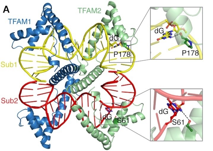 Fig. 1 Overall structure of the asymmetric unit of TFAM-NS2.1
Key structural properties of TFAM:
- Double HMG-box domains constitute the core DNA binding unit
- Flexible junction area gives conformation plasticity
- Basic amino acid rich regions mediate nonspecific DNA winding
Functions of TFAM
The core function of the TFAM gene is to regulate the replication and transcription of mitochondrial DNA. In addition, it is also involved in multiple cellular physiological activities, including maintaining genomic stability and regulating apoptosis.
| Function | Description |
| Transcriptional activation | Binding to the promoter region of mitochondrial DNA, RNA polymerases are recruited to form transcription initiation complexes. |
| Genome compression | Wrap and bend mitochondrial DNA in a sequence-independent manner, compressing it into a nucleoid structure. |
| Damage protection | Protect the fragile mitochondrial DNA from free radical attacks through physical coverage and maintain genomic integrity. |
| Apoptosis regulation | During cellular stress, the release of TFAM can act as a signaling molecule and participate in the initiation of the apoptotic signaling pathway. |
| Energy metabolism hub | By controlling the transcription of oxidative phosphorylation-related subunits, the level of cellular energy production is directly regulated. |
Unlike typical transcription factors, TFAM regulates mitochondrial gene expression in a "dose-dependent" manner - the number of its binding to DNA directly determines the level of transcriptional activity. This unique regulatory mechanism makes it a key molecular hub connecting nuclear gene expression and mitochondrial function.
Applications of TFAM and TFAM Antibody in Literature
1. Zhao, Meng, et al. "Mitochondrial ROS promote mitochondrial dysfunction and inflammation in ischemic acute kidney injury by disrupting TFAM-mediated mtDNA maintenance." Theranostics 11.4 (2021): 1845. https://doi.org/10.7150/thno.50905
The article indicates that in renal ischemia-reperfusion injury, mitochondrial reactive oxygen species inhibit the expression of the transcription factor TFAM and promote its degradation, leading to mitochondrial DNA damage, energy metabolism disorders and intensified inflammatory responses. TFAM deficiency is a key mechanism promoting kidney injury.
2. Kozhukhar, Natalya, and Mikhail F. Alexeyev. "35 years of TFAM research: old protein, new puzzles." Biology 12.6 (2023): 823. https://doi.org/10.3390/biology12060823
The article indicates that TFAM is a key protein for the maintenance and expression of mitochondrial DNA. This article reviews its 35-year research and points out that its functions are complex and diverse, and some of the conclusions are even contradictory. The latest structural research has brought new insights and raised new questions, which urgently need in-depth exploration to promote the development of the field.
3. Chen, Sisi, et al. "Depressed TFAM promotes acetaminophen-induced hepatotoxicity regulated by DDX3X–PGC1α–NRF2 signaling pathway." Molecular Medicine 30.1 (2024): 246. https://doi.org/10.1186/s10020-024-01017-0
The article indicates that in drug-induced liver injury, DDX3X inhibits the expression of TFAM through the PGC-1α/NRF-2 signaling pathway. The absence of TFAM leads to mitochondrial dysfunction, while increasing the level of TFAM can effectively alleviate liver injury, revealing its key protective effect.
4. Del Rey, Manuel J., et al. "TFAM-deficient mouse skin fibroblasts–an ex vivo model of mitochondrial dysfunction." Disease Models & Mechanisms 14.8 (2021): dmm048995. https://doi.org/10.1242/dmm.048995
Researchers specifically knocked out TFAM in skin fibroblasts and successfully established a mitochondrial dysfunction model. TFAM deficiency leads to impaired mtDNA expression, reduction of respiratory chain complexes and mitochondrial failure, and triggers cellular senescence and pro-inflammatory phenotypes.
5. Bruno, Giuseppina, et al. "TRAP1 modulates mitochondrial biogenesis via PGC-1α/TFAM signalling pathway in colorectal cancer cells." Journal of Molecular Medicine 102.10 (2024): 1285-1296. https://doi.org/10.1007/s00109-024-02479-9
The article indicates that in colorectal cancer, the molecular chaperone TRAP1 negatively regulates mitochondrial biosynthesis by inhibiting the PGC-1α/TFAM signaling pathway. Silencing TRAP1 can enhance mitochondrial quality and DNA copy number, promoting the transformation of cellular metabolism to oxidative phosphorylation.
Creative Biolabs: TFAM Antibodies for Research
Creative Biolabs specializes in the production of high-quality TFAM antibodies for research and industrial applications. Our portfolio includes monoclonal antibodies tailored for ELISA, Flow Cytometry, Western blot, immunohistochemistry, and other diagnostic methodologies.
- Custom TFAM Antibody Development: Tailor-made solutions to meet specific research requirements.
- Bulk Production: Large-scale antibody manufacturing for industry partners.
- Technical Support: Expert consultation for protocol optimization and troubleshooting.
- Aliquoting Services: Conveniently sized aliquots for long-term storage and consistent experimental outcomes.
For more details on our TFAM antibodies, custom preparations, or technical support, contact us at email.
Reference
- Choi, Woo Suk, and Miguel Garcia-Diaz. "A minimal motif for sequence recognition by mitochondrial transcription factor A (TFAM)." Nucleic Acids Research 50.1 (2022): 322-332. https://doi.org/10.1093/nar/gkab1230
Anti-TFAM antibodies
 Loading...


Hot products 
-
Mouse Anti-C1QC Recombinant Antibody (CBFYC-0600) (CBMAB-C0654-FY)

-
Mouse Anti-AKR1C3 Recombinant Antibody (V2-12560) (CBMAB-1050-CN)

-
Mouse Anti-BBS2 Recombinant Antibody (CBYY-0253) (CBMAB-0254-YY)

-
Mouse Anti-CD164 Recombinant Antibody (CBFYC-0077) (CBMAB-C0086-FY)

-
Mouse Anti-CD24 Recombinant Antibody (ALB9) (CBMAB-0176CQ)

-
Mouse Anti-CD83 Recombinant Antibody (HB15) (CBMAB-C1765-CQ)

-
Rat Anti-EMCN Recombinant Antibody (28) (CBMAB-E0280-FY)

-
Mouse Anti-CD46 Recombinant Antibody (CBFYC-0076) (CBMAB-C0085-FY)

-
Mouse Anti-CDK7 Recombinant Antibody (CBYY-C1783) (CBMAB-C3221-YY)

-
Mouse Anti-AKT1/AKT2/AKT3 (Phosphorylated T308, T309, T305) Recombinant Antibody (V2-443454) (PTM-CBMAB-0030YC)

-
Mouse Anti-ALB Recombinant Antibody (V2-55272) (CBMAB-H0819-FY)

-
Mouse Anti-CARD11 Recombinant Antibody (CBFYC-0811) (CBMAB-C0866-FY)

-
Mouse Anti-CCT6A/B Recombinant Antibody (CBXC-0168) (CBMAB-C5570-CQ)

-
Mouse Anti-ARIH1 Recombinant Antibody (C-7) (CBMAB-A3563-YC)

-
Mouse Anti-CALR Recombinant Antibody (CBFYC-0763) (CBMAB-C0818-FY)

-
Mouse Anti-CTNND1 Recombinant Antibody (CBFYC-2414) (CBMAB-C2487-FY)

-
Mouse Anti-BRD3 Recombinant Antibody (CBYY-0801) (CBMAB-0804-YY)

-
Mouse Anti-GFAP Recombinant Antibody (5) (CBMAB-G0346-LY)

-
Mouse Anti-GDF5 Recombinant Antibody (1F4) (CBMAB-G2740-LY)

-
Mouse Anti-AAV9 Recombinant Antibody (V2-634029) (CBMAB-AP023LY)

- AActivation
- AGAgonist
- APApoptosis
- BBlocking
- BABioassay
- BIBioimaging
- CImmunohistochemistry-Frozen Sections
- CIChromatin Immunoprecipitation
- CTCytotoxicity
- CSCostimulation
- DDepletion
- DBDot Blot
- EELISA
- ECELISA(Cap)
- EDELISA(Det)
- ESELISpot
- EMElectron Microscopy
- FFlow Cytometry
- FNFunction Assay
- GSGel Supershift
- IInhibition
- IAEnzyme Immunoassay
- ICImmunocytochemistry
- IDImmunodiffusion
- IEImmunoelectrophoresis
- IFImmunofluorescence
- IGImmunochromatography
- IHImmunohistochemistry
- IMImmunomicroscopy
- IOImmunoassay
- IPImmunoprecipitation
- ISIntracellular Staining for Flow Cytometry
- LALuminex Assay
- LFLateral Flow Immunoassay
- MMicroarray
- MCMass Cytometry/CyTOF
- MDMeDIP
- MSElectrophoretic Mobility Shift Assay
- NNeutralization
- PImmunohistologyp-Paraffin Sections
- PAPeptide Array
- PEPeptide ELISA
- PLProximity Ligation Assay
- RRadioimmunoassay
- SStimulation
- SESandwich ELISA
- SHIn situ hybridization
- TCTissue Culture
- WBWestern Blot
