
TSHR Antibodies
Background
The TSHR gene encodes a G protein-coupled receptor, which is mainly distributed on the surface of thyroid cell membranes. The protein expressed by this gene regulates the growth and hormone synthesis function of thyroid cells by specifically binding to thyroid stimulating hormone and activating the cAMP signaling pathway. As a core regulatory factor of thyroid function, mutations in the TSHR gene can lead to various diseases: activating mutations cause autonomous functional nodules in the thyroid, while inactivating mutations result in thyroid dysplasia or hormone resistance syndrome. This gene was successfully cloned in 1989. The analysis of its molecular mechanism not only revealed the occurrence mechanism of thyroid diseases but also promoted the clinical application of antibody detection technology targeting TSHR and receptor antagonists, providing a molecular basis for the precise diagnosis and treatment of autoimmune thyroid diseases such as Graves' disease.
Structure of TSHR
TSHR is a G protein-coupled receptor with a molecular weight of approximately 85-90 kDa. This value may fluctuate due to the degree of glycosylation modification and species differences.
| Species | Human | Mouse | Rat | Pig |
| Molecular Weight (kDa) | 87.5 | 86.2 | 86.8 | 88.1 |
| Primary Structural Differences | Typical structure with 764 amino acids | There is a variation at the N-glycosylation site | Extracellular region sequence is highly conserved | Across the membrane area and primate highly homologous |
This receptor is composed of 764 amino acids, and its polypeptide chain forms a characteristic seven-transmembrane topological structure. The most notable structural feature of TSHR lies in its possession of a vast extracellular domain (approximately 400 amino acids), which maintains conformational stability through multiple disulfide bonds and is specifically responsible for the specific recognition and binding to thyroid stimulating hormone. The transmembrane domain constitutes the core functional unit of the receptor, among which the conformational change of the sixth transmembrane helix plays a key role in the activation of G proteins. The intracellular domain contains specific sites coupled with Gs proteins. When the receptor is activated, conformational rearrangement occurs in this region, thereby triggering the downstream cAMP signaling cascade reaction.
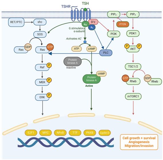 Fig. 1 Signaling pathways mediated by TSHR.1
Key structural properties of TSHR:
- Typical conformation of the seven-fold transmembrane helix topology
- Large extracellular domains form hormone-binding pockets
- Specific disulfide bond networks maintain receptor stability
Functions of TSHR
The core function of the protein encoded by the TSHR gene is to mediate thyroid stimulating hormone signal transduction. Its specific functional mechanisms include:
| Function | Description |
| Hormone signal recognition | Structure of extracellular domain specificity identification combined with thyroid stimulating hormone, hormone receptor complexes. |
| cAMP signal activation | The activation of adenylate cyclase through Gs protein promotes the large-scale production of cAMP. |
| Regulation of thyroid growth | Continuous signal stimulation guides the proliferation and differentiation of thyroid follicular epithelial cells. |
| Hormone synthesis promotion | Up-regulate the expression of key enzymes such as thyroid peroxidase and accelerate the synthesis of thyroid hormones. |
| Regulation of receptor activity | Negative feedback regulation of signal pathways is achieved through internalization mechanisms and phosphorylation. |
This receptor exhibits dual characteristics of basal activity and hormonal activation, and its signal intensity is jointly regulated by the G protein-coupling state and the recruitment of Arrestin protein. Some mutants can cause persistent activation of the receptor conformation, and even without hormone binding, they can trigger autonomic hyperfunction.
Applications of TSHR and TSHR Antibody in Literature
1. Sun, Weihua, et al. "Correlation of TSHR and CTLA‐4 Single Nucleotide Polymorphisms with Graves Disease." International journal of genomics 2019.1 (2019): 6982623. https://doi.org/10.1155/2019/6982623
This study explored the association between the polymorphisms of TSHR and CTLA-4 genes and Graves' disease. Research has found that loci such as rs179247 of TSHR and specific haplotypes are significantly associated with the risk of GD, and the AAA haplotype of CTLA-4 also increases the risk of GD.
2. Cui, Xuejiao, Futao Wang, and Cong Liu. "A review of TSHR-and IGF-1R-related pathogenesis and treatment of Graves' orbitopathy." Frontiers in Immunology 14 (2023): 1062045. https://doi.org/10.3389/fimmu.2023.1062045
This study explored that the pathogenesis of Graves' ophthalmopathy is closely related to orbital fibroblasts and two target antigens, TSHR and IGF-1R. Therapies for TSHR have become a research hotspot, and Teprotumumab has emerged as the latest second-line treatment drug.
3. Xu, Shaojie, et al. "TSHR in thyroid cancer: bridging biological insights to targeted strategies." European Thyroid Journal 14.4 (2025). https://doi.org/10.1530/ETJ-24-0369
This study explored that, in addition to promoting iodine absorption, TSHR plays a key role in the progression of thyroid cancer, especially providing a new potential therapeutic target for radioactive iodine-refractory differentiated thyroid cancer (RAIR-DTC).
4. Zhou, Jing, et al. "Signaling dynamics of TSHR-specific CAR-T cells revealed by FRET-based biosensors." Frontiers in Cell and Developmental Biology 10 (2022): 845319. https://doi.org/10.3389/fcell.2022.845319
The construction of CAR-T cells targeting TSHR was studied, and it was confirmed that they were effective for advanced thyroid cancer. Observing cell activation signals through FRET biosensors provides a new approach for optimizing CAR design to enhance therapeutic efficacy.
5. Klimaitė, Raimonda, et al. "The role of TSHR, PTEN and RASSF1A promoters' methylation status for non-invasive detection of papillary thyroid carcinoma." Journal of Clinical Medicine 11.16 (2022): 4917. https://doi.org/10.3390/jcm11164917
Research has found that the methylation level of the TSHR gene in the plasma and cancer tissues of patients with papillary thyroid carcinoma is significantly elevated and is related to tumor size and metastasis. It decreases after surgery and is expected to become a new minimally invasive diagnostic marker.
Creative Biolabs: TSHR Antibodies for Research
Creative Biolabs specializes in the production of high-quality TSHR antibodies for research and industrial applications. Our portfolio includes monoclonal antibodies tailored for ELISA, Flow Cytometry, Western blot, immunohistochemistry, and other diagnostic methodologies.
- Custom TSHR Antibody Development: Tailor-made solutions to meet specific research requirements.
- Bulk Production: Large-scale antibody manufacturing for industry partners.
- Technical Support: Expert consultation for protocol optimization and troubleshooting.
- Aliquoting Services: Conveniently sized aliquots for long-term storage and consistent experimental outcomes.
For more details on our TSHR antibodies, custom preparations, or technical support, contact us at email.
Reference
- Xu, Shaojie, et al. "TSHR in thyroid cancer: bridging biological insights to targeted strategies." European Thyroid Journal 14.4 (2025). https://doi.org/10.1530/ETJ-24-0369
Anti-TSHR antibodies
 Loading...
 Compare




Hot products 
- AActivation
- AGAgonist
- APApoptosis
- BBlocking
- BABioassay
- BIBioimaging
- CImmunohistochemistry-Frozen Sections
- CIChromatin Immunoprecipitation
- CTCytotoxicity
- CSCostimulation
- DDepletion
- DBDot Blot
- EELISA
- ECELISA(Cap)
- EDELISA(Det)
- ESELISpot
- EMElectron Microscopy
- FFlow Cytometry
- FNFunction Assay
- GSGel Supershift
- IInhibition
- IAEnzyme Immunoassay
- ICImmunocytochemistry
- IDImmunodiffusion
- IEImmunoelectrophoresis
- IFImmunofluorescence
- IGImmunochromatography
- IHImmunohistochemistry
- IMImmunomicroscopy
- IOImmunoassay
- IPImmunoprecipitation
- ISIntracellular Staining for Flow Cytometry
- LALuminex Assay
- LFLateral Flow Immunoassay
- MMicroarray
- MCMass Cytometry/CyTOF
- MDMeDIP
- MSElectrophoretic Mobility Shift Assay
- NNeutralization
- PImmunohistologyp-Paraffin Sections
- PAPeptide Array
- PEPeptide ELISA
- PLProximity Ligation Assay
- RRadioimmunoassay
- SStimulation
- SESandwich ELISA
- SHIn situ hybridization
- TCTissue Culture
- WBWestern Blot
