CCR8 Antibodies
Background
The CCR8 gene encodes a cell membrane protein belonging to the G protein-coupled receptor family, which is mainly expressed on the surface of T lymphocytes, especially regulatory T cells. This gene participates in the regulation of immune cell migration and inflammatory responses by binding to its ligands such as CCL1 and chemokines, and at the same time mediates the immune escape mechanism in the tumor microenvironment. In 1996, scientists first discovered CCR8 in activated T cells, and its gene is located in the 3p22 region of the human chromosome. As a key molecule in immune regulation, CCR8 has become an important target for the treatment of autoimmune diseases and tumor immunotherapy. Its specific expression characteristics are being widely applied in the field of targeted drug research and development.
Structure of CCR8
The CCR8 gene encodes a G protein-coupled receptor with a molecular weight of approximately 41 kDa. This molecular weight shows subtle differences among different mammals, mainly due to interspecific variations in the amino acid sequences of the extracellular region.
| Species | Human | Mouse | Rat | Rhesus monkey |
| Molecular Weight (kDa) | 41.2 | 40.8 | 40.9 | 41.1 |
| Primary Structural Differences | The seven across a membrane structure with 367 amino acids | There is a variation at the N-terminal glycosylation site | The second extracellular loop sequence is species-specific | Highly homologous to the human receptor sequence |
The CCR8 protein is composed of 367 amino acids and forms a typical seven-transmembrane topological structure. Its N-terminal region contains conserved glycosylation sites, and the third intracellular loop is responsible for G protein-coupled signal transduction. The second extracellular loop constitutes the chemokine binding pocket, and the key aspartic acid residue directly participates in ligand recognition. The activation mechanism of receptors involves conformational rearrangement of transmembrane helices, thereby triggering downstream signaling pathways.
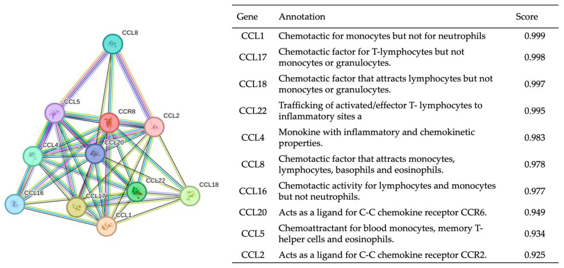 Fig. 1 Protein–protein interaction with CCR8.1
Key structural properties of CCR8:
- Typical configuration of the seven-fold transmembrane helix topology
- Conserved asparty-tyrosine structural motifs are used for signal transduction
- The second extracellular loop forms chemokine-specific binding pockets
Functions of CCR8
The core function of the CCR8 gene is to regulate the migration of immune cells and inflammatory responses, and it also participates in the immune regulation process in the tumor microenvironment.
| Function | Description |
| Chemotaxis of immune cells | Guide T lymphocytes to migrate directionally to the inflammatory site by recognizing chemokines such as CCL1. |
| Maintenance of immune tolerance | Promote the aggregation and immunosuppressive function of regulatory T cells in the tumor microenvironment. |
| Regulation of inflammatory response | Mediates Th2-type immune responses and participates in the pathological processes of allergic diseases and parasitic infections. |
| Angiogenic influence | Through indirect signaling pathways involved in the formation of tumor associated angiogenesis regulation. |
| Targeted therapy for diseases | As an immune checkpoint molecule, it provides new targets for the treatment of autoimmune diseases and tumors. |
The ligand binding curve of CCR8 exhibits typical GPCR characteristics. Its high affinity binding to CCL1 enables it to initiate signal transduction even in a low-concentration chemokine environment, a feature that differs from the broad-spectrum binding patterns of other chemokine receptors.
Applications of CCR8 and CCR8 Antibody in Literature
1. Van Damme, Helena, et al. "Therapeutic depletion of CCR8+ tumor-infiltrating regulatory T cells elicits antitumor immunity and synergizes with anti-PD-1 therapy." Journal for immunotherapy of cancer 9.2 (2021): e001749. https://doi.org/10.1136/jitc-2020-001749
This study found that CCR8 is a specific marker for tumor-infiltrating Treg cells. By constructing CCR8-targeted nanobodies, Treg cells within tumors can be specifically eliminated under the mediation of NK cells, and in combination with anti-PD-1 therapy, tumor growth can be inhibited without affecting peripheral Treg cells, demonstrating good therapeutic effects and safety.
2. Sun, Ting, et al. "Oxamate enhances the efficacy of CAR-T therapy against glioblastoma via suppressing ectonucleotidases and CCR8 lactylation." Journal of Experimental & Clinical Cancer Research 42.1 (2023): 253. https://doi.org/10.1186/s13046-023-02815-w
Research has found that lactic acid produced by tumor cell metabolism directly upregulates the expression of immunosuppressive genes such as CCR8 by inducing histone H3K18 lacylation. This promoted the increase of tumor-infiltrating Treg cells during CAR-T treatment, revealing that the lactate-CCR8 axis is a key mechanism for shaping the immunosuppressive microenvironment.
3. Korbecki, Jan, et al. "CC chemokines in a tumor: a review of pro-cancer and anti-cancer properties of receptors CCR5, CCR6, CCR7, CCR8, CCR9, and CCR10 ligands." International journal of molecular sciences 21.20 (2020): 7619. https://doi.org/10.3390/ijms21207619
Research has found that CCR8 is one of the chemokine receptors for CC, and its ligand is CCL1. In tumors, CCR8 not only affects the proliferation and migration of cancer cells but also promotes tumor progression by recruiting regulatory T cells and other immune cells, shaping an immunosuppressive microenvironment. It is an important potential target for cancer treatment.
4. Tian, Binle, et al. "CCR8 antagonist suppresses liver cancer progression via turning tumor-infiltrating Tregs into less immunosuppressive phenotype." Journal of Experimental & Clinical Cancer Research 44.1 (2025): 113. https://doi.org/10.1186/s13046-025-03286-x
This study confirmed that CCR8 is highly expressed in Treg cells infiltrating liver cancer. The use of the self-developed CCR8 antagonist IPG0521m can effectively weaken the immunosuppressive function of Treg cells, thereby activating the anti-tumor activity of CD8+ T cells and NK cells, significantly inhibiting the growth of liver cancer, and providing a new basis for tumor treatment targeting CCR8.
5. Moser, Bernhard. "Chemokine receptor-targeted therapies: special case for CCR8." Cancers 14.3 (2022): 511. https://doi.org/10.3390/cancers14030511
This study indicates that CCR8, as a chemokine receptor highly expressed on tumor-suppressive Treg cells, is a highly promising new target for cancer treatment. Targeting CCR8 can specifically eliminate immunosuppressive cells within tumors, thereby enhancing anti-tumor immunity and is expected to improve the efficacy and safety of existing immune checkpoint treatments.
Creative Biolabs: CCR8 Antibodies for Research
Creative Biolabs specializes in the production of high-quality CCR8 antibodies for research and industrial applications. Our portfolio includes monoclonal antibodies tailored for ELISA, Flow Cytometry, Western blot, immunohistochemistry, and other diagnostic methodologies.
- Custom CCR8 Antibody Development: Tailor-made solutions to meet specific research requirements.
- Bulk Production: Large-scale antibody manufacturing for industry partners.
- Technical Support: Expert consultation for protocol optimization and troubleshooting.
- Aliquoting Services: Conveniently sized aliquots for long-term storage and consistent experimental outcomes.
For more details on our CCR8 antibodies, custom preparations, or technical support, contact us at email.
Reference
- Kim, Nari, et al. "CCR8 as a therapeutic novel target: omics-integrated comprehensive analysis for systematically prioritizing indications." Biomedicines 11.11 (2023): 2910. https://doi.org/10.3390/biomedicines11112910
Anti-CCR8 antibodies
 Loading...


Hot products 
-
Rat Anti-CD300A Recombinant Antibody (172224) (CBMAB-C0423-LY)

-
Mouse Anti-CCS Recombinant Antibody (CBFYC-1093) (CBMAB-C1150-FY)

-
Mouse Anti-AKT1 (Phosphorylated S473) Recombinant Antibody (V2-505430) (PTM-CBMAB-0067LY)

-
Mouse Anti-CAPZB Recombinant Antibody (CBYY-C0944) (CBMAB-C2381-YY)

-
Mouse Anti-BCL6 Recombinant Antibody (CBYY-0442) (CBMAB-0445-YY)

-
Rabbit Anti-Acetyl-Histone H4 (Lys16) Recombinant Antibody (V2-623415) (CBMAB-CP1021-LY)

-
Mouse Anti-CARTPT Recombinant Antibody (113612) (CBMAB-C2450-LY)

-
Mouse Anti-CFL1 (Phospho-Ser3) Recombinant Antibody (CBFYC-1770) (CBMAB-C1832-FY)

-
Rabbit Anti-CAMK2A Recombinant Antibody (BA0032) (CBMAB-0137CQ)

-
Mouse Anti-ADIPOR2 Recombinant Antibody (V2-179983) (CBMAB-A1369-YC)

-
Rat Anti-4-1BB Recombinant Antibody (V2-1558) (CBMAB-0953-LY)

-
Mouse Anti-ARSA Recombinant Antibody (CBYC-A799) (CBMAB-A3679-YC)

-
Mouse Anti-AAV9 Recombinant Antibody (V2-634029) (CBMAB-AP023LY)

-
Mouse Anti-FPR2 Recombinant Antibody (1D6) (CBMAB-F2628-CQ)

-
Mouse Anti-ADAM29 Recombinant Antibody (V2-179787) (CBMAB-A1149-YC)

-
Rabbit Anti-Acetyl-Histone H3 (Lys36) Recombinant Antibody (V2-623395) (CBMAB-CP0994-LY)

-
Mouse Anti-ALB Recombinant Antibody (V2-55272) (CBMAB-H0819-FY)

-
Mouse Anti-CALR Recombinant Antibody (CBFYC-0763) (CBMAB-C0818-FY)

-
Mouse Anti-CD63 Recombinant Antibody (CBXC-1200) (CBMAB-C1467-CQ)

-
Rabbit Anti-ATF4 Recombinant Antibody (D4B8) (CBMAB-A3872-YC)

- AActivation
- AGAgonist
- APApoptosis
- BBlocking
- BABioassay
- BIBioimaging
- CImmunohistochemistry-Frozen Sections
- CIChromatin Immunoprecipitation
- CTCytotoxicity
- CSCostimulation
- DDepletion
- DBDot Blot
- EELISA
- ECELISA(Cap)
- EDELISA(Det)
- ESELISpot
- EMElectron Microscopy
- FFlow Cytometry
- FNFunction Assay
- GSGel Supershift
- IInhibition
- IAEnzyme Immunoassay
- ICImmunocytochemistry
- IDImmunodiffusion
- IEImmunoelectrophoresis
- IFImmunofluorescence
- IGImmunochromatography
- IHImmunohistochemistry
- IMImmunomicroscopy
- IOImmunoassay
- IPImmunoprecipitation
- ISIntracellular Staining for Flow Cytometry
- LALuminex Assay
- LFLateral Flow Immunoassay
- MMicroarray
- MCMass Cytometry/CyTOF
- MDMeDIP
- MSElectrophoretic Mobility Shift Assay
- NNeutralization
- PImmunohistologyp-Paraffin Sections
- PAPeptide Array
- PEPeptide ELISA
- PLProximity Ligation Assay
- RRadioimmunoassay
- SStimulation
- SESandwich ELISA
- SHIn situ hybridization
- TCTissue Culture
- WBWestern Blot
