CD96 Antibodies
Background
CD96 is a type I transmembrane glycoprotein that is mainly expressed on the surface of immune cells (such as T cells and NK cells). This protein participates in the regulation of immune responses by mediating cell adhesion. It can bind to the ligand CD155 to promote the activation of immune cells, and can also transduce inhibitory signals under specific conditions to maintain immune homeostasis. Its function was first discovered in the 1990s through the screening of monoclonal antibodies. Subsequent studies have revealed that CD96 has a dual regulatory role in tumor immunity, infection defense, and autoimmune diseases. As a member of the immune checkpoint protein family, the structural characteristics and signaling mechanisms of this molecule have become important research directions in the field of immunotherapy, providing potential targets for the development of new tumor immunotherapies.
Structure of CD96
CD96 is a type I transmembrane glycoprotein with a molecular weight of approximately 70 kDa. Its actual measured value may fluctuate within the range of 65-75 kDa due to glycosylation modifications and different splicing variants.
| Species | Human | Mouse | Rat |
| Molecular Weight (kDa) | ~70 | ~70 | ~70 |
| Primary Structural Differences | Containing 3 extracellular Ig-like domains (V-V-C2) and intracellular ITIM motif | Highly similar to humans, with similar ligand binding characteristics | The core structure is conserved, with specific sequence variations at the species level. |
This protein is composed of approximately 580 amino acid residues and exhibits a typical immunoglobulin superfamily folding conformation. Its core function relies on the three immunoglobulin-like domains in the extracellular segment, which together form a ligand (such as CD155) binding pocket. Its secondary structure is mainly composed of antiparallel β-sheet, forming a stable "sandwich" structure. The FG ring region within the first V-type domain is the key functional site. The conformation and specific amino acid residues (such as charged lysine and aspartic acid) in this ring directly mediate the specific interaction with the ligand, thereby regulating the adhesion and signal transduction between T cells and NK cells.
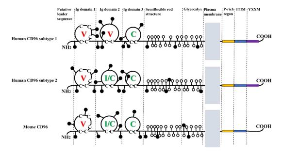 Fig. 1 Molecular structure of CD96.1
Key structural properties of CD96:
- Typical folded structure of the immunoglobulin superfamily
- Extracellular section is made up of three immune globulin structure domain (V - V - type C2) in series
- Contains a conserved ligand binding interface (located in the FG ring region of the V-shaped domain)
- Intracellular segment containing the immunoreceptor tyrosine inhibitory motif (ITIM)
Functions of CD96
The core function of CD96 is to act as an immune checkpoint molecule, regulating the activation and inhibition of T cells and NK cells. However, it is also involved in various immune-related processes, including tumor immune surveillance, infection defense, and autoimmune pathology.
| Function | Description |
| Immunological Costimulation | After binding to the ligand CD155, it can transmit activation signals under certain circumstances (such as in naive T cells), promoting cell proliferation and cytokine production. |
| Immune Co-Inhibition | In effector T cells or within the tumor microenvironment, it mainly transmits inhibitory signals, downregulating the immune response through its intracellular ITIM domain and maintaining self-tolerance. |
| Intercellular Adhesion | It mediates the specific adhesion between immune cells and target cells expressing CD155 (such as tumor cells and virus-infected cells). |
| Tumor Immune Evasion | In the tumor microenvironment, the inhibitory signaling pathway of CD96 is overutilized, leading to the exhaustion of T cells/NK cells' functions and promoting immune evasion. |
| Immune Regulation of Infection | During the infection by viruses (such as cytomegalovirus), the killing activity of NK cells is regulated to achieve a balance between immune clearance and tissue damage. |
The affinity of CD96 for its ligand CD155 is significantly higher than that of other co-receptors (such as CD226 and TIGIT). However, its signal output depends on cell type, activation status, and microenvironment, presenting a unique "context-dependent" feature, which enables it to play a precise dual regulatory role in immune homeostasis.
Applications of CD96 and CD96 Antibody in Literature
1. Feng, Shikai, et al. "CD96 as a potential immune regulator in cancers." International Journal of Molecular Sciences 24.2 (2023): 1303. https://doi.org/10.3390/ijms24021303
The article indicates that CD96, as a member of the NECTIN receptor family, is an emerging target for tumor immunotherapy. This article reviews its basic biological characteristics and research progress, and explores its potential application as an immune checkpoint inhibitor.
2. Georgiev, Hristo, et al. "Coming of age: CD96 emerges as modulator of immune responses." Frontiers in immunology 9 (2018): 1072. https://doi.org/10.3389/fimmu.2018.01072
The article indicates that CD96 is an immunoglobulin superfamily protein expressed on T/NK cells. Its binding to CD155 can inhibit the function of mouse NK cells. The mechanism of action is not yet fully understood, and there may be differences in function between human and mouse sources. Further exploration is needed within the CD155/TIGIT/CD226 regulatory network.
3. Li, Jiang, et al. "Tumor Cell‐Intrinsic CD96 Mediates Chemoresistance and Cancer Stemness by Regulating Mitochondrial Fatty Acid β‐Oxidation." Advanced Science 10.7 (2023): 2202956. https://doi.org/10.1002/advs.202202956
The research has found that the CD96 expressed by breast cancer cells is associated with poor prognosis of patients. It enhances mitochondrial fatty acid oxidation through the CD155-Src-Stat3-Opa1 pathway and promotes the drug resistance of tumor stem cells. Targeting the intrinsic CD96 of tumor cells can enhance the efficacy of chemotherapy.
4. Zhou, Wangying, Xiaobin Cai, and Feng Liu. "CD96 as a potential diagnostic biomarker and new target for skin cutaneous melanoma." Contrast Media & Molecular Imaging 2022.1 (2022): 6409376. https://doi.org/10.1155/2022/6409376
The research has found that CD96 is significantly overexpressed in skin melanoma, presenting a stark contrast to normal tissues. It influences the tumor progression by regulating pathways such as JAK-STAT and PI3K-Akt, and can potentially serve as a novel biomarker and therapeutic target.
5. Ye, Wenrui, et al. "CD96 correlates with immune infiltration and impacts patient prognosis: a pan-cancer analysis." Frontiers in oncology 11 (2021): 634617. https://doi.org/10.3389/fonc.2021.634617
The study found that CD96 shows significant differences in expression across various cancers and affects patient prognosis. In LGG, its high expression is associated with poor survival, while in SKCM, the opposite is true. It is widely associated with the expression of immune checkpoints and the infiltration levels of various immune cells, and is a potential biomarker for prognosis and immune infiltration.
Creative Biolabs: CD96 Antibodies for Research
Creative Biolabs specializes in the production of high-quality CD96 antibodies for research and industrial applications. Our portfolio includes monoclonal antibodies tailored for ELISA, Flow Cytometry, Western blot, immunohistochemistry, and other diagnostic methodologies.
- Custom CD96 Antibody Development: Tailor-made solutions to meet specific research requirements.
- Bulk Production: Large-scale antibody manufacturing for industry partners.
- Technical Support: Expert consultation for protocol optimization and troubleshooting.
- Aliquoting Services: Conveniently sized aliquots for long-term storage and consistent experimental outcomes.
For more details on our CD96 antibodies, custom preparations, or technical support, contact us at email.
Reference
- Feng, Shikai, et al. "CD96 as a potential immune regulator in cancers." International Journal of Molecular Sciences 24.2 (2023): 1303. https://doi.org/10.3390/ijms24021303
Anti-CD96 antibodies
 Loading...


Hot products 
-
Mouse Anti-AKR1C3 Recombinant Antibody (V2-12560) (CBMAB-1050-CN)

-
Mouse Anti-ENO1 Recombinant Antibody (CBYC-A950) (CBMAB-A4388-YC)

-
Rat Anti-FABP3 Recombinant Antibody (CBXF-2299) (CBMAB-F1612-CQ)

-
Human Anti-SARS-CoV-2 S1 Monoclonal Antibody (CBFYR-0120) (CBMAB-R0120-FY)

-
Mouse Anti-BMI1 Recombinant Antibody (CBYC-P026) (CBMAB-P0108-YC)

-
Mouse Anti-DISP2 Monoclonal Antibody (F66A4B1) (CBMAB-1112CQ)

-
Mouse Anti-ARHGDIA Recombinant Antibody (CBCNA-009) (CBMAB-R0415-CN)

-
Mouse Anti-ACVR1C Recombinant Antibody (V2-179685) (CBMAB-A1041-YC)

-
Mouse Anti-CSPG4 Recombinant Antibody (CBFYM-1050) (CBMAB-M1203-FY)

-
Rat Anti-AChR Recombinant Antibody (V2-12500) (CBMAB-0990-CN)

-
Mouse Anti-B2M Recombinant Antibody (CBYY-0050) (CBMAB-0050-YY)

-
Mouse Anti-AOC3 Recombinant Antibody (CBYY-0014) (CBMAB-0014-YY)

-
Mouse Anti-BACE1 Recombinant Antibody (CBLNB-121) (CBMAB-1180-CN)

-
Mouse Anti-AMH Recombinant Antibody (5/6) (CBMAB-A2527-YC)

-
Mouse Anti-DES Monoclonal Antibody (440) (CBMAB-AP1857LY)

-
Mouse Anti-FLI1 Recombinant Antibody (CBXF-0733) (CBMAB-F0435-CQ)

-
Mouse Anti-CD19 Recombinant Antibody (CBXC-1224) (CBMAB-C1491-CQ)

-
Mouse Anti-AMIGO2 Recombinant Antibody (CBYY-C0756) (CBMAB-C2192-YY)

-
Rabbit Anti-ALOX5AP Recombinant Antibody (CBXF-1219) (CBMAB-F0750-CQ)

-
Mouse Anti-ASTN1 Recombinant Antibody (H-9) (CBMAB-1154-CN)

- AActivation
- AGAgonist
- APApoptosis
- BBlocking
- BABioassay
- BIBioimaging
- CImmunohistochemistry-Frozen Sections
- CIChromatin Immunoprecipitation
- CTCytotoxicity
- CSCostimulation
- DDepletion
- DBDot Blot
- EELISA
- ECELISA(Cap)
- EDELISA(Det)
- ESELISpot
- EMElectron Microscopy
- FFlow Cytometry
- FNFunction Assay
- GSGel Supershift
- IInhibition
- IAEnzyme Immunoassay
- ICImmunocytochemistry
- IDImmunodiffusion
- IEImmunoelectrophoresis
- IFImmunofluorescence
- IGImmunochromatography
- IHImmunohistochemistry
- IMImmunomicroscopy
- IOImmunoassay
- IPImmunoprecipitation
- ISIntracellular Staining for Flow Cytometry
- LALuminex Assay
- LFLateral Flow Immunoassay
- MMicroarray
- MCMass Cytometry/CyTOF
- MDMeDIP
- MSElectrophoretic Mobility Shift Assay
- NNeutralization
- PImmunohistologyp-Paraffin Sections
- PAPeptide Array
- PEPeptide ELISA
- PLProximity Ligation Assay
- RRadioimmunoassay
- SStimulation
- SESandwich ELISA
- SHIn situ hybridization
- TCTissue Culture
- WBWestern Blot
