
GPX2 Antibodies
Background
The GPX2 gene encodes a selenoprotein belonging to the glutathione peroxidase family, which is mainly specifically expressed in epithelial tissues. This enzyme effectively protects cells from oxidative stress damage by catalyzing the reduction reaction of hydrogen peroxide and organic hydrogen peroxides, especially playing a core role in the antioxidant defense of the gastrointestinal mucosa. GPX2 was first identified in 1993. Due to its significant function in maintaining the stability of the intestinal environment, it was also named "gastrointestinal specific glutathione peroxidase". Its unique expression pattern and precise antioxidant mechanism provide key molecular targets for the study of cellular REDOX regulation, inflammation-related diseases and tumor development.
Structure of GPX2
The protein encoded by the GPX2 gene is a selenoprotein with a molecular weight of approximately 22kDa. This molecular weight remains highly stable in different mammals due to the conservation of the selenocysteine insertion mechanism.
| Species | Human | Mouse | Rat | Pig |
| Molecular Weight (kDa) | 22.0 | 21.9 | 22.0 | 22.1 |
| Primary Structural Differences | Selenium-containing cysteine active site | Active center highly conservative | High homology with human sequence | As the typical glutathione peroxidase domain structure |
The GPX2 protein is composed of 190 amino acid residues and exhibits a typical thioredoxin folding conformation. Its active center is a key selenocysteine residue, which directly participates in the catalytic reduction reaction of peroxides. The quaternary structure of proteins is a homotetramer, with each monomer containing a complete catalytic active site, which maintains its antioxidant function through precise spatial conformation.
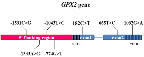 Fig. 1 Genetic information for the pig GPX2 gene.1
Key structural properties of GPX2:
- Typical thioredoxin folding architecture
- The tetramer interface forms stable active centers
- Key selenocysteine sites are responsible for catalytic reduction
- Glutathione binding domains specifically recognize substrates
Functions of GPX2
The core function of the GPX2 gene is to serve as an important component of the cellular antioxidant defense system. Its main physiological functions include:
| Function | Description |
| Peroxide removal | Specific catalytic hydrogen peroxide and the organic reduction reaction of hydrogen peroxide, prevent the generation of harmful material such as hydroxyl radicals. |
| Defense against oxidative stress | Maintain intracellular REDOX balance in rapidly renewing tissues such as gastrointestinal epithelium to protect cells from reactive oxygen species damage. |
| Intestinal barrier protection | By eliminating peroxides produced during food digestion, the integrity and functional stability of the intestinal mucosa can be maintained. |
| Inflammatory regulation | Interaction and nf-kappa B signaling pathway and regulating inflammation, prevent excessive tissue damage caused by oxidative stress. |
| Regulation of cell proliferation | Regulating the level of reactive oxygen species to influence the cell cycle process is particularly crucial in the intestinal crypt stem cell region. |
The catalytic efficiency of GPX2 for substrates exhibits typical saturation kinetics characteristics. Its specific dependence on glutathione enables it to occupy a unique position in the antioxidant network, especially playing a key role in responding to fluctuations in the intestinal environment.
Applications of GPX2 and GPX2 Antibody in Literature
1. Yang, Ming, et al. "GPX2 predicts recurrence-free survival and triggers the Wnt/β-catenin/EMT pathway in prostate cancer." PeerJ 10 (2022): e14263. https://doi.org/10.7717/peerj.14263
This study focused on the risk of prostate cancer recurrence and identified GPX2 as the core biomarker. Experiments have confirmed that GPX2 is highly expressed in cancer tissues. It induces epithelial-mesenchymal transition by activating the Wnt/β-catenin pathway and promotes the proliferation and invasion of cancer cells. Inhibiting GPX2 can reverse the above-mentioned malignant phenotype, suggesting it as a potential therapeutic target.
2. Pu, Lei, et al. "GPX2 Gene Affects Feed Efficiency of Pigs by Inhibiting Fat Deposition and Promoting Muscle Development." Animals 12.24 (2022): 3528. https://doi.org/10.3390/ani12243528
Research has found that the GPX2 gene in pigs can effectively improve feed efficiency. Duroc pigs with high expression of GPX2 have thinner backfat and higher feed conversion rate. The mechanism lies in that this gene can inhibit the generation and deposition of fat cells, while promoting the proliferation and differentiation of muscle cells, thereby achieving the effect of "reducing fat and gaining weight".
3. Lu X, Liu R, et al. "ACVRL1 drives resistance to multitarget tyrosine kinase inhibitors in colorectal cancer by promoting USP15-mediated GPX2 stabilization." BMC medicine 21.1 (2023): 366. https://doi.org/10.1186/s12916-023-03066-4
This study reveals a new mechanism of resistance to multi-target kinase inhibitors (mTKIs) in colorectal cancer. Research has found that ACVRL1 stabilizes the GPX2 protein through the deubiquitinating enzyme USP15, reduces the accumulation of reactive oxygen species (ROS) in cells and inhibits apoptosis, thereby triggering drug resistance. Targeting this pathway is expected to overcome mTKIs resistance.
4. Li, Yu-peng, et al. "The effect of GPX2 on the prognosis of lung adenocarcinoma diagnosis and proliferation, migration, and epithelial mesenchymal transition." Journal of oncology 2022.1 (2022): 7379157. https://doi.org/10.1155/2022/7379157
This study confirmed that GPX2 is highly expressed in lung adenocarcinoma and is associated with a poor prognosis for patients. Experiments show that overexpression of GPX2 can promote the proliferation, migration, invasion of A549 cells and inhibit apoptosis, while regulating the expression of EMT-related proteins. The results show that GPX2 promotes the progression of lung cancer through the EMT/β-catenin pathway and is expected to become a new target for diagnosis and treatment.
5. Yang, Xu, et al. "N6-methyladenosine-modified GPX2 impacts cancer cell stemness and TKI resistance through regulating of redox metabolism." Cell Death & Disease 16.1 (2025): 458. https://doi.org/10.1038/s41419-025-07764-0
This study reveals that GPX2 is the key factor leading to resistance to EGFR-targeted drugs in non-small cell lung cancer. It triggers drug resistance by activating the Hedgehog signaling pathway and enhancing the characteristics of tumor stem cells. Mettl14-mediated m6A modification can reduce the stability of GPX2. In vivo experiments have confirmed that inhibiting GPX2 can effectively enhance the efficacy of targeted drugs, indicating that it is a potential precision therapeutic target.
Creative Biolabs: GPX2 Antibodies for Research
Creative Biolabs specializes in the production of high-quality GPX2 antibodies for research and industrial applications. Our portfolio includes monoclonal antibodies tailored for ELISA, Flow Cytometry, Western blot, immunohistochemistry, and other diagnostic methodologies.
- Custom GPX2 Antibody Development: Tailor-made solutions to meet specific research requirements.
- Bulk Production: Large-scale antibody manufacturing for industry partners.
- Technical Support: Expert consultation for protocol optimization and troubleshooting.
- Aliquoting Services: Conveniently sized aliquots for long-term storage and consistent experimental outcomes.
For more details on our GPX2 antibodies, custom preparations, or technical support, contact us at email.
Reference
- Pu, Lei, et al. "GPX2 Gene Affects Feed Efficiency of Pigs by Inhibiting Fat Deposition and Promoting Muscle Development." Animals 12.24 (2022): 3528. https://doi.org/10.3390/ani12243528
Anti-GPX2 antibodies
 Loading...
 Compare




Hot products 
- AActivation
- AGAgonist
- APApoptosis
- BBlocking
- BABioassay
- BIBioimaging
- CImmunohistochemistry-Frozen Sections
- CIChromatin Immunoprecipitation
- CTCytotoxicity
- CSCostimulation
- DDepletion
- DBDot Blot
- EELISA
- ECELISA(Cap)
- EDELISA(Det)
- ESELISpot
- EMElectron Microscopy
- FFlow Cytometry
- FNFunction Assay
- GSGel Supershift
- IInhibition
- IAEnzyme Immunoassay
- ICImmunocytochemistry
- IDImmunodiffusion
- IEImmunoelectrophoresis
- IFImmunofluorescence
- IGImmunochromatography
- IHImmunohistochemistry
- IMImmunomicroscopy
- IOImmunoassay
- IPImmunoprecipitation
- ISIntracellular Staining for Flow Cytometry
- LALuminex Assay
- LFLateral Flow Immunoassay
- MMicroarray
- MCMass Cytometry/CyTOF
- MDMeDIP
- MSElectrophoretic Mobility Shift Assay
- NNeutralization
- PImmunohistologyp-Paraffin Sections
- PAPeptide Array
- PEPeptide ELISA
- PLProximity Ligation Assay
- RRadioimmunoassay
- SStimulation
- SESandwich ELISA
- SHIn situ hybridization
- TCTissue Culture
- WBWestern Blot
