OTOA Antibodies
Background
OTOA is a transport protein present on cell membranes, mainly distributed in tissues such as the liver, kidneys and intestines. It is responsible for mediating the transmembrane transport of various endogenous and exogenous organic anions, including bile acids, hormones and drugs, etc., and plays a key role in metabolic regulation and detoxification. The expression level of OTOA in diving mammals is relatively high, which may be related to their need to adapt to the efficient clearance of metabolic products under high-pressure conditions. This protein family was first identified by Hagenbuch and Meier in 1994. Its unique 12-transmembrane domain characteristics provide an important model for the study of membrane transport proteins. The discovery of OTOA has greatly advanced our understanding of pharmacokinetics, liver and kidney functions, and transmembrane transport mechanisms. It remains one of the important targets for drug development and disease treatment at present.
Structure of OTOA
OTOA is a transmembrane transport protein with a molecular weight of approximately 80-90 kDa, and its precise molecular weight varies slightly depending on the species and subtype.
| Species | Human (OATP1B1) | Rat (Oatp1a1) | Mouse (Oatp1a4) | Rhesus monkey (OATP1B3) |
| Molecular Weight (kDa) | ~84 | ~82 | ~83 | ~85 |
| Primary Structural Differences | 12 transmembrane domains, extracellular glycosylation sites | It is related to the liver uptake function of rats | Express significant blood brain barrier | It is highly homologous to the human OATP1B3 |
OTOA is composed of approximately 650 to 700 amino acids, and its core structural feature is 12 α-helical transmembrane domains (TM1-TM12), which form transport channels through hydrophobic interactions. Both the N-terminal and C-terminal are located intracellular, with the TM4-TM6 region forming the substrate binding pocket. Key amino acid residues (such as positively charged Arg/Lys) are responsible for recognizing organic anions. The extracellular loop 2 (ECL2) in the structure contains conserved cysteine residues, which stabilize the spatial conformation through disulfide bonds. The TMH3-TMH5 helical cluster, which is directly related to the transport function, achieves transmembrane transport of substrates through conformational changes, and its mechanism is similar to the "alternating pathway model".
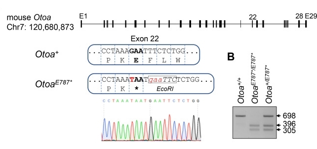 Fig. 1 Construction and functional verification of a mouse model with nonsense mutation of Otoa gene p.GL787*.1
Key structural properties of OTOA:
- 12 transmembrane α-helical domains (TM1-TM12)
- The positively charged substrate binding pocket (located in the TM4-TM6 region)
- Conserved disulfide bonds in extracellular loop 2 (ECL2)
- Key histidine/arginine residues (such as His41/Arg580)
- Intracellular ATP-binding motif (atypical)
Functions of OTOA
The core function of OTOA is to mediate the transmembrane transport of organic anions, and it also plays a key role in drug metabolism and the maintenance of homeostasis in vivo.
| Function | Description |
| Drug transport | Mediating the uptake of statins, antibiotics and other drugs by hepatocytes directly affects pharmacokinetics (such as the liver clearance rate of rosuvastatin). |
| Bile acid cycle | Participates in the reabsorption of conjugated bile acids (such as taurocholate) in enterohepatic circulation and maintains the balance of cholesterol metabolism. |
| Hormone transport | Transport thyroid hormones (T4/T3) and steroid hormone metabolites, and regulate endocrine function. |
| Detoxification function | Promoting the intrahepatic clearance of endogenous toxins such as bilirubin and prostaglandins, the serum bilirubin level is negatively correlated with OTOA activity. |
| Pathological regulation | Gene polymorphisms (such as SLCO1B1*5) lead to transport function defects and are directly related to simvastatin myotoxicity and hyperbilirubinemia. |
The transport kinetics of OTOA exhibit the characteristics of the subsubstrate dependent Mie equation. Unlike the synergistic binding of hemoglobin, its transport efficiency depends on the membrane potential and pH gradient (ΔpH). This electrically neutral exchange mechanism is particularly suitable for the physiological environment of the hepatic sinusoidal space. The special expression pattern of OTOA subtypes in the liver of diving mammals may be related to the adaptive evolution of metabolite clearance under high-pressure conditions.
Applications of OTOA and OTOA Antibody in Literature
1. Sugiyama, Kenjiro, et al. "Mid-frequency hearing loss is characteristic clinical feature of OTOA-associated hearing loss." Genes 10.9 (2019): 715. https://doi.org/10.3390/genes10090715
This study for the first time found that copy number variations (CNVs) of the OTOA gene accounted for 0.6%(14 cases) among 2,262 Japanese patients with autosomal recessive deafness, among which 7 cases were diagnosed as OTOA-related deafness (0.3%). The clinical feature of mid-frequency hearing loss in patients with deafness caused by OTOA was reported for the first time.
2. Ortore, Rocco Pio, et al. "Compound heterozygosity for OTOA truncating variant and genomic rearrangement cause autosomal recessive sensorineural hearing loss in an Italian family." Audiology Research 11.3 (2021): 443-451. https://doi.org/10.3390/audiolres11030041
In this study, A brother and sister in Italy suffered from moderate to severe sensorineural deafness. Genetic testing revealed compound heterozygous mutations in the OTOA gene: a de novo nonsense mutation from the mother (c.2223G>A) and a deletion of approximately 150Kb from the father. Studies have confirmed that OTOA is an important pathogenic gene for deafness in common cryptoafrican syndrome, and a diagnosis needs to be made by combining sequencing and genomic analysis.
3. Danquah, Bright D., et al. "Mass Spectrometric analysis of antibody—Epitope peptide complex dissociation: Theoretical concept and practical procedure of binding strength characterization." Molecules 25.20 (2020): 4776.https://doi.org/10.3390/ijms252413471
This study found that homozygous deletion of the OTOA gene on chromosome 16 led to sensorineural deafness in the children of a consanguineous couple, and at the same time, there was a novel homozygous missense variation of the ERCC4 gene. The research emphasizes that whole exome sequencing needs to be combined with deep phenotypic analysis to improve diagnostic accuracy.
4. Kim, Ju Ang, et al. "In vivo consequences of varying degrees of OTOA alteration elucidated using knock-in mouse models and pseudogene contamination-free long-read sequencing." Genes & Diseases 12.3 (2025): 101533.https://doi.org/10.1016/j.gendis.2025.101533
This study, by constructing an Otoa gene knock-in mouse model, confirmed that the C-terminal GPI anchoring site of the OTOA protein is crucial for maintaining the attachment of the cochlear cap membrane. The P.GLul787 * variant discovered by short-read sequencing was actually a pseudogene (OTOAP1) sequence. Whole-genome sequencing of the patient revealed an inversion variation at the 3' end of OTOA, revealing the limitations of short-read sequencing.
5. Kaneda, Toshihiko, et al. "Massive digital gene expression analysis reveals different predictive profiles for immune checkpoint inhibitor therapy between adenocarcinoma and squamous cell carcinoma of advanced lung cancer." BMC cancer 22.1 (2022): 154.https://doi.org/10.1186/s12885-022-09264-2
This study found through RNA expression analysis that the expression of the OTOA gene was enhanced in patients with lung adenocarcinoma who responded to nivolumab treatment. The study revealed the differentiated gene prediction patterns of anti-PD-1 antibody treatment for non-small cell lung cancer of different histological types.
Creative Biolabs: OTOA Antibodies for Research
Creative Biolabs specializes in the production of high-quality OTOA antibodies for research and industrial applications. Our portfolio includes monoclonal antibodies tailored for ELISA, Flow Cytometry, Western blot, immunohistochemistry, and other diagnostic methodologies.
- Custom OTOA Antibody Development: Tailor-made solutions to meet specific research requirements.
- Bulk Production: Large-scale antibody manufacturing for industry partners.
- Technical Support: Expert consultation for protocol optimization and troubleshooting.
- Aliquoting Services: Conveniently sized aliquots for long-term storage and consistent experimental outcomes.
For more details on our OTOA antibodies, custom preparations, or technical support, contact us at email.
Reference
- Kim, Ju Ang, et al. "In vivo consequences of varying degrees of OTOA alteration elucidated using knock-in mouse models and pseudogene contamination-free long-read sequencing." Genes & Diseases 12.3 (2025): 101533.https://doi.org/10.1016/j.gendis.2025.101533
Anti-OTOA antibodies
 Loading...


Hot products 
-
Mouse Anti-AAV8 Recombinant Antibody (V2-634028) (CBMAB-AP022LY)

-
Mouse Anti-C5b-9 Recombinant Antibody (aE11) (CBMAB-AO138LY)

-
Mouse Anti-CD24 Recombinant Antibody (2Q1282) (CBMAB-C1624-CN)

-
Rat Anti-CD63 Recombinant Antibody (7G4.2E8) (CBMAB-C8725-LY)

-
Mouse Anti-CTNND1 Recombinant Antibody (CBFYC-2414) (CBMAB-C2487-FY)

-
Mouse Anti-DISP2 Monoclonal Antibody (F66A4B1) (CBMAB-1112CQ)

-
Mouse Anti-CD164 Recombinant Antibody (CBFYC-0077) (CBMAB-C0086-FY)

-
Mouse Anti-AMOT Recombinant Antibody (CBYC-A564) (CBMAB-A2552-YC)

-
Human Anti-SARS-CoV-2 Spike Recombinant Antibody (CBC05) (CBMAB-CR005LY)

-
Mouse Anti-DHFR Recombinant Antibody (D0821) (CBMAB-D0821-YC)

-
Rat Anti-C5AR1 Recombinant Antibody (8D6) (CBMAB-C9139-LY)

-
Armenian hamster Anti-CD40 Recombinant Antibody (HM40-3) (CBMAB-C10365-LY)

-
Mouse Anti-CSPG4 Recombinant Antibody (CBFYM-1050) (CBMAB-M1203-FY)

-
Mouse Anti-ADGRL2 Recombinant Antibody (V2-58519) (CBMAB-L0166-YJ)

-
Mouse Anti-CCT6A/B Recombinant Antibody (CBXC-0168) (CBMAB-C5570-CQ)

-
Mouse Anti-CD8 Recombinant Antibody (C1083) (CBMAB-C1083-LY)

-
Mouse Anti-AOC3 Recombinant Antibody (CBYY-0014) (CBMAB-0014-YY)

-
Mouse Anti-ARHGDIA Recombinant Antibody (CBCNA-009) (CBMAB-R0415-CN)

-
Mouse Anti-FPR2 Recombinant Antibody (1D6) (CBMAB-F2628-CQ)

-
Mouse Anti-CAT Recombinant Antibody (724810) (CBMAB-C8431-LY)

- AActivation
- AGAgonist
- APApoptosis
- BBlocking
- BABioassay
- BIBioimaging
- CImmunohistochemistry-Frozen Sections
- CIChromatin Immunoprecipitation
- CTCytotoxicity
- CSCostimulation
- DDepletion
- DBDot Blot
- EELISA
- ECELISA(Cap)
- EDELISA(Det)
- ESELISpot
- EMElectron Microscopy
- FFlow Cytometry
- FNFunction Assay
- GSGel Supershift
- IInhibition
- IAEnzyme Immunoassay
- ICImmunocytochemistry
- IDImmunodiffusion
- IEImmunoelectrophoresis
- IFImmunofluorescence
- IGImmunochromatography
- IHImmunohistochemistry
- IMImmunomicroscopy
- IOImmunoassay
- IPImmunoprecipitation
- ISIntracellular Staining for Flow Cytometry
- LALuminex Assay
- LFLateral Flow Immunoassay
- MMicroarray
- MCMass Cytometry/CyTOF
- MDMeDIP
- MSElectrophoretic Mobility Shift Assay
- NNeutralization
- PImmunohistologyp-Paraffin Sections
- PAPeptide Array
- PEPeptide ELISA
- PLProximity Ligation Assay
- RRadioimmunoassay
- SStimulation
- SESandwich ELISA
- SHIn situ hybridization
- TCTissue Culture
- WBWestern Blot
