SHANK3 Antibodies
Background
SHANK3 protein, as a scaffold protein, is mainly distributed in the postsynaptic dense regions of the central nervous system. This protein forms a macromolecular complex by assembling multiple interacting proteins, which not only maintains the stability of the synaptic cytoskeleton but also mediates neurotransmitter receptor signal transduction, thereby regulating the synaptic plasticity process. Patients with autism spectrum disorder often carry SHANK3 gene mutations, which are closely related to social behavior deficits caused by abnormal synaptic function. This gene was first reported by German scientist Thomas Breder in 1999. The PDZ domain encoded by it was decoded in 2011 as the first crystal structure precisely depicting the assembly mechanism of neuronal scaffolds, promoting a deeper understanding of the molecular pathology of neurodevelopmental disorders. The modular feature of this multi-domain protein has become an important model for studying the association between synaptic protein networks and neuropsychiatric diseases.
Structure of SHANK3
SHANK3 is a large postsynaptic scaffold protein. Its molecular weight varies significantly among different splicing isomers, with the main functional subtype being approximately 190 kDa.
| Species | Human | Mouse | Rat | Non-human primates |
| Molecular Weight (kDa) | 190 | 188 | 189 | 190 |
| Primary Structural Differences | Contains the complete array of ANK-SH3-PDZ-SAM domains | Splice variants exist in the C-terminal SAM domain | The PDZ domain has sequence polymorphism | Highly conserved with human SHANK3 |
This protein is composed of approximately 1,740 amino acids, and its primary structure contains multiple characteristic functional domains: the N-terminal anchor protein repeat sequence mediates Spectrin cytoskeletal binding, the central SH3 domain regulates dynamic protein interactions, the PDZ domain recognizes the C-terminal motif of synaptic receptors, and the C-terminal SAM domain drives self-assembly to form a three-dimensional network. The core architecture is composed of protein binding interfaces dominated by α -helices and extended helical junction regions in the secondary structure. Among them, the GLGF characteristic sequence of the PDZ domain forms a typical β -folding -α -helical -β -folding topology, providing a structural basis for ligand binding. The key histidine residues (such as His-1280) and conserved cysteine clusters jointly maintain the metal ion coordination of the zinc finger domain, ensuring the structural integrity of the scaffold complex, thereby coordinating the spatial organization and signal transduction of multiple neurotransmitter receptors in excitational synapses.
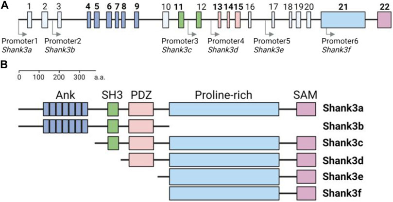 Fig. 1 Structure of mouse Shank3 gene and protein domain composition of Shank3 isoforms.1
Key structural properties of SHANK3:
- Multi-domain modular architecture
- 3D mesh assembly in the postsynaptic dense region
- SAM domain-mediated helical oligomerization
Functions of SHANK3
The core function of the SHANK3 protein is to serve as a molecular scaffold for excitatory synapses. However, it also plays a role in various neural development processes, including synaptic maturation, signal transduction and behavioral regulation.
| Function | Description |
| Synaptic scaffold assembly | The postsynaptic protein network was constructed through the multi-domain array (ANK-SH3-PDZ-SAM) to provide a structural platform for receptors and signaling molecules. |
| Neurotransmitter receptor anchoring | The PDZ domain specifically binds to the C-terminal sequence of glutamate receptors (such as mGluR5), regulating the aggregation and stability of receptors on the postsynaptic membrane. |
| Morphological regulation of dendritic spines | Coordinate the cytoskeletal recombination of actin and affect the structural plasticity of dendritic spines. Its absence leads to abnormal spine density and maturity. |
| Signal path integration | The SH3 domain mediates the assembly of Ras-MAPK and Homer Shank signaling modules to achieve transsynaptic signal transmission from membrane receptors to nuclear gene expression. |
| Social behavior regulation | By maintaining the homeostasis of excitatory synapses in the prefrontal cortex - striatum circuit, it affects advanced neural functions such as social interaction, learning and memory. |
The scaffold efficacy of SHANK3 shows a typical dose-dependent pattern. Its haploid insufficiency leads to a significant reduction in synaptic density, which contrasts with the synergistic oxygenation curve of hemoglobin and reflects the nonlinear characteristics of scaffold proteins in maintaining the homeostasis of neural networks.
Applications of SHANK3 and SHANK3 Antibody in Literature
1. Zhang, Linlin, et al. "SHANK3 in vagal sensory neurons regulates body temperature, systemic inflammation, and sepsis." Frontiers in Immunology 14 (2023): 1124356. https://doi.org/10.3389/fimmu.2023.1124356
The article indicates that SHANK3 gene defect can increase the inflammatory response and the risk of sepsis in mice. Research has found that SHANK3 in vagus nerve sensory neurons participates in temperature regulation and systemic inflammation control by regulating the expression of Trpm2. This mechanism provides a new perspective for understanding inflammatory abnormalities in autism spectrum disorder.
2. Huang, Min, Qi Qi, and Tao Xu. "Targeting Shank3 deficiency and paresthesia in autism spectrum disorder: A brief review." Frontiers in Molecular Neuroscience 16 (2023): 1128974. https://doi.org/10.3389/fnmol.2023.1128974
The article indicates that SHANK3 gene defect can increase the inflammatory response and the risk of sepsis in mice. Research has found that SHANK3 in vagus nerve sensory neurons participates in temperature regulation and systemic inflammation control by regulating the expression of Trpm2. This mechanism provides a new perspective for understanding inflammatory abnormalities in autism spectrum disorder.
3. Kim, Yoonhee, et al. "The emerging roles of Shank3 in cardiac function and dysfunction." Frontiers in Cell and Developmental Biology 11 (2023): 1191369. https://doi.org/10.3389/fcell.2023.1191369
The article indicates that SHANK3 is not only a key scaffold protein of neural synapses, but its function also extends to the cardiac domain. Studies have shown that SHANK3 participates in regulating cardiac signal transduction by interacting with proteins in myocardial cells and is associated with myocardial infarction and age-related cardiac function changes.
4. Pagano, Jessica, et al. "Shank3 deletion in PV neurons is associated with abnormal behaviors and neuronal functions that are rescued by increasing GABAergic signaling." Molecular autism 14.1 (2023): 28. https://doi.org/10.1186/s13229-023-00557-2
The article indicates that haploidy deficiency of SHANK3 can disrupt the excitation/inhibition balance of the cerebral cortex, leading to excessive excitation of the cortex and related behavioral abnormalities. Research has found that specific knockout of SHANK3 in PV inhibitory neurons is sufficient to trigger these defects, and the GABAA receptor modulator Ganaxolone can effectively alleviate symptoms, providing a potential therapeutic direction for Phelan-McDermid syndrome.
5. Wu, Shanshan, et al. "Shank3 deficiency elicits autistic-like behaviors by activating p38α in hypothalamic AgRP neurons." Molecular Autism 15.1 (2024): 14. https://doi.org/10.1186/s13229-024-00595-4
The article indicates that the deletion of the SHANK3 gene can trigger stereotyped behaviors and social disorders in mice by activating the p38α signal in hypothalamic AgRP neurons. Studies have shown that specifically inhibiting the p38α activity of AgRP neurons (rather than POMC neurons) can effectively improve these autism-like behaviors, revealing a potential therapeutic target for autism related to SHANK3 mutations.
Creative Biolabs: SHANK3 Antibodies for Research
Creative Biolabs specializes in the production of high-quality SHANK3 antibodies for research and industrial applications. Our portfolio includes monoclonal antibodies tailored for ELISA, Flow Cytometry, Western blot, immunohistochemistry, and other diagnostic methodologies.
- Custom SHANK3 Antibody Development: Tailor-made solutions to meet specific research requirements.
- Bulk Production: Large-scale antibody manufacturing for industry partners.
- Technical Support: Expert consultation for protocol optimization and troubleshooting.
- Aliquoting Services: Conveniently sized aliquots for long-term storage and consistent experimental outcomes.
For more details on our SHANK3 antibodies, custom preparations, or technical support, contact us at email.
Reference
- Kim, Yoonhee, et al. "The emerging roles of Shank3 in cardiac function and dysfunction." Frontiers in Cell and Developmental Biology 11 (2023): 1191369. https://doi.org/10.3389/fcell.2023.1191369
Anti-SHANK3 antibodies
 Loading...


Hot products 
-
Mouse Anti-AQP2 Recombinant Antibody (G-3) (CBMAB-A3359-YC)

-
Mouse Anti-A2M Recombinant Antibody (V2-178822) (CBMAB-A0036-YC)

-
Mouse Anti-CD83 Recombinant Antibody (HB15) (CBMAB-C1765-CQ)

-
Mouse Anti-CAPZB Recombinant Antibody (CBYY-C0944) (CBMAB-C2381-YY)

-
Mouse Anti-CCN2 Recombinant Antibody (CBFYC-2383) (CBMAB-C2456-FY)

-
Mouse Anti-GFP Recombinant Antibody (28) (CBMAB-G3038-LY)

-
Armenian hamster Anti-CD40 Recombinant Antibody (HM40-3) (CBMAB-C10365-LY)

-
Rat Anti-ABCC11 Recombinant Antibody (V2-179001) (CBMAB-A0236-YC)

-
Mouse Anti-FLI1 Recombinant Antibody (CBXF-0733) (CBMAB-F0435-CQ)

-
Rat Anti-ADGRE4 Recombinant Antibody (V2-160163) (CBMAB-F0011-CQ)

-
Mouse Anti-ARSA Recombinant Antibody (CBYC-A799) (CBMAB-A3679-YC)

-
Mouse Anti-CD63 Recombinant Antibody (CBXC-1200) (CBMAB-C1467-CQ)

-
Mouse Anti-AKT1 Recombinant Antibody (V2-180546) (CBMAB-A2070-YC)

-
Mouse Anti-ESR1 Recombinant Antibody (Y31) (CBMAB-1208-YC)

-
Mouse Anti-BRCA2 Recombinant Antibody (CBYY-0790) (CBMAB-0793-YY)

-
Rabbit Anti-ENO2 Recombinant Antibody (BA0013) (CBMAB-0272CQ)

-
Mouse Anti-ADV Recombinant Antibody (V2-503423) (CBMAB-V208-1364-FY)

-
Mouse Anti-ENO1 Recombinant Antibody (8G8) (CBMAB-E1329-FY)

-
Mouse Anti-CHRNA9 Recombinant Antibody (8E4) (CBMAB-C9161-LY)

-
Mouse Anti-ENO2 Recombinant Antibody (85F11) (CBMAB-0276CQ)

- AActivation
- AGAgonist
- APApoptosis
- BBlocking
- BABioassay
- BIBioimaging
- CImmunohistochemistry-Frozen Sections
- CIChromatin Immunoprecipitation
- CTCytotoxicity
- CSCostimulation
- DDepletion
- DBDot Blot
- EELISA
- ECELISA(Cap)
- EDELISA(Det)
- ESELISpot
- EMElectron Microscopy
- FFlow Cytometry
- FNFunction Assay
- GSGel Supershift
- IInhibition
- IAEnzyme Immunoassay
- ICImmunocytochemistry
- IDImmunodiffusion
- IEImmunoelectrophoresis
- IFImmunofluorescence
- IGImmunochromatography
- IHImmunohistochemistry
- IMImmunomicroscopy
- IOImmunoassay
- IPImmunoprecipitation
- ISIntracellular Staining for Flow Cytometry
- LALuminex Assay
- LFLateral Flow Immunoassay
- MMicroarray
- MCMass Cytometry/CyTOF
- MDMeDIP
- MSElectrophoretic Mobility Shift Assay
- NNeutralization
- PImmunohistologyp-Paraffin Sections
- PAPeptide Array
- PEPeptide ELISA
- PLProximity Ligation Assay
- RRadioimmunoassay
- SStimulation
- SESandwich ELISA
- SHIn situ hybridization
- TCTissue Culture
- WBWestern Blot
