TFRC Antibodies
Background
The TFRC gene encodes transferrin receptor 1 (CD71), a type II transmembrane glycoprotein widely expressed on the cell membrane, which is mainly responsible for mediating iron uptake by transferrin to maintain intracellular iron homeostasis. This protein transfers the iron-transferrin complex into cells through the endocytosis pathway, providing essential iron for key physiological processes such as hemoglobin synthesis and DNA replication. This gene was simultaneously identified by two independent research teams in 1984. Its promoter region contains the classic iron response element (IRE), which can be precisely regulated by intracellular iron concentration through the IRP/IRE system. As a core regulatory factor for cell proliferation and iron metabolism, TFRC not only provides a molecular basis for the study of iron metabolism diseases, but its expression level has also become an important biomarker for evaluating cell proliferation activity, and it holds significant value in cancer research and hematopoietic system disease research.
Structure of TFRC
The transferrin receptor protein (CD71) encoded by the TFRC gene is a type II transmembrane glycoprotein with a molecular weight of approximately 85 kDa. Its actual molecular weight fluctuates within the range of 84-95 kDa depending on the degree of glycosylation modification.
| Species | Human | Mouse | Rat | Hamster | Rhesus monkey |
| Molecular Weight (kDa) | 84.9 | 85.2 | 84.8 | 85.1 | 84.7 |
| Primary Structural Differences | 760 amino acids containing the internalized signal of YTRF | 76% sequence identity with human | Extracellular domain structure of highly conservative | Sequence differences in transmembrane regions | 98% homology with human sequence |
This protein is composed of 760 amino acids and forms a typical topological structure of type II membrane proteins. Its extracellular C-terminal region presents a spherical conformation and forms transferrin binding sites through a large number of β -folds. A single transmembrane region is composed of 22 hydrophobic amino acids. The intracellular N-terminal fragment contains the key internalized signal sequence of YTRF. The secondary structure of this receptor is dominated by β -folding, a feature that significantly distinguishes it from myoglobin, which is mainly composed of α -helix, in terms of structural classification.
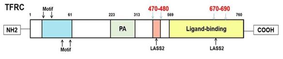 Fig. 1 Predict the structure of TFRC through UniProt.1
Key structural properties of TFRC:
- A heterodimer transmembrane structure composed of α and β subunits
- Extracellular into tapered transferrin combined cavity
- Intracellular domains of iron-containing responsive elements
- A stable ligand binding interface with disulfide bonds
Functions of TFRC
The transferrin receptor encoded by the TFRC gene mainly mediates iron uptake in cells and is also involved in the regulation of various cellular physiological processes.
| Function | Description |
| Iron ion transport | The transferrin - iron complex is transferred into the cell through the endocytosis pathway to maintain intracellular iron homeostasis. |
| Regulation of cell proliferation | For DNA synthesis in rapidly dividing cells increased, and energy metabolism to provide necessary source of iron. |
| Immune regulation | Iron metabolism reprogramming involved in the activation process of T cells affects the efficacy of immune responses. |
| Adaptation to hypoxia | Regulated by HIF-1α, it is upregulated in expression under hypoxic conditions to enhance the iron acquisition ability of cells. |
| Metabolic reprogramming | By regulating iron availability, it affects mitochondrial function and cellular metabolic status. |
The iron uptake kinetics of this receptor exhibits typical ligand-dependent endocytosis characteristics, which are different from the linear kinetics of free iron diffusion, indicating its precise regulatory role in maintaining iron homeostasis. Its expression is regulated by the negative feedback of intracellular iron levels through the IRP/IRE system. This mechanism has similar physiological significance to the oxygen-binding property of myoglobin in maintaining metabolic balance.
Applications of TFRC and TFRC Antibody in Literature
1. Yi, Lu, et al. "TFRC upregulation promotes ferroptosis in CVB3 infection via nucleus recruitment of Sp1." Cell death & disease 13.7 (2022): 592. https://doi.org/10.1038/s41419-022-05027-w
This study is the first to confirm that CVB3 infection can induce ferroptosis and reveal its mechanism: the transcription factor Sp1 binds to the TFRC promoter, upregulates TFRC expression and promotes its nuclear translocation, exacerbating intracellular iron overload and lipid peroxidation, and ultimately leading to ferroptosis. The Sp1/TFRC/Fe axis may become a new therapeutic target for CVB3.
2. Li, Rong, et al. "FTO deficiency in older livers exacerbates ferroptosis during ischaemia/reperfusion injury by upregulating ACSL4 and TFRC." Nature Communications 15.1 (2024): 4760. https://doi.org/10.1038/s41467-024-49202-3
Research has revealed the mechanism by which the liver in the elderly is prone to ischemia-reperfusion injury: down-regulation of the FTO gene, enhanced stability of ACSL4 and TFRC mRNA through the m6A method, and promotion of ferroptosis. Supplementing NMN can activate FTO, inhibit ferroptosis, and provide a new target for improving donor liver function in the elderly.
3. Zhou, Xing, et al. "RAB17 promotes endometrial cancer progression by inhibiting TFRC-dependent ferroptosis." Cell Death & Disease 15.9 (2024): 655. https://doi.org/10.1038/s41419-024-07013-w
Research has revealed that RAB17 promotes tumor progression in endometrial cancer by ubiquitinating and degrading TFRC protein, inhibiting ferroptosis. Especially under conditions of glucose deficiency, the expression of RAB17 is upregulated, enhancing the survival ability of cancer cells through this axis.
4. Wang, Xinyuan, et al. "Knockdown of ANXA10 induces ferroptosis by inhibiting autophagy-mediated TFRC degradation in colorectal cancer." Cell death & disease 14.9 (2023): 588. https://doi.org/10.1038/s41419-023-06114-2
Studies have revealed that ANXA10 is highly expressed in colorectal cancer and is associated with a poor prognosis. It promotes the survival and metastasis of cancer cells by inhibiting autophagy-mediated TFRC degradation and preventing intracellular ferroptosis. ANXA10 may become a potential therapeutic target.
5. Wei, Xue-biao, et al. "Exosome-derived lncRNA NEAT1 exacerbates sepsis-associated encephalopathy by promoting ferroptosis through regulating miR-9-5p/TFRC and GOT1 axis." Molecular Neurobiology 59.3 (2022): 1954-1969. https://doi.org/10.1007/s12035-022-02738-1
Studies have found that sepsis highly expresses NEAT1 in serum exosomes, which acts as ceRNA to adsorb miR-9-5p, thereby upregulating the expression of TFRC and GOT1, inducing ferroptosis and exacerbating sepsis-related encephalopathy. Targeting this axis may be a new therapeutic strategy.
Creative Biolabs: TFRC Antibodies for Research
Creative Biolabs specializes in the production of high-quality TFRC antibodies for research and industrial applications. Our portfolio includes monoclonal antibodies tailored for ELISA, Flow Cytometry, Western blot, immunohistochemistry, and other diagnostic methodologies.
- Custom TFRC Antibody Development: Tailor-made solutions to meet specific research requirements.
- Bulk Production: Large-scale antibody manufacturing for industry partners.
- Technical Support: Expert consultation for protocol optimization and troubleshooting.
- Aliquoting Services: Conveniently sized aliquots for long-term storage and consistent experimental outcomes.
For more details on our TFRC antibodies, custom preparations, or technical support, contact us at email.
Reference
- Huang, Yunfei, et al. "LASS2 suppresses metastasis in multiple cancers by regulating the ferroptosis signalling pathway through interaction with TFRC." Cancer Cell International 24.1 (2024): 87. https://doi.org/10.1186/s12935-024-03275-8
Anti-TFRC antibodies
 Loading...


Hot products 
-
Mouse Anti-GFAP Recombinant Antibody (5) (CBMAB-G0346-LY)

-
Mouse Anti-A2M Recombinant Antibody (V2-178822) (CBMAB-A0036-YC)

-
Mouse Anti-AAV8 Recombinant Antibody (V2-634028) (CBMAB-AP022LY)

-
Mouse Anti-CD8 Recombinant Antibody (C1083) (CBMAB-C1083-LY)

-
Mouse Anti-ARID1B Recombinant Antibody (KMN1) (CBMAB-A3546-YC)

-
Mouse Anti-EGR1 Recombinant Antibody (CBWJZ-100) (CBMAB-Z0289-WJ)

-
Mouse Anti-BRD3 Recombinant Antibody (CBYY-0801) (CBMAB-0804-YY)

-
Mouse Anti-BSN Recombinant Antibody (219E1) (CBMAB-1228-CN)

-
Mouse Anti-ATG5 Recombinant Antibody (9H197) (CBMAB-A3945-YC)

-
Mouse Anti-4-Hydroxynonenal Recombinant Antibody (V2-502280) (CBMAB-C1055-CN)

-
Mouse Anti-BANF1 Recombinant Antibody (3F10-4G12) (CBMAB-A0707-LY)

-
Mouse Anti-AKR1B1 Antibody (V2-2449) (CBMAB-1001CQ)

-
Mouse Anti-ADAM29 Recombinant Antibody (V2-179787) (CBMAB-A1149-YC)

-
Mouse Anti-CHRNA9 Recombinant Antibody (8E4) (CBMAB-C9161-LY)

-
Mouse Anti-CD59 Recombinant Antibody (CBXC-2097) (CBMAB-C4421-CQ)

-
Mouse Anti-ADRB2 Recombinant Antibody (V2-180026) (CBMAB-A1420-YC)

-
Mouse Anti-AMH Recombinant Antibody (5/6) (CBMAB-A2527-YC)

-
Mouse Anti-CCDC25 Recombinant Antibody (CBLC132-LY) (CBMAB-C9786-LY)

-
Mouse Anti-ACTG1 Recombinant Antibody (V2-179597) (CBMAB-A0916-YC)

-
Mouse Anti-BLK Recombinant Antibody (CBYY-0618) (CBMAB-0621-YY)

- AActivation
- AGAgonist
- APApoptosis
- BBlocking
- BABioassay
- BIBioimaging
- CImmunohistochemistry-Frozen Sections
- CIChromatin Immunoprecipitation
- CTCytotoxicity
- CSCostimulation
- DDepletion
- DBDot Blot
- EELISA
- ECELISA(Cap)
- EDELISA(Det)
- ESELISpot
- EMElectron Microscopy
- FFlow Cytometry
- FNFunction Assay
- GSGel Supershift
- IInhibition
- IAEnzyme Immunoassay
- ICImmunocytochemistry
- IDImmunodiffusion
- IEImmunoelectrophoresis
- IFImmunofluorescence
- IGImmunochromatography
- IHImmunohistochemistry
- IMImmunomicroscopy
- IOImmunoassay
- IPImmunoprecipitation
- ISIntracellular Staining for Flow Cytometry
- LALuminex Assay
- LFLateral Flow Immunoassay
- MMicroarray
- MCMass Cytometry/CyTOF
- MDMeDIP
- MSElectrophoretic Mobility Shift Assay
- NNeutralization
- PImmunohistologyp-Paraffin Sections
- PAPeptide Array
- PEPeptide ELISA
- PLProximity Ligation Assay
- RRadioimmunoassay
- SStimulation
- SESandwich ELISA
- SHIn situ hybridization
- TCTissue Culture
- WBWestern Blot
