WDR5 Antibodies
Background
WDR5 is a widely expressed WD40 repeat protein that mainly serves as a key scaffold component in the assembly of various core chromatin complexes, including the histone methyltransferase complex MLL/SET1. This protein directly regulates the epigenetic modification process by specifically recognizing the unmethylated state of histone H3 lysine 4 (H3K4), thereby influencing gene transcriptional activation. As a cell fate determinant, WDR5 plays a core role in embryonic development and tumorigenesis through mechanisms such as maintaining stem cell pluripotency and coordinating the progress of the cell cycle. Its highly conserved helical β -helical tertiary structure was analyzed by X-ray crystallography in 2006, providing a paradigm for revealing the molecular mechanism of protein interactions mediated by the WD40 domain, and having significant theoretical value in the fields of epigenetic regulatory networks and targeted drug development.
Structure of WDR5
WDR5 is a scaffold protein with a molecular weight of approximately 36 kDa. Its exact weight may fluctuate slightly due to post-translational modifications such as acetylation or phosphorylation.
| Species | Human | Mouse | Zebrafish | Yeast |
| Molecular Weight (kDa) | ~36 | ~36 | ~35 | ~34 |
| Primary Structural Differences | Containing the WD40 repeating domain, it is highly conservative | High homology with humans | The core domains are similar | Homologous substances have differentiated functions and simplified structures |
The WDR5 protein is composed of 334 amino acids and folds into a typical β-helical tertiary structure. The core of this structure is its seven sheet-like WD40 repeat sequence units, which form a rigid and highly conserved "annular β-helix" platform. This platform, as a key protein-protein interaction interface, can specifically recognize and bind to core partner proteins such as histone H3 and MLL. A distinct "arginine binding pocket" on its surface is crucial for identifying protein ligands containing the "ARA" motif, directly regulating the assembly and function of histone methyltransferase complexes.
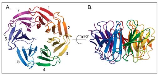 Fig. 1 WDR5 is a seven bladed β-propeller protein.1
Key structural properties of WDR5:
- Conservative seven-leaf β-helical conformation
- Rigid protein interface to form the central channel
- Arginine-binding pockets mediate specific molecular recognition
Functions of WDR5
The core function of WDR5 is to serve as a molecular scaffold for epigenetic regulation. However, this protein is also involved in a variety of cellular processes, including cell cycle regulation and stem cell maintenance.
| Function | Description |
| Epigenetic scaffold | As a core component of complexes such as MLL/COMPASS, it precisely recruits histone methyltransferases to specific genomic loci. |
| Transcriptional regulation | The transcriptional process of the target gene is directly activated by mediating the trimethylation modification of lysine at position 4 of histone H3. |
| Cell fate determination | Maintaining the self-renewal and pluripotency of stem cells, the imbalance of their expression is closely related to abnormal cell differentiation and tumorigenesis. |
| Ribosome biosynthesis | By interacting with the rDNA promoter, it participates in regulating the transcription of ribosomal RNA and affects the cell growth rate. |
| Mitotic regulation | In the cell cycle and centromere protein interaction, guarantee the accuracy of the chromosome separation and genomic stability. |
WDR5 interacts with multiple protein ligands through its highly conserved WD40 domain. This multivalent binding property enables it to integrate different upstream signals and ultimately coordinate chromatin states with cellular functions.
Applications of WDR5 and WDR5 Antibody in Literature
1. Wang, Da, et al. "MBD2 regulates the progression and chemoresistance of cholangiocarcinoma through interaction with WDR5." Journal of Experimental & Clinical Cancer Research 43.1 (2024): 272. https://doi.org/10.1186/s13046-024-03188-4
Research has found that MBD2 in cholangiocarcinoma activates the expression of the ABCB1 gene by binding to WDR5, thereby leading to chemotherapy resistance. The inhibitor MM-102 can block this process and enhance the efficacy of cisplatin.
2. Guarnaccia, Alissa duPuy, and William Patrick Tansey. "Moonlighting with WDR5: a cellular multitasker." Journal of clinical medicine 7.2 (2018): 21. https://doi.org/10.3390/jcm7020021
Research has found that WDR5 is a multifunctional protein, not only serving as the core scaffold for histone methylation but also playing a key role in various cellular processes. Small molecule inhibitors developed for its structure have provided a new direction for anti-cancer treatment.
3. Chodisetty, Swathi, et al. "MLL/WDR5 complex recruits centriolar satellite protein Cep72 to regulate microtubule nucleation and spindle formation." Science Advances 10.50 (2024): eadn0086.https://doi.org/10.1126/sciadv.adn0086
Studies have found that the MLL/WDR5 protein complex is located in the centrosome and regulates the assembly of γ-TuRCs by recruiting proteins such as Cep72, thereby affecting microtubule nucleation and spindle formation. This provides new insights into the mechanism by which MLL absence leads to microcephaly.
4. Liu, Lulu, et al. "Wdr5 is essential for fetal erythropoiesis and hematopoiesis." Experimental hematology & oncology 12.1 (2023): 39. https://doi.org/10.1186/s40164-023-00385-3
Research has found that Wdr5 is crucial for red blood cell production during the process of embryonic hematopoiesis. Specific knockout of Wdr5 can lead to embryonic death and completely disrupt the function of hematopoietic stem and progenitor cells and their c-Kit phenotype.
5. Aho, Erin R., et al. "Targeting WDR5: A WINning anti-cancer strategy?." Epigenetics insights 12 (2019): 2516865719865282. https://doi.org/10.1177/2516865719865282
Research has found that inhibitors targeting the WIN site of the WDR5 protein do not work through epigenetic mechanisms but rather by stripping it from chromatin to inhibit protein synthesis, thereby inducing nucleolar stress and killing MLL cancer cells.
Creative Biolabs: WDR5 Antibodies for Research
Creative Biolabs specializes in the production of high-quality WDR5 antibodies for research and industrial applications. Our portfolio includes monoclonal antibodies tailored for ELISA, Flow Cytometry, Western blot, immunohistochemistry, and other diagnostic methodologies.
- Custom WDR5 Antibody Development: Tailor-made solutions to meet specific research requirements.
- Bulk Production: Large-scale antibody manufacturing for industry partners.
- Technical Support: Expert consultation for protocol optimization and troubleshooting.
- Aliquoting Services: Conveniently sized aliquots for long-term storage and consistent experimental outcomes.
For more details on our WDR5 antibodies, custom preparations, or technical support, contact us at email.
Reference
- Guarnaccia, Alissa duPuy, and William Patrick Tansey. "Moonlighting with WDR5: a cellular multitasker." Journal of clinical medicine 7.2 (2018): 21. https://doi.org/10.3390/jcm7020021
Anti-WDR5 antibodies
 Loading...


Hot products 
-
Mouse Anti-C1QC Recombinant Antibody (CBFYC-0600) (CBMAB-C0654-FY)

-
Mouse Anti-DHFR Recombinant Antibody (D0821) (CBMAB-D0821-YC)

-
Rabbit Anti-ALOX5AP Recombinant Antibody (CBXF-1219) (CBMAB-F0750-CQ)

-
Rabbit Anti-DLK1 Recombinant Antibody (9D8) (CBMAB-D1061-YC)

-
Mouse Anti-AOC3 Recombinant Antibody (CBYY-0014) (CBMAB-0014-YY)

-
Mouse Anti-ATP1A2 Recombinant Antibody (M7-PB-E9) (CBMAB-A4013-YC)

-
Mouse Anti-GLP1R Recombinant Antibody (4F3) (CBMAB-G0521-LY)

-
Mouse Anti-EMP3 Recombinant Antibody (CBFYE-0100) (CBMAB-E0207-FY)

-
Mouse Anti-FN1 Monoclonal Antibody (D6) (CBMAB-1240CQ)

-
Mouse Anti-FOXA3 Recombinant Antibody (2A9) (CBMAB-0377-YC)

-
Mouse Anti-AQP2 Recombinant Antibody (G-3) (CBMAB-A3359-YC)

-
Mouse Anti-CFL1 (Phospho-Ser3) Recombinant Antibody (CBFYC-1770) (CBMAB-C1832-FY)

-
Mouse Anti-BSN Recombinant Antibody (219E1) (CBMAB-1228-CN)

-
Mouse Anti-AGO2 Recombinant Antibody (V2-634169) (CBMAB-AP203LY)

-
Mouse Anti-EPO Recombinant Antibody (CBFYR0196) (CBMAB-R0196-FY)

-
Mouse Anti-ALX1 Recombinant Antibody (96k) (CBMAB-C0616-FY)

-
Mouse Anti-ADIPOR2 Recombinant Antibody (V2-179983) (CBMAB-A1369-YC)

-
Mouse Anti-GDF5 Recombinant Antibody (1F4) (CBMAB-G2740-LY)

-
Rabbit Anti-ADRA1A Recombinant Antibody (V2-12532) (CBMAB-1022-CN)

-
Mouse Anti-AKT1 (Phosphorylated S473) Recombinant Antibody (V2-505430) (PTM-CBMAB-0067LY)

- AActivation
- AGAgonist
- APApoptosis
- BBlocking
- BABioassay
- BIBioimaging
- CImmunohistochemistry-Frozen Sections
- CIChromatin Immunoprecipitation
- CTCytotoxicity
- CSCostimulation
- DDepletion
- DBDot Blot
- EELISA
- ECELISA(Cap)
- EDELISA(Det)
- ESELISpot
- EMElectron Microscopy
- FFlow Cytometry
- FNFunction Assay
- GSGel Supershift
- IInhibition
- IAEnzyme Immunoassay
- ICImmunocytochemistry
- IDImmunodiffusion
- IEImmunoelectrophoresis
- IFImmunofluorescence
- IGImmunochromatography
- IHImmunohistochemistry
- IMImmunomicroscopy
- IOImmunoassay
- IPImmunoprecipitation
- ISIntracellular Staining for Flow Cytometry
- LALuminex Assay
- LFLateral Flow Immunoassay
- MMicroarray
- MCMass Cytometry/CyTOF
- MDMeDIP
- MSElectrophoretic Mobility Shift Assay
- NNeutralization
- PImmunohistologyp-Paraffin Sections
- PAPeptide Array
- PEPeptide ELISA
- PLProximity Ligation Assay
- RRadioimmunoassay
- SStimulation
- SESandwich ELISA
- SHIn situ hybridization
- TCTissue Culture
- WBWestern Blot
