Protein Concentration & Concentration Determination
As an expert in the field of antibody development, Creative Biolabs is not only committed to recombinant antibody production, we also offer protein concentration & concentration determination service using our first-class technology platform and high-quality products. Our special Antibody Concentration Kit can meet your protein concentration requirement. We provide a range of total protein quantification methods, such as the absorbance at 280, Bradford or BCA assays and quantitative amino acid analysis.
Introduction
Sometimes, antibodies are only available at low concentrations and usually contain some substances of low molecular weight that interfere with the labeling of enzymes, biotin, streptavidin and fluorophores. Thus, antibodies should be concentrated to increase the concentration. Some methods or kits presently are used to achieve the purpose.
Protein concentration determination is often essential before processing protein samples for isolation, separation and analysis by chromatographic, electrophoretic and immunochemical techniques. Based on the accuracy required and the amount and purity of the protein available, different methods are suitable for determining protein concentration.
At present, the simplest and most direct assay method for protein quantitation is to measure the absorbance at 280 nm (UV range). Amino acids exhibit strong UV-light absorption with aromatic side chains (i.e., tyrosine, tryptophan and phenylalanine). Proteins absorb UV-light commonly in direct proportion to their aromatic amino acid content and total concentration. Another analytical method is to use high-performance liquid chromatography (HPLC) to label all primary amines (N-terminal and side chains of lysine residues) with colored or fluorescent dyes (such as ninhydrin or o-phthalaldehyde (OPA)). However, due to the different aromatic amino acid content of different proteins, the absorption characteristics are different, so the absorption effect of ultraviolet absorption method on protein mixture is not ideal. Additionally, any non-protein content that absorbs UV light also can disturb the measurements.
Therefore, several colorimetric and fluorescent, reagent-based protein assay techniques have been developed to overcome the above disadvantages. Simply, protein samples are added to the reagent, producing a color change or increased fluorescence in proportion to the amount added. Then the protein concentration is determined by reference to a standard curve based on known concentrations of a purified reference protein.
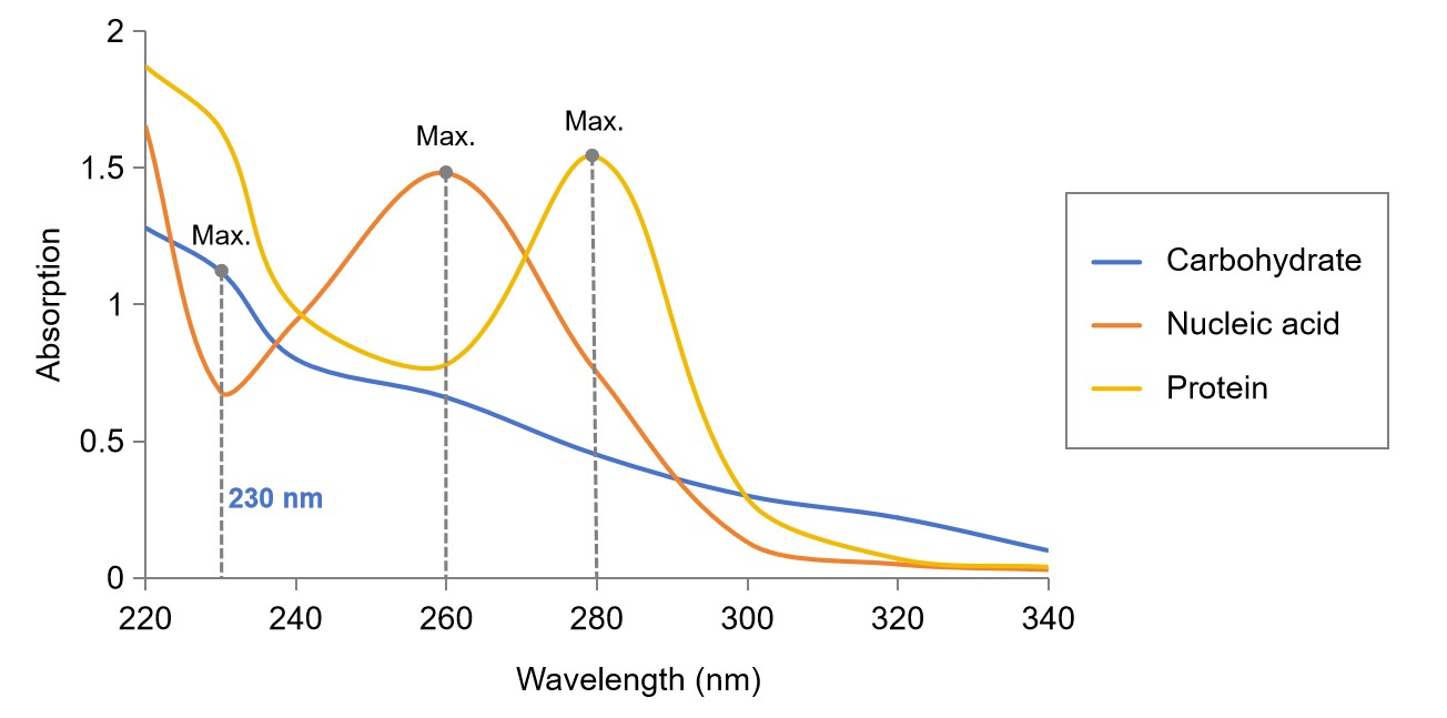 Fig.1 Protein, nucleic acid and carbohydrate quantification.
Method of Protein Concentration
Using an Antibody Concentration Kit can easily and quickly remove the excess buffer from the antibody to obtain a more concentrated antibody solution.
- Add antibody to spin cartridge.
- Spin for 1-3 minutes in a microfuge at max speed to reduce the buffer volume. It is best not to spin dry the antibody, because the reconstitution of the antibody will be difficult and significant antibody loss and degradation may occur.
- Repeat steps 1 through 2 as many times to process the entire antibody to the desired concentration. It may be necessary to discard the extra buffers collected in the collection pipe between rotations
- Recover the concentrated antibody from the spin cartridge.
Protein Concentration Determination
- Protein concentration determination using absorbance at 280 nm
- Warm up the UV lamp (approximately 15 min).
- Adjust wavelength to 280 nm.
- Calibrate to zero absorbance with buffer solution.
- Measure absorbance of the protein solution.
- Adjust wavelength to 260 nm.
- Calibrate to zero absorbance with buffer solution.
- Measure absorbance of the protein solution.
- Protein concentration determination using BCA Assays
- Reagent preparation.
- Standard curve preparation.
- Dilute samples.
- Pipette standards or samples into duplicate wells in a clear bottom 96 well plate.
- Add working solution into each well that contains the standard or samples.
- Shake gently to mix. Incubate for 30-90 min at 37-60°C. Cool to room temperature.
- Measure OD at 562 nm.
- Protein concentration determination using Bradford Assays
- Make a dilution series of the chosen model protein.
- Mix the samples, standards and a blank (buffer, no protein) with Bradford Reagent.
- Read absorbance at 595 nm.
- Subtract the average 595 nm measurement of the blank from the 595 nm measurements of all other individual standards and unknown samples. Plot the mean blank corrected 595 nm measurement for each standard and concentration. The slope of this standard curve is used to estimate the protein concentration of unknown samples.
For more information about protein concentration & concentration determination, please contact us.
What Can We Do For You?
As your experienced partner for antibody development, our expertise in antibody and protein contents can accelerate development timelines by delivering accurate and robust analytical services. Currently, we offer protein concentration & concentration determination expertise to help you to meet and exceed quality, safety and regulatory standards. Please feel free to contact us, and see how we can help to address your problems.
Work flow of rAb production includes:
- Gene Cloning
- Transformation of Expression Plasmids
- Small-scale Expression
- Large-scale Expression & Bacterial Disruption
- Purification of Fusion Proteins
- Protein Concentration & Concentration Determination
- Detection Assays
Reference
- Degaim, Z. D.; et al. IMMUNIZATION MODEL OF RATS TREATED WITH TWO ANTIGENS FROM MRSA BACTERIA ISOLATED FROM BURN PATIENTS. European Journal of Biology and Medical Science Research. 2016, 4(1): 26-36.