ACPP Antibodies
Background
The ACPP gene encodes a secreted glycoprotein called prostate acid phosphatase (PAP), which is highly expressed in prostate epithelial cells. This enzyme regulates cell growth by hydrolyzing phosphate compounds and plays a tumor-suppressing role in prostate cancer. Its mechanism involves dephosphorylation regulation of the ErbB-2 signaling pathway. The gene is located on chromosome 3q21 and its expression is regulated by transcription factors such as NF-κB, affecting the sensitivity of tumor cells to androgens. Since the protein product of this gene was first identified in 1938, the research on ACPP has continued to deepen. In recent years, it has been discovered that this gene is also involved in the regulation of pain signals in the nervous system and plays a role in brain diseases such as Alzheimer's disease, providing an important foundation for understanding the mechanism of cancer occurrence and exploring biomarkers for related diseases.
Structure of ACPP
The prostate acid phosphatase (PAP) encoded by the ACPP gene has a relative molecular mass of approximately 100 kDa and is composed of two subunits. PAP from different species varies in molecular structure and enzymatic properties, mainly manifested as subtle changes in glycosylation degree and amino acid sequence.
| Species | Human | Rat | Mouse | Dog |
| Molecular Weight (kDa) | About 100 | About 96 | About 95 | About 98 |
| Primary Structural Differences | Sugar modification, bimolecular | Lower degree of glycosylation | Highly homologous to human sequence | Expression with tissue specificity |
The PAP protein contains approximately 800 amino acid residues (about 400 for each subunit), presenting a typical spherical glycoprotein structure. Its active center is located at the interface of the two subunits, containing highly conserved acidic amino acid residues that are responsible for catalyzing the hydrolysis of phosphate ester bonds. The three-dimensional structure of this enzyme molecule is composed of alternating α-helices and β-sheets, forming a stable hydrophobic core and maintaining conformational stability through disulfide bonds. The histidine residues in the active site directly participate in the transfer of phosphate groups, while the aspartic acid residues are responsible for maintaining the catalytic efficiency in an acidic pH environment. The glycosylation modification of this enzyme not only affects its secretion efficiency but also regulates its binding specificity with substrate molecules.
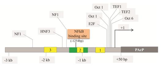 Fig. 1 The schematic representation of the PAcP gene promoter.1
The key structural characteristics of the protein encoded by the ACPP gene:
- Homologous dimeric glycoprotein: Two approximately 50 kDa subunits are non-covalently bound
- Acidic active center: Contains conserved histidine and aspartic acid, with the optimal pH range of 4-6
- Disulfide bond stabilizes structure: Maintains the enzyme conformation and activity
- Sulfation modification: Affects protein folding and substrate recognition
- Substrate binding pocket: Selectively recognizes phosphate compounds
Functions of ACPP
The main function of the protein encoded by the ACPP gene:
| Function | Description |
| Hydrolysis of Phosphate Compounds | Catalyzes the hydrolysis of substrates such as phosphatidylcholine, releasing phosphate ions and choline, and participating in the biochemical metabolism of semen. |
| Tumor Suppression Regulation | Inhibits the proliferation of prostate cancer cells and tumor formation by dephosphorylating ErbB-2/HER-2. |
| Regulation of Androgen Sensitivity | Interacts with ErbB-2 to maintain the androgen-dependent nature of prostate cancer cells. |
| Regulation of Pain Signal | Exerts an anti-harmful sensation effect in the dorsal root ganglion and participates in the regulation of chronic pain. |
| Potential Neuroprotective Function | Expressed in brain tissue, possibly involved in the pathological process of neurodegenerative diseases. |
This enzyme exhibits the highest activity under acidic conditions (pH 4-6), has strong substrate specificity, and mainly hydrolyzes small molecular phosphate compounds rather than large molecular phosphate proteins. Its bimolecular structure and glycosylation modification contribute to its high stability in body fluids. The serum PAP level has been used as a clinical marker for prostate cancer metastasis.
Applications of ACPP and ACPP Antibody in Literature
1. Hanley, Peter J. "Elusive physiological role of prostatic acid phosphatase (PAP): generation of choline for sperm motility via auto-and paracrine cholinergic signaling." Frontiers in Physiology 14 (2023): 1327769. https://doi.org/10.3389/fphys.2023.1327769
Proposed prostatic acid phosphatase (ACPP gene encoding) by new functions: the phosphoric acid hydrolysis after ejaculation sperm choline generated choline, and then into the acetylcholine, through cholinergic signal to enhance the vitality of the sperm and may promote the female genital tract contraction.
2. Muniyan, Sakthivel, et al. "Human prostatic acid phosphatase: structure, function and regulation." International journal of molecular sciences 14.5 (2013): 10438-10464. https://doi.org/10.3390/ijms140510438
The article indicates that prostate acid phosphatase (encoded by the ACPP gene) inhibits the proliferation of prostate cancer cells by dephosphorylating ErbB-2 and maintaining their sensitivity to androgens. The expression of this enzyme is regulated by NF-κB, and its loss of function is associated with the occurrence of castration-resistant prostate cancer.
3. Alpert, Evgenia, et al. "Multifunctionality of prostatic acid phosphatase in prostate cancer pathogenesis." Bioscience reports 41.10 (2021): BSR20211646. https://doi.org/10.1042/BSR20211646
By using the signal sequence substitution technique, it was discovered that prostate acid phosphatase (encoded by the ACPP gene) has three distinct functional subtypes. Among them, the PLPAcP subtype shows a specific increase in early prostate cancer tissues and may become a new therapeutic target.
4. Xu, Huan, et al. "Prostatic acid phosphatase (PAP) predicts prostate cancer progress in a population‐based study: the renewal of PAP?." Disease markers 2019.1 (2019): 7090545. https://doi.org/10.1155/2019/7090545
Based on the analysis of data from 5184 patients with positive prostate acid phosphatase (encoded by the ACPP gene) in the SEER database, it was found that these patients had a higher rate of tumor invasion and metastasis, worse pathological grading, and shorter survival periods. PAP can be used as an effective indicator for evaluating the prognosis of prostate cancer.
5. Muniyan, Sakthivel, et al. "Human prostatic acid phosphatase: structure, function and regulation." International journal of molecular sciences 14.5 (2013): 10438-10464. https://doi.org/10.3390/ijms140510438
The article indicates that prostate acid phosphatase (encoded by the ACPP gene) inhibits tumor growth by dephosphorylating ErbB-2 and maintains the sensitivity of cancer cells to androgens. Its expression is regulated by NF-κB, and loss of function can lead to castration-resistant prostate cancer.
Creative Biolabs: ACPP Antibodies for Research
Creative Biolabs specializes in the production of high-quality ACPP antibodies for research and industrial applications. Our portfolio includes monoclonal and polyclonal antibodies tailored for ELISA, Flow Cytometry, Western blot, immunohistochemistry, and other diagnostic methodologies.
- Custom ACPP Antibody Development: Tailor-made solutions to meet specific research requirements.
- Bulk Production: Large-scale antibody manufacturing for industry partners.
- Technical Support: Expert consultation for protocol optimization and troubleshooting.
- Aliquoting Services: Conveniently sized aliquots for long-term storage and consistent experimental outcomes.
For more details on our ACPP antibodies, custom preparations, or technical support, contact us at email.
Reference
- Muniyan, Sakthivel, et al. "Human prostatic acid phosphatase: structure, function and regulation." International journal of molecular sciences 14.5 (2013): 10438-10464. Distributed under Open Access license CC BY 3.0, without modification. https://doi.org/10.3390/ijms140510438
Anti-ACPP antibodies
 Loading...


Hot products 
-
Mouse Anti-GGT1 Recombinant Antibody (1F9) (CBMAB-G3273-LY)

-
Rabbit Anti-ABL1 (Phosphorylated Y185) Recombinant Antibody (V2-443434) (PTM-CBMAB-0001YC)

-
Mouse Anti-AGK Recombinant Antibody (V2-258056) (CBMAB-M0989-FY)

-
Mouse Anti-CD24 Recombinant Antibody (HIS50) (CBMAB-C10123-LY)

-
Mouse Anti-ALOX5 Recombinant Antibody (33) (CBMAB-1890CQ)

-
Mouse Anti-CORO1A Recombinant Antibody (4G10) (V2LY-1206-LY806)

-
Mouse Anti-AAV8 Recombinant Antibody (V2-634028) (CBMAB-AP022LY)

-
Mouse Anti-ACTN4 Recombinant Antibody (V2-6075) (CBMAB-0020CQ)

-
Mouse Anti-ADGRE2 Recombinant Antibody (V2-261270) (CBMAB-C0813-LY)

-
Rabbit Anti-ALDOA Recombinant Antibody (D73H4) (CBMAB-A2314-YC)

-
Mouse Anti-ARHGAP5 Recombinant Antibody (54/P190-B) (CBMAB-P0070-YC)

-
Mouse Anti-CCL18 Recombinant Antibody (64507) (CBMAB-C7910-LY)

-
Rat Anti-CD63 Recombinant Antibody (7G4.2E8) (CBMAB-C8725-LY)

-
Rabbit Anti-CCN1 Recombinant Antibody (CBWJC-3580) (CBMAB-C4816WJ)

-
Mouse Anti-ARHGDIA Recombinant Antibody (CBCNA-009) (CBMAB-R0415-CN)

-
Mouse Anti-ARID3A Antibody (A4) (CBMAB-0128-YC)

-
Mouse Anti-FN1 Monoclonal Antibody (D6) (CBMAB-1240CQ)

-
Human Anti-SARS-CoV-2 S1 Monoclonal Antibody (CBFYR-0120) (CBMAB-R0120-FY)

-
Mouse Anti-CCND2 Recombinant Antibody (DCS-3) (CBMAB-G1318-LY)

-
Rat Anti-(1-5)-α-L-Arabinan Recombinant Antibody (V2-501861) (CBMAB-XB0003-YC)

- AActivation
- AGAgonist
- APApoptosis
- BBlocking
- BABioassay
- BIBioimaging
- CImmunohistochemistry-Frozen Sections
- CIChromatin Immunoprecipitation
- CTCytotoxicity
- CSCostimulation
- DDepletion
- DBDot Blot
- EELISA
- ECELISA(Cap)
- EDELISA(Det)
- ESELISpot
- EMElectron Microscopy
- FFlow Cytometry
- FNFunction Assay
- GSGel Supershift
- IInhibition
- IAEnzyme Immunoassay
- ICImmunocytochemistry
- IDImmunodiffusion
- IEImmunoelectrophoresis
- IFImmunofluorescence
- IGImmunochromatography
- IHImmunohistochemistry
- IMImmunomicroscopy
- IOImmunoassay
- IPImmunoprecipitation
- ISIntracellular Staining for Flow Cytometry
- LALuminex Assay
- LFLateral Flow Immunoassay
- MMicroarray
- MCMass Cytometry/CyTOF
- MDMeDIP
- MSElectrophoretic Mobility Shift Assay
- NNeutralization
- PImmunohistologyp-Paraffin Sections
- PAPeptide Array
- PEPeptide ELISA
- PLProximity Ligation Assay
- RRadioimmunoassay
- SStimulation
- SESandwich ELISA
- SHIn situ hybridization
- TCTissue Culture
- WBWestern Blot
