BCL2 Antibodies
Background
BCL2 is an important anti-apoptotic protein, mainly existing on the outer mitochondrial membrane, endoplasmic reticulum and nuclear membrane of vertebrate cells. This protein prevents programmed cell death by inhibiting the release of mitochondrial cytochrome c, thereby maintaining cell survival and participating in the regulation of tissue homeostasis. Cancer cells often utilize the overexpression of BCL2 to evade apoptosis, which makes it an important target for cancer treatment. This protein family was first discovered by Tsujimoto and Croce in 1985 when they were studying B-cell lymphoma, and its name is precisely derived from the abbreviation of B-cell lymphoma 2. The unique BH domain of the BCL2 protein family and its interaction mechanism with pro-apoptotic proteins provide a key model for the study of cell death regulation and have had a profound impact on the research of oncology, immunology and neurodegenerative diseases.
Structure of BCL2
BCL2 is an anti-apoptotic protein with a molecular weight of approximately 26 kDa. Its size may vary slightly due to different subtypes (such as BCL2α, BCL2β) and post-translational modifications.
| Species | Human | Mouse | Rats | Bovine |
| Molecular Weight (kDa) | 26 | 25.8 | 26.1 | 25.9 |
| Primary Structural Differences | Containing 4 BH domains (BH1-BH4) | Highly conserved and similar to humans | Tiny amino acid differences | Highly conserved function |
BCL2 is an anti-apoptotic protein composed of approximately 239 amino acids. Its three-dimensional structure is mainly made up of α -helices and maintains stability through a hydrophobic core. The key structural features of this protein include: The BH domain (BH1-BH4) mediates the interaction with pro-apoptotic proteins (such as BAX, BAK) to regulate cell survival; The transmembrane domain (TM) anchors it to the outer mitochondrial membrane, endoplasmic reticulum and nuclear membrane, determining its subcellular localization. And the flexible ring region located at the N-terminal can regulate the activity of BCL2 through phosphorylation modification. By inhibiting the release of mitochondrial cytochrome c, BCL2 can effectively prevent apoptosis. This unique structural feature makes it an important target in the field of cancer treatment.
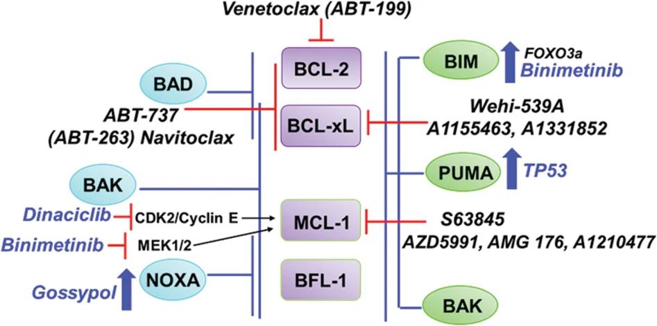 Fig. 1 Structural changes of myoglobin.1
Functions of BCL2
BCL2 protein's main function is to regulate apoptosis, maintain cells survive, also involved in many physiological and pathological process, including cancer and immune regulation.
| Function | Description |
| Anti-apoptotic effect | By inhibiting the activation of pro-apoptotic proteins such as BAX/BAK and preventing the release of mitochondrial cytochrome c, cell survival is maintained. |
| Tumorigenesis promotion | Overexpressed in various cancers, it helps tumor cells evade programmed death, promotes tumor growth and drug resistance. |
| Immune regulation | Regulate the survival of lymphocytes, affect the homeostasis of the immune system, and participate in autoimmune diseases and infection responses. |
| Oxidative stress response | By regulating the stability of the mitochondrial membrane, it affects the production of reactive oxygen species (ROS) and participates in the REDOX balance of cells. |
| Developmental regulation | Play a key role in embryonic development and organization steady state, such as nerve cells survive and hematopoietic system maintain. |
The mechanism of action of BCL2 protein depends on its interaction with other members of the BCL2 family, forming a complex regulatory network, and its expression level directly affects the fate of cells. Compared with the rapid activation of pro-apoptotic proteins (such as BAX), the role of BCL2 is more inclined to maintain cell survival in the long term, making it an important target in cancer treatment and cell engineering.
Applications of BCL2 and BCL2 Antibody in Literature
1. Ito, Takahiko, et al. "Bcl-2 phosphorylation required for anti-apoptosis function." Journal of Biological Chemistry 272.18 (1997): 11671-11673. https://doi.org/10.1074/jbc.272.18.11671
This article indicates that BCL2 exerts an anti-apoptotic effect through phosphorylation at the Ser70 site, and this process depends on the activation of PKC. Studies have found that the phosphorylation of BCL2 induced by growth factors such as IL-3 is crucial for its inhibition of apoptosis, and the heterodimerization of BCL2-Bax is not sufficient to fully activate its function.
2. Bennett, Rory, Ella Thompson, and Constantine Tam. "SOHO state of the art updates and next questions| mechanisms of resistance to BCL2 inhibitor therapy in chronic lymphocytic leukemia and potential future therapeutic directions." Clinical Lymphoma Myeloma and Leukemia 22.11 (2022): 795-804. https://doi.org/10.1016/j.clml.2022.07.013
This article indicates that the overexpression of BCL2 protein in chronic lymphocytic leukemia (CLL) leads to the dysregulation of cell apoptosis. BCL2 inhibitors (such as venetoclax) significantly improve clinical efficacy by targeting and inhibiting the BCL2 protein, and are not affected by traditional genetic abnormalities. However, continuous medication may trigger secondary drug resistance mechanisms such as BCL2 variations and abnormal BCL2 family proteins. Current research focuses on new strategies for overcoming drug resistance.
3. Renner, Wilfried, et al. "BCL2 genotypes and prostate cancer survival." Strahlentherapie Und Onkologie 193.6 (2017): 466. https://doi.org/10.1007/s00066-017-1126-9
Studies have found that the -938CC genotype of the BCL2 gene is significantly associated with a poor survival prognosis in patients with prostate cancer. In the multivariate analysis, this genotype was an independent risk factor for cancer-specific survival and overall survival, suggesting that the polymorphism of the BCL2 gene may become an important molecular marker for the prognosis assessment of prostate cancer.
4. Callagy, Grace M., et al. "Meta-analysis confirms BCL2 is an independent prognostic marker in breast cancer." BMC cancer 8 (2008): 1-10. https://doi.org/10.1186/1471-2407-8-153
Prognostic studies of breast cancer have confirmed that the expression level of BCL2 protein is an independent prognostic factor. Meta-analysis showed that patients with high expression of BCL2 had significantly prolonged disease-free survival and overall survival, and this predictive value was not affected by other factors such as lymph node status and tumor size.
5. Hsu, Yi-Te, and Richard J. Youle. "Nonionic detergents induce dimerization among members of the Bcl-2 family." Journal of Biological Chemistry 272.21 (1997): 13829-13834. https://doi.org/10.1074/jbc.272.21.13829
Studies have found that BCL2 family proteins (BCL2, BCL-XL and Bax) regulate apoptosis through conformational changes. The specific monoclonal antibody 6A7 revealed that Bax presented a concealed conformation in the physiological state. After treatment with a detergent, the epitope was exposed and a dimer was formed. These antibodies provide new tools for studying the interactions of BCL2 family proteins.
Creative Biolabs: BCL2 Antibodies for Research
Creative Biolabs specializes in the production of high-quality BCL2 antibodies for research and industrial applications. Our portfolio includes monoclonal antibodies tailored for ELISA, Flow Cytometry, Western blot, immunohistochemistry, and other diagnostic methodologies.
- Custom BCL2 Antibody Development: Tailor-made solutions to meet specific research requirements.
- Bulk Production: Large-scale antibody manufacturing for industry partners.
- Technical Support: Expert consultation for protocol optimization and troubleshooting.
- Aliquoting Services: Conveniently sized aliquots for long-term storage and consistent experimental outcomes.
For more details on our BCL2 antibodies, custom preparations, or technical support, contact us at info@creative-biolabs.com.
Reference
- Kapoor, Isha, et al. "Targeting BCL-2 in B-cell malignancies and overcoming therapeutic resistance." Cell death & disease 11.11 (2020): 941. https://doi.org/10.1038/s41419-020-03144-y
Anti-BCL2 antibodies
 Loading...


Hot products 
-
Mouse Anti-AMOT Recombinant Antibody (CBYC-A564) (CBMAB-A2552-YC)

-
Rabbit Anti-CCL5 Recombinant Antibody (R0437) (CBMAB-R0437-CN)

-
Rabbit Anti-CCN1 Recombinant Antibody (CBWJC-3580) (CBMAB-C4816WJ)

-
Mouse Anti-GFAP Recombinant Antibody (5) (CBMAB-G0346-LY)

-
Mouse Anti-CIITA Recombinant Antibody (CBLC160-LY) (CBMAB-C10987-LY)

-
Mouse Anti-APP Recombinant Antibody (5C2A1) (CBMAB-A3314-YC)

-
Mouse Anti-AKT1/AKT2/AKT3 (Phosphorylated T308, T309, T305) Recombinant Antibody (V2-443454) (PTM-CBMAB-0030YC)

-
Mouse Anti-CCT6A/B Recombinant Antibody (CBXC-0168) (CBMAB-C5570-CQ)

-
Mouse Anti-AFDN Recombinant Antibody (V2-58751) (CBMAB-L0408-YJ)

-
Mouse Anti-ARHGDIA Recombinant Antibody (CBCNA-009) (CBMAB-R0415-CN)

-
Mouse Anti-BHMT Recombinant Antibody (CBYY-0547) (CBMAB-0550-YY)

-
Rat Anti-CCR2 Recombinant Antibody (475301) (CBMAB-C1338-LY)

-
Mouse Anti-CAT Recombinant Antibody (724810) (CBMAB-C8431-LY)

-
Rabbit Anti-BRCA2 Recombinant Antibody (D9S6V) (CBMAB-CP0017-LY)

-
Mouse Anti-CASP7 Recombinant Antibody (10-01-62) (CBMAB-C2005-LY)

-
Mouse Anti-APCS Recombinant Antibody (CBYC-A663) (CBMAB-A3054-YC)

-
Rabbit Anti-ADRA1A Recombinant Antibody (V2-12532) (CBMAB-1022-CN)

-
Mouse Anti-CASP8 Recombinant Antibody (CBYY-C0987) (CBMAB-C2424-YY)

-
Mouse Anti-CD24 Recombinant Antibody (ALB9) (CBMAB-0176CQ)

-
Rabbit Anti-ABL1 (Phosphorylated Y185) Recombinant Antibody (V2-443434) (PTM-CBMAB-0001YC)

- AActivation
- AGAgonist
- APApoptosis
- BBlocking
- BABioassay
- BIBioimaging
- CImmunohistochemistry-Frozen Sections
- CIChromatin Immunoprecipitation
- CTCytotoxicity
- CSCostimulation
- DDepletion
- DBDot Blot
- EELISA
- ECELISA(Cap)
- EDELISA(Det)
- ESELISpot
- EMElectron Microscopy
- FFlow Cytometry
- FNFunction Assay
- GSGel Supershift
- IInhibition
- IAEnzyme Immunoassay
- ICImmunocytochemistry
- IDImmunodiffusion
- IEImmunoelectrophoresis
- IFImmunofluorescence
- IGImmunochromatography
- IHImmunohistochemistry
- IMImmunomicroscopy
- IOImmunoassay
- IPImmunoprecipitation
- ISIntracellular Staining for Flow Cytometry
- LALuminex Assay
- LFLateral Flow Immunoassay
- MMicroarray
- MCMass Cytometry/CyTOF
- MDMeDIP
- MSElectrophoretic Mobility Shift Assay
- NNeutralization
- PImmunohistologyp-Paraffin Sections
- PAPeptide Array
- PEPeptide ELISA
- PLProximity Ligation Assay
- RRadioimmunoassay
- SStimulation
- SESandwich ELISA
- SHIn situ hybridization
- TCTissue Culture
- WBWestern Blot
