CD79A Antibodies
Background
The protein encoded by the CD79A gene is an important component of the B-cell antigen-receptor complex and is mainly expressed on the cell membrane surface of B lymphocytes. This protein forms a heterodimer with CD79B and jointly participates in the assembly and signal transduction process of BCR, playing a key role in the development, differentiation and immune response of B cells. This gene was first identified in 1989 and is the first confirmed B-cell-specific signaling molecule. Its discovery has promoted major breakthroughs in the research of the mechanisms of immune deficiency diseases and B-cell malignancies. The characteristic of this gene transmitting signals through the tyrosine activation motif of immune receptors has become a classic model for understanding the signal transduction mechanism of immune cells, providing an important theoretical basis for the research and development of monoclonal antibody therapy and targeted drugs.
Structure of CD79A
CD79A is a transmembrane glycoprotein with a molecular weight of approximately 40-45 kDa. The actual measured value may vary among different cell types due to the degree of glycosylation modification.
| Species | Human | Mouse | Rat | Rhesus monkey |
| Molecular Weight (kDa) | 40-45 | 42-44 | 41-43 | 40-44 |
| Primary Structural Differences | Containing 228 amino acids, intracellular area have ITAM die body | High homology with human and conserved ITAM sequence | Amino acids in the transmembrane region are highly consistent | Glycosylation sites similar to human height |
This protein is composed of 228 amino acids, and its polypeptide chain folds to form a typical immunoglobulin-like domain. CD79A transmits B-cell receptor signals through the immune receptor tyrosine activating motif (ITAM) in the intracellular region. The conserved tyrosine residues in this motif can provide anchoring sites for downstream signaling molecules after phosphorylation. It forms a stable heterodimer with CD79B through a disulfide bond in the extracellular region, jointly constituting the signal transduction module of the B-cell antigen receptor.
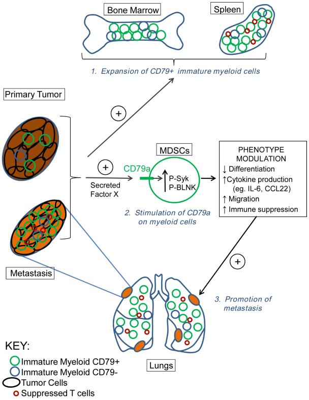 Fig. 1 CD79a mediates immature myeloid-tumor cell interactions.1
Key structural properties of CD79A:
- Immunoglobulin-like (Ig-like) extracellular domains
- Heterodimers are formed with CD79B through disulfide bonds
- Intracellular region containing immune receptor tyrosine activation motif (ITAM)
- Conserved tyrosine residues are the structural basis for B-cell signal transduction
Functions of CD79A
The core function of CD79A is to participate in the assembly and signal transduction of B-cell receptor (BCR) complexes. In addition, this gene also plays a key role in the immune response of B cells, apoptosis regulation, and the establishment of immune tolerance.
| Function | Description |
| BCR assembly | With CD79B transmembrane heterologous dimers, complete BCR expressed in the stability of the membrane surface. |
| Signal transduction | The downstream signaling pathway is initiated through tyrosine phosphorylation of the intracellular ITAM domain. |
| Activation of B cells | Mediate the primary signal transduction after antigen recognition, triggering the proliferation and differentiation of B cells. |
| Immune tolerance | Participate in the cloning, deletion and receptor editing process of autoreactive B cells. |
| Lymphoma occurrence | The abnormal signal is closely related to the pathogenesis of various B-cell malignant tumors. |
The signal intensity mediated by CD79A is negatively regulated by the immune receptor tyrosine inhibitory motif (ITIM), a characteristic similar to the synergistic effect of hemoglobin, jointly maintaining the dynamic balance of B cell activation and inhibition.
Applications of CD79A and CD79A Antibody in Literature
1. Julamanee, Jakrawadee, et al. "Composite CD79A/CD40 co-stimulatory endodomain enhances CD19CAR-T cell proliferation and survival." Molecular Therapy 29.9 (2021): 2677-2690. https://doi.org/10.1016/j.ymthe.2021.04.038
In this study, a novel CD79A/CD40 complex costimulatory domain was designed, which can effectively activate the NF-κB and p38 signaling pathways. CD19 CAR-T cells equipped with this structure demonstrated stronger proliferation ability and anti-tumor effect both in vitro and in mouse models, outperforming the traditional CD28 or 4-1BB structures.
2. Lenk, Lennart, et al. "CD79a promotes CNS-infiltration and leukemia engraftment in pediatric B-cell precursor acute lymphoblastic leukemia." Communications biology 4.1 (2021): 73. https://doi.org/10.1038/s42003-020-01591-z
The current diagnostic methods for central nervous system leukemia have limitations. The research has proposed a new hypothesis: the preBCR signaling complex composed of CD79A and others may be directly related to the central infiltration of B-lineage acute lymphocytic leukemia cells, providing a new direction for diagnosis and targeted therapy.
3. Yao, Shucong, et al. "CD79A work as a potential target for the prognosis of patients with OSCC: analysis of immune cell infiltration in oral squamous cell carcinoma based on the CIBERSORTx deconvolution algorithm." BMC Oral Health 23.1 (2023): 411. https://doi.org/10.1186/s12903-023-02936-w
This study found through the analysis of oral squamous cell carcinoma samples that an increase in B lymphocyte infiltration in the tumor microenvironment can improve the prognosis of patients. Among them, those with low expression of CD79A have a significantly better prognosis, indicating that CD79A can serve as a potential new target for predicting the prognosis of oral squamous cell carcinoma.
4. Tran Nguyen Truc, Linh, et al. "Mechanism of cystogenesis by Cd79a-driven, conditional mTOR activation in developing mouse nephrons." Scientific Reports 13.1 (2023): 508. https://doi.org/10.1038/s41598-023-27766-2
This study utilized Cd79a-Cre; The Tsc1ff mouse model reveals the key role of mTORC1 signaling in kidney development. Excessive activation of mTORC1 driven by the Cd79a promoter can disrupt the normal morphology of cells in the distal renal tubules, such as planar polarity, and eventually lead to the occurrence of polycystic kidney disease.
5. Suhren, Jan-Theile, et al. "CD20/CD79a/PAX5/CD3-negative post-transplant lymphoma with aberrant actin and desmin co-expression—a potential differential diagnostic pitfall between PTLD and PTSMT." Annals of Hematology 101.4 (2022): 881-883. https://doi.org/10.1007/s00277-021-04394-2
This research report presents a rare case of lymphoproliferative disorder after liver transplantation. Tumor cells do not express B-cell markers such as CD20 and CD79A, but there are immunoglobulin light chain limitations. The negative result of CD79A increased the difficulty of diagnosis. Eventually, it was confirmed as plasmoblastic PTLD through gene rearrangement analysis.
Creative Biolabs: CD79A Antibodies for Research
Creative Biolabs specializes in the production of high-quality CD79A antibodies for research and industrial applications. Our portfolio includes monoclonal antibodies tailored for ELISA, Flow Cytometry, Western blot, immunohistochemistry, and other diagnostic methodologies.
- Custom CD79A Antibody Development: Tailor-made solutions to meet specific research requirements.
- Bulk Production: Large-scale antibody manufacturing for industry partners.
- Technical Support: Expert consultation for protocol optimization and troubleshooting.
- Aliquoting Services: Conveniently sized aliquots for long-term storage and consistent experimental outcomes.
For more details on our CD79A antibodies, custom preparations, or technical support, contact us at email.
Reference
- Luger, Dror, et al. "Expression of the B-cell receptor component CD79a on immature myeloid cells contributes to their tumor promoting effects." PLoS One 8.10 (2013): e76115. https://doi.org/10.1371/journal.pone.0076115
Anti-CD79A antibodies
 Loading...


Hot products 
-
Mouse Anti-FLT1 Recombinant Antibody (11) (CBMAB-V0154-LY)

-
Mouse Anti-AOC3 Recombinant Antibody (CBYY-0014) (CBMAB-0014-YY)

-
Mouse Anti-GIPC2 Recombinant Antibody (10) (CBMAB-G0476-LY)

-
Mouse Anti-ACO2 Recombinant Antibody (V2-179329) (CBMAB-A0627-YC)

-
Mouse Anti-CAT Recombinant Antibody (724810) (CBMAB-C8431-LY)

-
Mouse Anti-AAV9 Recombinant Antibody (V2-634029) (CBMAB-AP023LY)

-
Mouse Anti-ACTG1 Recombinant Antibody (V2-179597) (CBMAB-A0916-YC)

-
Mouse Anti-CTNND1 Recombinant Antibody (CBFYC-2414) (CBMAB-C2487-FY)

-
Mouse Anti-ATG5 Recombinant Antibody (9H197) (CBMAB-A3945-YC)

-
Rabbit Anti-Acetyl-Histone H3 (Lys36) Recombinant Antibody (V2-623395) (CBMAB-CP0994-LY)

-
Mouse Anti-AHCYL1 Recombinant Antibody (V2-180270) (CBMAB-A1703-YC)

-
Mouse Anti-ATP1B1 Recombinant Antibody (E4) (CBMAB-0463-LY)

-
Mouse Anti-AGK Recombinant Antibody (V2-258056) (CBMAB-M0989-FY)

-
Mouse Anti-ARG1 Recombinant Antibody (CBYCL-103) (CBMAB-L0004-YC)

-
Human Anti-SARS-CoV-2 Spike Recombinant Antibody (CR3022) (CBMAB-CR014LY)

-
Mouse Anti-CD164 Recombinant Antibody (CBFYC-0077) (CBMAB-C0086-FY)

-
Mouse Anti-ADGRE5 Recombinant Antibody (V2-360335) (CBMAB-C2088-CQ)

-
Mouse Anti-ACTN4 Recombinant Antibody (V2-6075) (CBMAB-0020CQ)

-
Mouse Anti-AMIGO2 Recombinant Antibody (CBYY-C0756) (CBMAB-C2192-YY)

-
Mouse Anti-FOXL1 Recombinant Antibody (CBXF-0845) (CBMAB-F0462-CQ)

- AActivation
- AGAgonist
- APApoptosis
- BBlocking
- BABioassay
- BIBioimaging
- CImmunohistochemistry-Frozen Sections
- CIChromatin Immunoprecipitation
- CTCytotoxicity
- CSCostimulation
- DDepletion
- DBDot Blot
- EELISA
- ECELISA(Cap)
- EDELISA(Det)
- ESELISpot
- EMElectron Microscopy
- FFlow Cytometry
- FNFunction Assay
- GSGel Supershift
- IInhibition
- IAEnzyme Immunoassay
- ICImmunocytochemistry
- IDImmunodiffusion
- IEImmunoelectrophoresis
- IFImmunofluorescence
- IGImmunochromatography
- IHImmunohistochemistry
- IMImmunomicroscopy
- IOImmunoassay
- IPImmunoprecipitation
- ISIntracellular Staining for Flow Cytometry
- LALuminex Assay
- LFLateral Flow Immunoassay
- MMicroarray
- MCMass Cytometry/CyTOF
- MDMeDIP
- MSElectrophoretic Mobility Shift Assay
- NNeutralization
- PImmunohistologyp-Paraffin Sections
- PAPeptide Array
- PEPeptide ELISA
- PLProximity Ligation Assay
- RRadioimmunoassay
- SStimulation
- SESandwich ELISA
- SHIn situ hybridization
- TCTissue Culture
- WBWestern Blot
