GSTP1 Antibodies
Background
The GSTP1 gene encodes a small molecule glutathione S-transferase P1 protein, which is mainly present in the cytoplasm of mammalian cells. This protein catalyzes detoxification reactions of various electrophilic substances by binding to glutathione and participates in the regulatory mechanisms of cellular antioxidant stress and signal transduction. In tumor cells, GSTP1 often undergoes epigenetic silencing through promoter methylation, which is closely related to the formation of chemotherapy drug resistance. This gene was first identified in 1985, and its protein crystal structure was resolved by X-ray diffraction in the 1990s, becoming an important model for studying enzyme catalytic mechanisms and carcinogen metabolism. Its highly conserved functional domain provides a key molecular basis for understanding the substrate specificity of phase II metabolic enzymes, cellular defense pathways, and disease-related mutations.
Structure of GSTP1
GSTP1 is a relatively small protein with a molecular weight of approximately 23.2 kDa. This molecular weight may vary slightly among different species, mainly due to variations in the amino acid sequence encoded by the gene.
| Species | Human | Rat | Mouse |
| Molecular Weight (kDa) | 23.2 | 23.5 | 23.4 |
| Primary Structural Differences | Highly conservative catalytic domain | High sequence similarity and consistent active sites | Highly similar to humans, often used in model research |
This protein is composed of 209 amino acids and forms a compact spherical homodimer through its primary structure folding. The three-dimensional structure of GSTP1 contains a hydrophilic glutathione binding site (G-site) and a hydrophobic substrate binding pocket (H-site). This structure enables it to catalyze various electrophilic poison-glutathione binding reactions. The key tyrosine residue in its active center is crucial for promoting the deprotonation of glutathione and initiating nucleophilic reactions. The secondary structure of the protein is mainly composed of α-helices and β-sheets, which jointly maintain the stable conformation required for its catalytic function.
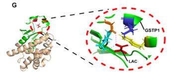 Fig. 1 Modeled structure of parts of GSTP1.1
Key structural properties of GSTP1:
- Typical thioredoxin folding structure
- Hydrophobic/hydrophilic compatible substrate binding pocket
- Active site tyrosine residues mediated catalytic reaction
- Homologous dimer interface through hydrogen bonding network stable conformation
Functions of GSTP1
The main function of the GSTP1 protein is to catalyze the binding of various electrophilic toxins with reduced glutathione (GSH), thereby enabling the detoxification of cells. Additionally, it is deeply involved in regulating the cellular redox balance, signal transduction, and drug metabolism, among other physiological and pathological processes.
| Function | Description |
| Cellular Detoxification | Catalyzes the conjugation of GSH with reactive substrates (such as environmental toxins, carcinogens, and chemotherapy drugs) to increase their water solubility for excretion. |
| Antioxidant Defense | By combining and eliminating reactive electrophilic molecules such as lipid peroxidation end products, it protects cells from oxidative damage. |
| Signal Regulation | Interacts with kinases such as JNK, regulates the apoptotic and proliferation signaling pathways, and influences tumor occurrence and development. |
| Drug Metabolism and Resistance | Various drugs are metabolized in the liver and tumor tissues. The overexpression of these enzymes is one of the important mechanisms leading to chemotherapy resistance in tumors. |
| Disease Biomarkers | Epigenetic silencing (such as high methylation of promoters) is frequently observed in various tumors including prostate cancer and lung cancer, and can serve as potential diagnostic and prognostic biomarkers. |
Unlike many enzymatic reactions, the catalytic action of GSTP1 on GSH follows the "ping-pong" mechanism. That is, GSH first binds and undergoes deprotonation before the enzyme-glutathione complex combines with the hydrophobic substrate to complete the reaction. This enables it to efficiently handle a wide variety of toxic substances.
Applications of GSTP1 and GSTP1 Antibody in Literature
1. Sun, Yandi, et al. "A GSTP1-mediated lactic acid signaling promotes tumorigenesis through the PPP oxidative branch." Cell Death & Disease 14.7 (2023): 463. https://doi.org/10.1038/s41419-023-05998-4
Studies have shown that the tumor metabolite lactic acid inhibits the phosphorylation of G6PD by binding to GSTP1, enhances the generation of NADPH in the pentose phosphate pathway, and thereby maintains redox homeostasis and promotes breast cancer growth. This mechanism is particularly significant in certain breast cancer subtypes.
2. Lei, Xiao, et al. "GSTP1 as a novel target in radiation induced lung injury." Journal of translational medicine 19.1 (2021): 297. https://doi.org/10.1186/s12967-021-02978-0
Studies have shown that GSTP1 is the main antioxidant enzyme in lung tissue and is associated with the risk of radiation-induced lung injury. It participates in the disease process by regulating oxidative stress and cell apoptosis. Specific gene polymorphisms can affect susceptibility.
3. Santric, Veljko, et al. "GSTP1 rs1138272 polymorphism affects prostate cancer risk." Medicina 56.3 (2020): 128. https://doi.org/10.3390/medicina56030128
The study found that two specific mutations (rs1695 and rs1138272) of the GSTP1 gene were significantly associated with the risk of prostate cancer. Especially when both mutations occurred simultaneously, the risk of the disease increased by 5.46 times. The co-occurrence of multiple GST gene mutations would further synergistically increase the risk.
4. Fang, Cheng, et al. "Aberrant GSTP1 promoter methylation is associated with increased risk and advanced stage of breast cancer: a meta-analysis of 19 case-control studies." BMC cancer 15.1 (2015): 920. https://doi.org/10.1186/s12885-015-1926-1
The meta-analysis confirmed that abnormal methylation of the GSTP1 gene promoter is significantly associated with the risk of breast cancer (OR = 7.85), and it is more common in advanced patients. This change exists in different ethnic groups and can serve as a potential biomarker for breast cancer diagnosis.
5. Miao, Li-Feng, Xiang-Hua Ye, and Xiao-Feng He. "Individual and combined effects of GSTM1, GSTT1, and GSTP1 polymorphisms on breast cancer risk: a meta-analysis and re-analysis of systematic meta-analyses." PloS one 15.3 (2020): e0216147. https://doi.org/10.1371/journal.pone.0216147
This study, through a meta-analysis of 101 articles, indicates that the evidence linking the polymorphisms of the GSTM1, GSTT1 and GSTP1 genes (including both individual and combined effects) to an increased risk of breast cancer is limited. In high-quality studies, the reliability of most positive results is relatively low.
Creative Biolabs: GSTP1 Antibodies for Research
Creative Biolabs specializes in the production of high-quality GSTP1 antibodies for research and industrial applications. Our portfolio includes monoclonal and polyclonal antibodies tailored for ELISA, Flow Cytometry, Western blot, immunohistochemistry, and other diagnostic methodologies.
- Custom GSTP1 Antibody Development: Tailor-made solutions to meet specific research requirements.
- Bulk Production: Large-scale antibody manufacturing for industry partners.
- Technical Support: Expert consultation for protocol optimization and troubleshooting.
- Aliquoting Services: Conveniently sized aliquots for long-term storage and consistent experimental outcomes.
For more details on our GSTP1 antibodies, custom preparations, or technical support, contact us at info@creative-biolabs.com.
Reference
- Sun, Yandi, et al. "A GSTP1-mediated lactic acid signaling promotes tumorigenesis through the PPP oxidative branch." Cell Death & Disease 14.7 (2023): 463. Distributed under Open Access license CC BY 4.0. Cropped from the original figure. https://doi.org/10.1038/s41419-023-05998-4
Anti-GSTP1 antibodies
 Loading...


Hot products 
-
Mouse Anti-CD24 Recombinant Antibody (SN3) (CBMAB-C1037-CQ)

-
Mouse Anti-CCDC25 Recombinant Antibody (CBLC132-LY) (CBMAB-C9786-LY)

-
Mouse Anti-ADV Recombinant Antibody (V2-503423) (CBMAB-V208-1364-FY)

-
Mouse Anti-ADAM12 Recombinant Antibody (V2-179752) (CBMAB-A1114-YC)

-
Mouse Anti-ABL2 Recombinant Antibody (V2-179121) (CBMAB-A0364-YC)

-
Mouse Anti-DMD Recombinant Antibody (D1190) (CBMAB-D1190-YC)

-
Rat Anti-EPO Recombinant Antibody (16) (CBMAB-E1578-FY)

-
Mouse Anti-BCL6 Recombinant Antibody (CBYY-0442) (CBMAB-0445-YY)

-
Mouse Anti-CD24 Recombinant Antibody (HIS50) (CBMAB-C10123-LY)

-
Mouse Anti-ENO1 Recombinant Antibody (8G8) (CBMAB-E1329-FY)

-
Mouse Anti-ATP1B1 Recombinant Antibody (E4) (CBMAB-0463-LY)

-
Mouse Anti-ALPL Antibody (B4-78) (CBMAB-1009CQ)

-
Mouse Anti-ATP5F1A Recombinant Antibody (51) (CBMAB-A4043-YC)

-
Mouse Anti-4-Hydroxynonenal Recombinant Antibody (V2-502280) (CBMAB-C1055-CN)

-
Mouse Anti-ESR1 Recombinant Antibody (Y31) (CBMAB-1208-YC)

-
Mouse Anti-CDK7 Recombinant Antibody (CBYY-C1783) (CBMAB-C3221-YY)

-
Mouse Anti-ARG1 Recombinant Antibody (CBYCL-103) (CBMAB-L0004-YC)

-
Mouse Anti-ADAM29 Recombinant Antibody (V2-179787) (CBMAB-A1149-YC)

-
Rabbit Anti-ATF4 Recombinant Antibody (D4B8) (CBMAB-A3872-YC)

-
Mouse Anti-CARD11 Recombinant Antibody (CBFYC-0811) (CBMAB-C0866-FY)

- AActivation
- AGAgonist
- APApoptosis
- BBlocking
- BABioassay
- BIBioimaging
- CImmunohistochemistry-Frozen Sections
- CIChromatin Immunoprecipitation
- CTCytotoxicity
- CSCostimulation
- DDepletion
- DBDot Blot
- EELISA
- ECELISA(Cap)
- EDELISA(Det)
- ESELISpot
- EMElectron Microscopy
- FFlow Cytometry
- FNFunction Assay
- GSGel Supershift
- IInhibition
- IAEnzyme Immunoassay
- ICImmunocytochemistry
- IDImmunodiffusion
- IEImmunoelectrophoresis
- IFImmunofluorescence
- IGImmunochromatography
- IHImmunohistochemistry
- IMImmunomicroscopy
- IOImmunoassay
- IPImmunoprecipitation
- ISIntracellular Staining for Flow Cytometry
- LALuminex Assay
- LFLateral Flow Immunoassay
- MMicroarray
- MCMass Cytometry/CyTOF
- MDMeDIP
- MSElectrophoretic Mobility Shift Assay
- NNeutralization
- PImmunohistologyp-Paraffin Sections
- PAPeptide Array
- PEPeptide ELISA
- PLProximity Ligation Assay
- RRadioimmunoassay
- SStimulation
- SESandwich ELISA
- SHIn situ hybridization
- TCTissue Culture
- WBWestern Blot
