MTBP Antibodies
Background
The MTBP gene encodes a key regulatory factor that binds and stabilizes the p53 protein, mainly distributed in the cell nucleus. This protein regulates the conformational stability and intracellular concentration of p53 through direct interaction with p53, thereby participating in critical biological processes such as DNA damage response, cell cycle regulation, and apoptosis. Studies have found that MTBP is abnormally expressed in various cancers, and its dysfunction may affect the tumor suppression mechanism. Since its identification in the early 2000s, MTBP has been continuously studied as an important regulatory node of the p53 signaling pathway, providing valuable molecular basis for understanding tumor occurrence, cellular stress response, and the development of anti-cancer drug targets.
Structure of MTBP
The protein molecule weight encoded by the MTBP gene is approximately 92 kDa. This value varies slightly among different mammals due to differences in amino acid sequences.
| Species | Human | Mouse | Rat |
| Molecular Weight (kDa) | 92 | 91 | 91.5 |
| Primary Structural Differences | Contains multiple domains, such as the nuclear localization sequence | Highly homologous, functionally conserved | Has an extremely high similarity to the human sequence |
This protein is composed of approximately 900 amino acids and forms a complex spatial structure. Its functional core region contains a conserved p53 binding domain, which achieves specific interaction with the p53 protein through a combination of specific α-helices and β-sheets. The key binding interface is composed of multiple hydrophobic amino acid residues, forming a stable binding pocket, thereby effectively regulating the conformation and stability of p53. This mechanism is the structural basis for its participation in cell cycle arrest and apoptosis regulation.
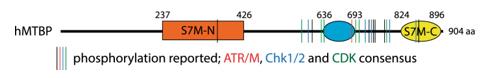 Fig. 1 Domain architecture of human MTBP with reported phosphorylation sites.1
Key structural properties of MTBP:
- Contains multiple domain structure, the core domain as the conservative p53 combining structure
- Forming a stable complex with p53 through the hydrophobic core and specific interfaces
- Have a sequence of check and ratify, ensure that the functions play a regulation in the nuclei
Functions of MTBP
The core function of the MTBP gene-encoded protein is to act as a crucial stabilizing and regulatory factor for the p53 protein. Additionally, it is involved in various cellular processes, including cell cycle regulation and maintenance of genomic stability.
| Function | Description |
| Stable p53 | Directly binds to and stabilizes the p53 protein, preventing it from being degraded by proteasomes mediated by E3 ubiquitin ligases such as MDM2, and maintaining the level of p53 protein within the cell. |
| DNA Damage Response | Under the influence of DNA damage signals, it promotes the activation and accumulation of p53, assisting in initiating the cell cycle checkpoint arrest and providing time for DNA repair. |
| Apoptosis Regulation | By enhancing the transcriptional activity of p53, it regulates the expression of downstream pro-apoptotic genes (such as PUMA and BAX), thereby participating in the determination of cell fate. |
| Tumor Suppression | Its dysfunction is associated with the occurrence and development of various cancers, and it plays an important role in tumor suppression through the aforementioned mechanisms. |
| Cell proliferation influence | Independent of the p53 pathway, it may directly affect the cell proliferation process by interacting with other proteins (such as MYC). |
The interaction with p53 exhibits a synergistic and tightly regulated mode. MTBP does not directly bind to DNA; instead, it acts as a crucial "molecular chaperone" and "allosteric regulator", precisely amplifying the response of p53 to various stress signals, ensuring that it is moderately activated at the appropriate time. This is in contrast to the simple linear activation patterns of many transcriptional cofactors.
Applications of MTBP and MTBP Antibody in Literature
1. Ferreira, Pedro, et al. "MTBP phosphorylation controls DNA replication origin firing." Scientific reports 11.1 (2021): 4242. https://doi.org/10.1038/s41598-021-83287-w
This study has confirmed that MTBP is a key platform for regulating the initiation of replication in eukaryotes, and it is subject to multiple phosphorylation modifications such as those by CDK and DNA damage checkpoint kinases. Phosphorylation at the CDK site mimics the mutation and promotes replication initiation, while phosphorylation at the damage checkpoint site inhibits this function, revealing the core regulatory role of MTBP in the stable replication of the genome.
2. Grieb, Brian C., and Christine M. Eischen. "MTBP and MYC: a dynamic duo in proliferation, cancer, and aging." Biology 11.6 (2022): 881. https://doi.org/10.3390/biology11060881
The article indicates that MTBP is identified as a key transcriptional co-activator of MYC, working in concert with MYC to promote cell proliferation and tumor formation in cancer. Its expression level is elevated in various cancers and is associated with poor prognosis of patients. Studies have shown that inhibiting MTBP can suppress the growth of cancer cells, making it a potential new target for cancer treatment.
3. Zaffar, Eman, et al. "The Role of MTBP as a Replication Origin Firing Factor." Biology 11.6 (2022): 827. https://doi.org/10.3390/biology11060827
The article indicates that MTBP is a key factor for initiating DNA replication in multicellular eukaryotes and is homologous to the yeast Sld7. It precisely regulates the site, timing and efficiency of DNA replication initiation through means such as receiving kinase signals, ensuring accurate and single genome replication. Its core role in replication remains to be elucidated.
4. Song, Yifu, et al. "MTBP regulates cell survival and therapeutic sensitivity in TP53 wildtype glioblastomas." Theranostics 9.20 (2019): 6019. https://doi.org/10.7150/thno.35747
The article indicates that in TP53 wild-type glioblastoma, high expression of MTBP is associated with poor prognosis. It regulates p53 through the MDM2-dependent pathway, promoting tumor cell survival and resistance to radiotherapy and chemotherapy. c-myc can positively regulate its expression. Targeting MTBP is expected to become a new therapeutic strategy.
5. Wang, Hongbo, et al. "MTBP enhances the activation of transcription factor ETS-1 and promotes the proliferation of hepatocellular carcinoma cells." Frontiers in Oncology 12 (2022): 985082. https://doi.org/10.3389/fonc.2022.985082
This study reveals that MTBP is a novel oncogene in hepatocellular carcinoma (HCC). Acting as a co-activator of the transcription factor ETS-1, it enhances the transcription of ETS-1 target genes, thereby driving the proliferation of HCC cells and providing a new potential target for liver cancer treatment.
Creative Biolabs: MTBP Antibodies for Research
Creative Biolabs specializes in the production of high-quality MTBP antibodies for research and industrial applications. Our portfolio includes monoclonal antibodies tailored for ELISA, Flow Cytometry, Western blot, immunohistochemistry, and other diagnostic methodologies.
- Custom MTBP Antibody Development: Tailor-made solutions to meet specific research requirements.
- Bulk Production: Large-scale antibody manufacturing for industry partners.
- Technical Support: Expert consultation for protocol optimization and troubleshooting.
- Aliquoting Services: Conveniently sized aliquots for long-term storage and consistent experimental outcomes.
For more details on our MTBP antibodies, custom preparations, or technical support, contact us at email.
Reference
- Ferreira, Pedro, et al. "MTBP phosphorylation controls DNA replication origin firing." Scientific reports 11.1 (2021): 4242. https://doi.org/10.1038/s41598-021-83287-w
Anti-MTBP antibodies
 Loading...


Hot products 
-
Mouse Anti-GGT1 Recombinant Antibody (1F9) (CBMAB-G3273-LY)

-
Mouse Anti-ASH1L Monoclonal Antibody (ASH5H03) (CBMAB-1372-YC)

-
Mouse Anti-FLT1 Recombinant Antibody (11) (CBMAB-V0154-LY)

-
Mouse Anti-BHMT Recombinant Antibody (CBYY-0547) (CBMAB-0550-YY)

-
Mouse Anti-ATP5F1A Recombinant Antibody (51) (CBMAB-A4043-YC)

-
Mouse Anti-ADV Recombinant Antibody (V2-503423) (CBMAB-V208-1364-FY)

-
Mouse Anti-CCL18 Recombinant Antibody (64507) (CBMAB-C7910-LY)

-
Mouse Anti-CRYAB Recombinant Antibody (A4345) (CBMAB-A4345-YC)

-
Mouse Anti-ALX1 Recombinant Antibody (96k) (CBMAB-C0616-FY)

-
Mouse Anti-ABCA3 Recombinant Antibody (V2-178911) (CBMAB-A0145-YC)

-
Mouse Anti-DLL4 Recombinant Antibody (D1090) (CBMAB-D1090-YC)

-
Rabbit Anti-ADRA1A Recombinant Antibody (V2-12532) (CBMAB-1022-CN)

-
Human Anti-SARS-CoV-2 S1 Monoclonal Antibody (CBFYR-0120) (CBMAB-R0120-FY)

-
Mouse Anti-CA9 Recombinant Antibody (CBXC-2079) (CBMAB-C0131-CQ)

-
Rabbit Anti-ABL1 (Phosphorylated Y245) Recombinant Antibody (V2-505716) (PTM-CBMAB-0465LY)

-
Mouse Anti-ALPL Antibody (B4-78) (CBMAB-1009CQ)

-
Mouse Anti-CD1C Recombinant Antibody (L161) (CBMAB-C2173-CQ)

-
Mouse Anti-AKT1 (Phosphorylated S473) Recombinant Antibody (V2-505430) (PTM-CBMAB-0067LY)

-
Mouse Anti-BAX Recombinant Antibody (CBYY-0216) (CBMAB-0217-YY)

-
Rat Anti-ADGRE4 Recombinant Antibody (V2-160163) (CBMAB-F0011-CQ)

- AActivation
- AGAgonist
- APApoptosis
- BBlocking
- BABioassay
- BIBioimaging
- CImmunohistochemistry-Frozen Sections
- CIChromatin Immunoprecipitation
- CTCytotoxicity
- CSCostimulation
- DDepletion
- DBDot Blot
- EELISA
- ECELISA(Cap)
- EDELISA(Det)
- ESELISpot
- EMElectron Microscopy
- FFlow Cytometry
- FNFunction Assay
- GSGel Supershift
- IInhibition
- IAEnzyme Immunoassay
- ICImmunocytochemistry
- IDImmunodiffusion
- IEImmunoelectrophoresis
- IFImmunofluorescence
- IGImmunochromatography
- IHImmunohistochemistry
- IMImmunomicroscopy
- IOImmunoassay
- IPImmunoprecipitation
- ISIntracellular Staining for Flow Cytometry
- LALuminex Assay
- LFLateral Flow Immunoassay
- MMicroarray
- MCMass Cytometry/CyTOF
- MDMeDIP
- MSElectrophoretic Mobility Shift Assay
- NNeutralization
- PImmunohistologyp-Paraffin Sections
- PAPeptide Array
- PEPeptide ELISA
- PLProximity Ligation Assay
- RRadioimmunoassay
- SStimulation
- SESandwich ELISA
- SHIn situ hybridization
- TCTissue Culture
- WBWestern Blot
