P2RY12 Antibodies
Background
The P2RY12 gene encodes a G protein-coupled receptor called the purinergic receptor P2Y12, which is mainly expressed on the surface of platelets. This receptor specifically binds to adenosine diphosphate (ADP), thereby activating platelet aggregation and thrombosis processes, and plays a key role in hemostasis and vascular repair. This gene was cloned and identified in 2001. The clarification of its function directly promoted the research and development of antiplatelet drugs such as clopidogrel and greatly improved the clinical treatment of cardiovascular diseases. The structure and signal transduction mechanism of the P2RY12 receptor have become an important model for the study of thrombotic diseases, deepening people's understanding of the physiological, pathological and drug target action mechanisms of platelets.
Structure of P2RY12
The P2RY12 gene encodes a G protein-coupled receptor with a molecular weight of approximately 39-44 kDa. The exact molecular weight varies among different species and subtypes (such as long/short isomers), mainly due to changes in the transmembrane domain and the amino acid sequence at the intracellular C-terminal.
| Species | Human | Mouse | Rat |
| Molecular Weight (kDa) | Approximately 39.8 (short isomer) | Approximately 44 (long isomer) | About 39.9 |
| Primary Structural Differences | Two functional isoforms, long/short, exist | Sequence is highly conserved across the membrane area, there are differences between the inner zone | With the human receptor homology is high, is commonly used experimental model |
This receptor protein is composed of 342 (short type) or 374 (long type) amino acids and has a typical seven-time transmembrane structure. Its ligand binding pocket is located in the hydrophobic core region formed by the transmembrane helix and can specifically recognize adenosine diphosphate (ADP). The intracellular second and third rings of this receptor and the C-terminal region are crucial for the coupling of G proteins (especially the Gi subtype), and they are the structural basis for initiating the downstream signaling pathway that inhibits adenylate cyclase.
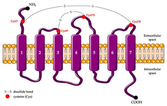 Fig. 1 Schematic secondary structure of P2RY12.1
Key structural properties of P2RY12:
- Typical sevenfold transmembrane domains
- Hydrophobic transmembrane core
- Conservative disulfide bond network
Functions of P2RY12
The P2Y12 receptor encoded by the P2RY12 gene has the core function of serving as the primary ADP receptor on platelets, mediating platelet activation and aggregation. However, it is also involved in many other physiological and pathological processes, including inflammatory responses and immune regulation.
| Function | Description |
| Platelet activation and aggregation | After binding to adenosine diphosphate (ADP), it initiates the Gi signaling pathway, inhibits adenylate cyclase, and reduces intracellular cAMP levels, thereby strongly promoting platelet activation, shape alteration, and irreversible aggregation. |
| Thrombus stability | By releasing and amplifying signals, it strengthens the initial hemostatic emboli and plays a key role in arterial thrombosis. |
| Inflammatory regulation | Microglia in the central nervous system and other immune cells express, involved in nerve inflammation, the activated state associated with the development of neurodegenerative diseases. |
| Atherosclerosis | In addition to the role of platelets, it also participates in the formation and development of atherosclerotic plaques by influencing inflammatory cells in the vascular wall. |
| Pharmacological targets | Is the clinical front line antiplatelet drugs, such as clopidogrel, on behalf of Greg los) directly targets, inhibit its function to be effective in preventing myocardial infarction and ischemic stroke. |
Unlike the rapid reversible characteristics of general GPCR signal transduction, when the P2Y12 receptor is inhibited by thiophenopyridine drugs (such as clopidogrel), its covalent binding mechanism leads to irreversible inactivation of its function throughout the platelet life cycle, which constitutes the unique pharmacological basis for it as a drug target.
Applications of P2RY12 and P2RY12 Antibody in Literature
- Gómez Morillas, Albert, Valérie C. Besson, and Dominique Lerouet. "Microglia and neuroinflammation: what place for P2RY12?." International Journal of Molecular Sciences 22.4 (2021): 1636. https://doi.org/10.3390/ijms22041636
This article focuses on the P2Y receptor P2RY12 on the surface of microglia, reviews its structure, function and research tools, and explores the key role of this receptor in neuroinflammation of acute and chronic brain diseases, aiming to clarify the mechanism of P2RY12 in mediating the migration of microglia and neuroinflammation.
- Škandík, Martin, et al. "Age-associated microglial transcriptome leads to diminished immunogenicity and dysregulation of MCT4 and P2RY12/P2RY13 related functions." Cell Death Discovery 11.1 (2025): 16. https://doi.org/10.1038/s41420-025-02295-1
This study utilized a long-term cultured BV-2 microglia model, combined with mouse and human datasets, and found that the immune response ability of senescent microglia was weakened. The abnormal expression of the key molecule MCT4 (SLC16A3) and the purine receptors P2RY12/P2RY13 jointly reveals its aging phenotypic characteristics.
- Bisht, Kanchan, et al. "Capillary-associated microglia regulate vascular structure and function through PANX1-P2RY12 coupling in mice." Nature communications 12.1 (2021): 5289. https://doi.org/10.1038/s41467-021-25590-8
Research has found that there is a special type of microglia (CAMs) around the capillaries in the brain, which sense the purine signals released by the PANX1 channel through the P2RY12 receptor, thereby regulating the structure and function of the neurovascular system.
- Palacios-Acedo, Ana Luisa, et al. "P2RY12-inhibitors reduce cancer-associated thrombosis and tumor growth in pancreatic cancers." Frontiers in Oncology 11 (2021): 704945. https://doi.org/10.3389/fonc.2021.704945
This study shows that in pancreatic cancer models, clopidogrel, which inhibits the P2RY12 receptor of platelets, is superior to aspirin in improving survival rates, inhibiting thrombosis, and slowing down tumor growth and metastasis. The expression of P2RY12 endows pancreatic cancer with a proliferation advantage, suggesting its potential as a therapeutic target.
- Uweru, Ogochukwu J., et al. "A P2RY12 deficiency results in sex-specific cellular perturbations and sexually dimorphic behavioral anomalies." Journal of Neuroinflammation 21.1 (2024): 95. https://doi.org/10.1186/s12974-024-03079-7
Research has found that there is a significant gender difference in the key receptor P2RY12 expressed by microglia: it is more expressed in adult females. The deletion of P2RY12 specifically leads to more severe cellular dysfunction and behavioral abnormalities in female mice, revealing that this receptor has gender-specific functions in neuroimmune interactions.
Creative Biolabs: P2RY12 Antibodies for Research
Creative Biolabs specializes in the production of high-quality P2RY12 antibodies for research and industrial applications. Our portfolio includes monoclonal antibodies tailored for ELISA, Flow Cytometry, Western blot, immunohistochemistry, and other diagnostic methodologies.
- Custom P2RY12 Antibody Development: Tailor-made solutions to meet specific research requirements.
- Bulk Production: Large-scale antibody manufacturing for industry partners.
- Technical Support: Expert consultation for protocol optimization and troubleshooting.
- Aliquoting Services: Conveniently sized aliquots for long-term storage and consistent experimental outcomes.
For more details on our P2RY12 antibodies, custom preparations, or technical support, contact us at email.
Reference
- Gómez Morillas, Albert, Valérie C. Besson, and Dominique Lerouet. "Microglia and neuroinflammation: what place for P2RY12?." International Journal of Molecular Sciences 22.4 (2021): 1636. https://doi.org/10.3390/ijms22041636
Anti-P2RY12 antibodies
 Loading...


Hot products 
-
Mouse Anti-BHMT Recombinant Antibody (CBYY-0547) (CBMAB-0550-YY)

-
Mouse Anti-ACLY Recombinant Antibody (V2-179314) (CBMAB-A0610-YC)

-
Mouse Anti-ATM Recombinant Antibody (2C1) (CBMAB-A3970-YC)

-
Mouse Anti-AMACR Recombinant Antibody (CB34A) (CBMAB-CA034LY)

-
Mouse Anti-CAPZB Recombinant Antibody (CBYY-C0944) (CBMAB-C2381-YY)

-
Mouse Anti-DHFR Recombinant Antibody (D0821) (CBMAB-D0821-YC)

-
Mouse Anti-CRYAB Recombinant Antibody (A4345) (CBMAB-A4345-YC)

-
Mouse Anti-APOA1 Monoclonal Antibody (CBFYR0637) (CBMAB-R0637-FY)

-
Mouse Anti-BIRC3 Recombinant Antibody (315304) (CBMAB-1214-CN)

-
Mouse Anti-AFM Recombinant Antibody (V2-634159) (CBMAB-AP185LY)

-
Mouse Anti-ELAVL4 Recombinant Antibody (6B9) (CBMAB-1132-YC)

-
Mouse Anti-BRCA2 Recombinant Antibody (CBYY-0790) (CBMAB-0793-YY)

-
Rat Anti-CD300A Recombinant Antibody (172224) (CBMAB-C0423-LY)

-
Mouse Anti-8-oxoguanine Recombinant Antibody (V2-7719) (CBMAB-1898CQ)

-
Rat Anti-CCR2 Recombinant Antibody (475301) (CBMAB-C1338-LY)

-
Mouse Anti-Acetyl SMC3 (K105/K106) Recombinant Antibody (V2-634053) (CBMAB-AP052LY)

-
Mouse Anti-ESR1 Recombinant Antibody (Y31) (CBMAB-1208-YC)

-
Rat Anti-ADGRE4 Recombinant Antibody (V2-160163) (CBMAB-F0011-CQ)

-
Mouse Anti-GFAP Recombinant Antibody (24) (CBMAB-G2927-LY)

-
Mouse Anti-NSUN6 Recombinant Antibody (D-5) (CBMAB-N3674-WJ)

- AActivation
- AGAgonist
- APApoptosis
- BBlocking
- BABioassay
- BIBioimaging
- CImmunohistochemistry-Frozen Sections
- CIChromatin Immunoprecipitation
- CTCytotoxicity
- CSCostimulation
- DDepletion
- DBDot Blot
- EELISA
- ECELISA(Cap)
- EDELISA(Det)
- ESELISpot
- EMElectron Microscopy
- FFlow Cytometry
- FNFunction Assay
- GSGel Supershift
- IInhibition
- IAEnzyme Immunoassay
- ICImmunocytochemistry
- IDImmunodiffusion
- IEImmunoelectrophoresis
- IFImmunofluorescence
- IGImmunochromatography
- IHImmunohistochemistry
- IMImmunomicroscopy
- IOImmunoassay
- IPImmunoprecipitation
- ISIntracellular Staining for Flow Cytometry
- LALuminex Assay
- LFLateral Flow Immunoassay
- MMicroarray
- MCMass Cytometry/CyTOF
- MDMeDIP
- MSElectrophoretic Mobility Shift Assay
- NNeutralization
- PImmunohistologyp-Paraffin Sections
- PAPeptide Array
- PEPeptide ELISA
- PLProximity Ligation Assay
- RRadioimmunoassay
- SStimulation
- SESandwich ELISA
- SHIn situ hybridization
- TCTissue Culture
- WBWestern Blot
