BrdU Antibodies
Background
BrdU is a synthetic nucleoside analogue whose structure is similar to that of thymidine, but the methyl group of thymidine is replaced by a bromine atom. This compound can integrate into newly synthesized DNA strands and replace the position of thymidine, and thus is often used as a marker of cell proliferation. BrdU has extensive applications in cell biology research, especially in detecting cell division and tracking cell lineages. It was first synthesized in the 1950s and quickly became an important tool for studying the cell cycle and DNA replication. By detecting BrDU-labeled cells with specific antibodies, scientists can study processes such as tissue regeneration, tumor growth and stem cell differentiation. The introduction of BrdU has greatly promoted the development of developmental biology, neuroscience and cancer research, providing crucial technical support for understanding cell dynamics.
Structure of BrdU
BrdU is a synthetic nucleoside analogue with a molecular weight of 307.1 Da. Its structure is similar to that of thymidine, but the methyl group of thymidine is replaced by a bromine atom. BrdU with different modified forms may have slight differences in molecular weight due to the different connecting groups.
| Compound | Molecular formula | Molecular weight (Da) | Structural features |
| BrdU | C9H₁₁BrN₂O₅ | 307.1 | Thymidine methyl bromide |
| BrdU-5' -triphosphate | C9H₁₄BrN₂O₁₄P₃ | 585.1 | Carrying three phosphate groups, can be directly involved in the DNA synthesis |
| Fluorescently labeled BrdU | In accordance with the markers | ~400-800 | Coupled with fluorescein /FITC, etc., for high-sensitivity detection |
BrdU integrates into the newly synthesized DNA strand through competitive substitution of thymidine, and its bromidine ring can be recognized by specific antibodies. This molecule has a planar structure. Its base part pairs with the complementary chain adenosine through hydrogen bonds, while the bromine atom enhances antigenicity. The detection of BrdU relies on anti-bromouridine antibodies, whose binding sites mainly target the stereoconformation of the pyrimidine ring. This technology is widely applied in cell cycle analysis, stem cell tracking and tumor proliferation research. Its detection sensitivity is significantly affected by DNA denaturation treatments (such as acid hydrolysis or thermal denaturation).
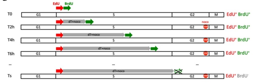 Fig. 1 The S-phase duration of cells was determined by EdU/BrdU double-labeled flow cytometry.1
Key structural properties of BrdU:
- Structure of thymidine analogues
- Planar conjugate system
- Phosphate diester bond integration
Functions of BrdU
The main function of BrdU is to serve as a marker for cell proliferation, used to track DNA replication and cell division activities. In addition, it plays a significant role in a variety of experimental applications, including cell cycle analysis, stem cell research, and tumor proliferation assessment.
| Function | Description |
| Cell proliferation marker | BrdU integrates into the newly synthesized DNA by replacing thymidine as direct evidence of cell division. |
| Cell cycle analysis | Cells in the S phase can be distinguished by pulse labeling, and the cell cycle dynamics can be analyzed. |
| Stem cell tracking | Long-term labeling of stem cells and their offspring for the study of tissue regeneration and differentiation lineages. |
| Tumor proliferation detection | To evaluate the proliferative activity of tumor cells and provide a basis for cancer research and clinical prognosis. |
| Research on DNA Damage and Repair | BrdU incorporation sites can be used as sensitive markers for DNA damage and repair. |
The detection of BrdU relies on anti-bromouridine antibodies, whose binding efficiency is affected by DNA denaturation treatment (such as acid or heat denaturation) to ensure that the antibodies can recognize BrdU embedded in the DNA strand. Compared with thymidine, the incorporation of BrdU does not significantly affect the normal function of DNA, but high concentrations may cause mild toxicity. This technology has become an important tool for cell proliferation research due to its high sensitivity and specificity.
Applications of BrdU and BrdU Antibody in Literature
1. Bartley, John, et al. "BrdU-positive cells in the neonatal mouse hippocampus following hypoxic-ischemic brain injury." BMC neuroscience 6.1 (2005): 15. https://doi.org/10.1186/1471-2202-6-15
The article shows that after neonatal hypoxic-ischemic brain injury, BrdU labeling indicates a significant increase in new cells in the hippocampus (especially the dentate gyrus), including microglia, endothelial cells and neurons. The peak of neurogenesis is later than that of other cells. The regeneration of nerves on the injured side is enhanced while the generation of oligodendrocytes is reduced, suggesting that the new compensatory mechanism may promote recovery.
2. Liboska, Radek, et al. "Most anti-BrdU antibodies react with 2'-deoxy-5-ethynyluridine-the method for the effective suppression of this cross-reactivity." PloS one 7.12 (2012): e51679. https://doi.org/10.1371/journal.pone.0051679
The article shows that most BrdU antibodies can cross-recognize EdU, and the residual EdU signal is difficult to be completely cleared. Although increasing the concentration of azide dyes can reduce the BrdU antibody signal, it will increase the background noise. Non-fluorescent azide molecules can effectively inhibit non-specific binding. Compared with hydrochloric acid treatment, the copper ion method for detecting BrdU can reduce background interference.
3. Danquah, Bright D., et al. "Mass Spectrometric analysis of antibody—Epitope peptide complex dissociation: Theoretical concept and practical procedure of binding strength characterization." Molecules 25.20 (2020): 4776. https://doi.org/10.1016/j.xpro.2021.100978
This study established a non-denatured immunofluorescence detection method based on BrdU labeling for the quantitative analysis of single-stranded DNA (ssDNA) produced by DNA terminal excision in mammalian cells. This method can accurately assess the degree of DNA terminal resection under different DNA damage conditions (such as ionizing radiation or hydroxyurea treatment) by specifically recognizing undenatated BrDU-labeled ssDNA through anti-BRDU antibodies, and is compatible with co-labeling analysis of cell cycle markers.
4. Bialic, Marta, et al. "Measuring S-phase duration from asynchronous cells using dual EdU-BrdU Pulse-chase labeling flow cytometry." Genes 13.3 (2022): 408. https://doi.org/10.3390/genes13030408
This study developed a flow cytometry method based on EdU/BrdU dual labeling combined with thymidine tracking, which can directly measure the duration of the S phase without cell synchronization. This method is applicable to multiple cell types and can detect the impact of drugs or mutations on the S phase. It has also been found that the S phase of some cancer cell lines is longer than that of normal cells.
5. Yahaya, B., G. McLachlan, and D. D. S. Collie. "BrdU pulse labelling in vivo to characterise cell proliferation during regeneration and repair following injury to the airway wall in sheep." The Scientific World Journal 2013.1 (2013): 871932. https://doi.org/10.1155/2013/871932
This study utilized BrdU labeling technology to track the cell proliferation dynamics during the airway injury repair process in sheep. The results showed that the number of BRDU-positive cells in the airway wall increased significantly 1-3 days after injury, and the proliferation activity decreased and was accompanied by airway remodeling 7 days later. This confirmed that the in vivo pulse labeling of BrdU could effectively trace the proliferation pattern of cells during the repair period.
Creative Biolabs: BrdU Antibodies for Research
Creative Biolabs specializes in the production of high-quality BrdU antibodies for research and industrial applications. Our portfolio includes monoclonal antibodies tailored for ELISA, Flow Cytometry, Western blot, immunohistochemistry, and other diagnostic methodologies.
- Custom BrdU Antibody Development: Tailor-made solutions to meet specific research requirements.
- Bulk Production: Large-scale antibody manufacturing for industry partners.
- Technical Support: Expert consultation for protocol optimization and troubleshooting.
- Aliquoting Services: Conveniently sized aliquots for long-term storage and consistent experimental outcomes.
For more details on our BrdU antibodies, custom preparations, or technical support, contact us at email.
Reference
- Bialic, Marta, et al. "Measuring S-phase duration from asynchronous cells using dual EdU-BrdU Pulse-chase labeling flow cytometry." Genes 13.3 (2022): 408. https://doi.org/10.3390/genes13030408
Anti-BrdU antibodies
 Loading...


Hot products 
-
Mouse Anti-ARID3A Antibody (A4) (CBMAB-0128-YC)

-
Mouse Anti-CORO1A Recombinant Antibody (4G10) (V2LY-1206-LY806)

-
Mouse Anti-ARHGAP5 Recombinant Antibody (54/P190-B) (CBMAB-P0070-YC)

-
Mouse Anti-ACTG1 Recombinant Antibody (V2-179597) (CBMAB-A0916-YC)

-
Mouse Anti-BCL6 Recombinant Antibody (CBYY-0435) (CBMAB-0437-YY)

-
Mouse Anti-CSPG4 Recombinant Antibody (CBFYM-1050) (CBMAB-M1203-FY)

-
Mouse Anti-DLG1 Monolconal Antibody (4F3) (CBMAB-0225-CN)

-
Mouse Anti-CFL1 Recombinant Antibody (CBFYC-1771) (CBMAB-C1833-FY)

-
Mouse Anti-CD33 Recombinant Antibody (6C5/2) (CBMAB-C8126-LY)

-
Mouse Anti-CCN2 Recombinant Antibody (CBFYC-2383) (CBMAB-C2456-FY)

-
Mouse Anti-GIPC2 Recombinant Antibody (10) (CBMAB-G0476-LY)

-
Mouse Anti-GLP1R Recombinant Antibody (4F3) (CBMAB-G0521-LY)

-
Mouse Anti-C5b-9 Recombinant Antibody (aE11) (CBMAB-AO138LY)

-
Rabbit Anti-ALDOA Recombinant Antibody (D73H4) (CBMAB-A2314-YC)

-
Mouse Anti-CEMIP Recombinant Antibody (3C12) (CBMAB-K0296-LY)

-
Mouse Anti-BANF1 Recombinant Antibody (3F10-4G12) (CBMAB-A0707-LY)

-
Mouse Anti-AKT1 (Phosphorylated S473) Recombinant Antibody (V2-505430) (PTM-CBMAB-0067LY)

-
Mouse Anti-APOH Recombinant Antibody (4D9A4) (CBMAB-A3249-YC)

-
Mouse Anti-ADAM12 Recombinant Antibody (V2-179752) (CBMAB-A1114-YC)

-
Mouse Anti-CCS Recombinant Antibody (CBFYC-1093) (CBMAB-C1150-FY)

- AActivation
- AGAgonist
- APApoptosis
- BBlocking
- BABioassay
- BIBioimaging
- CImmunohistochemistry-Frozen Sections
- CIChromatin Immunoprecipitation
- CTCytotoxicity
- CSCostimulation
- DDepletion
- DBDot Blot
- EELISA
- ECELISA(Cap)
- EDELISA(Det)
- ESELISpot
- EMElectron Microscopy
- FFlow Cytometry
- FNFunction Assay
- GSGel Supershift
- IInhibition
- IAEnzyme Immunoassay
- ICImmunocytochemistry
- IDImmunodiffusion
- IEImmunoelectrophoresis
- IFImmunofluorescence
- IGImmunochromatography
- IHImmunohistochemistry
- IMImmunomicroscopy
- IOImmunoassay
- IPImmunoprecipitation
- ISIntracellular Staining for Flow Cytometry
- LALuminex Assay
- LFLateral Flow Immunoassay
- MMicroarray
- MCMass Cytometry/CyTOF
- MDMeDIP
- MSElectrophoretic Mobility Shift Assay
- NNeutralization
- PImmunohistologyp-Paraffin Sections
- PAPeptide Array
- PEPeptide ELISA
- PLProximity Ligation Assay
- RRadioimmunoassay
- SStimulation
- SESandwich ELISA
- SHIn situ hybridization
- TCTissue Culture
- WBWestern Blot
