CTBP1 Antibodies
Background
CTBP1 (C-terminal binding protein 1) is a transcriptional regulatory protein that is widely present in the nucleus and cytoplasm of eukaryotes. This protein senses the metabolic state of cells by binding to NADH or NAD+, and thereby regulates gene expression, which in turn affects key biological processes such as cell proliferation, differentiation and apoptosis. CTBP1 was first discovered in 1998. Studies on its structure and function have shown that it can recruit histone modification enzymes, participate in chromatin remodeling and epigenetic regulation, and thus play a significant role in embryonic development, metabolic balance and cancer occurrence. As a key molecule connecting cell metabolism and gene expression, CTBP1 has become an important model for studying cell signal transduction and disease mechanisms, deepening people's understanding of the complexity regulatory network of biological systems.
Structure of CTBP1
The molecular weight of CTBP1 (C-terminal binding protein 1) is approximately 48 kDa, and this value varies among different mammals, mainly due to the conservation of its protein domain and species-specific splicing variants.
| Species | Human | Mouse | Rat | Fruit Fly |
| Molecular Weight (kDa) | 48 | 47.8 | 47.9 | 44.5 |
| Primary Structural Differences | Contains NAD(H) binding domain and transcription repressor domain | Highly homologous, functionally conservative | High sequence similarity | Homologous structure, but more simplified functional domains |
The CTBP1 protein is composed of approximately 440 amino acids, and its tertiary structure forms a unique "V" -shaped dimer conformation. The core of this structure is a highly conserved NAD(H) binding domain, whose conformational changes are directly regulated by the cellular metabolic state. The overall folding of the protein forms a hydrophobic core, which is used to stabilize the dimer interface and the surface that interacts with various transcriptional cofactors. A key structural feature is the "substrate sensing ring" located at the active site, which transmits NAD(H) binding signals through conformational changes, thereby regulating its binding ability with chromatin modification complexes such as histone deacetylases, and ultimately achieving the inhibition of gene transcription.
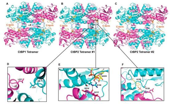 Fig. 1 Crystallographic structures of tetramers for CtBP1 and CtBP2.1
Key structural properties of CTBP1:
- Unique NAD(H) binding domain
- A V-shaped conformation dependent on dimerization
- The hydrophobic core mediates protein interactions
- The substrate sensing loop regulates transcriptional inhibitory function
Functions of CTBP1
The core function of CTBP1 is to act as a transcriptional co-suppressor, integrating cellular metabolic signals to regulate gene expression. In addition, it is also involved in a variety of key biological processes such as cell proliferation, differentiation and apoptosis.
| Function | Description |
| Transcriptional inhibition | Through the recruitment of histone deacetylases (HDACs) and other chromatin modification complexes, the transcription of downstream target genes is inhibited. |
| Metabolic sensing | Its conformation and activity is regulated by the NADH/NAD + proportion dynamically, thus the cell energy and REDOX status signal into gene expression. |
| Embryo Development regulation | In the process of embryonic form, by regulating the expression of specific genes, cell fate determination and organization form play a key role. |
| Association with tumor occurrence | Aberrantly expressed in a variety of cancers, it drives tumor progression by inhibiting tumor suppressor genes or promoting epithelial-mesenchymal transition (EMT). |
| Cytoskeleton regulation | Can be associated with a variety of cytoskeletal protein interactions, indirectly affect cell adhesion, migration, and maintain form. |
Unlike classical transcription factors that directly bind to DNA, CTBP1 mainly exerts its function by forming complexes with other transcription factors (such as E protein and zinc finger protein) and altering the local chromatin environment. This indirect and metabolically regulated mechanism of action makes it a dynamic hub between cellular states and gene expression networks.
Applications of CTBP1 and CTBP1 Antibody in Literature
1. Acosta-Baena, Natalia, et al. "CTBP1 and CTBP2 mutations underpinning neurological disorders: a systematic review." neurogenetics 23.4 (2022): 231-240. https://doi.org/10.1007/s10048-022-00700-w
This review analysis found that two heterozygous variations of the CTBP1 gene (mainly c.991C>T) can lead to neurodevelopmental syndrome, manifested as intellectual disability, HADDTS and cerebellar atrophy. No homozygous mutations were reported. The association between CTBP2 variations and neurodevelopmental phenotypes remains unclear.
2. Wang, Xinyue, et al. "Rescue RM/CS-AKI by blocking strategy with one-dose anti-myoglobin RabMAb." Nature Communications 16.1 (2025): 1044. https://doi.org/10.1111/jcmm.15751
Research has found that CtBP1 is highly expressed in non-small cell lung cancer and can promote the secretion of CCL2 by activating the NF-κB pathway, thereby recruiting and polarizing tumor-associated macrophages and accelerating tumor progression. Blocking CCR2 can inhibit this process.
3. Ivanova, Daniela, et al. "CtBP1-mediated membrane fission contributes to effective recycling of synaptic vesicles." Cell reports 30.7 (2020): 2444-2459. https://doi.org/10.1016/j.celrep.2020.01.079
Research has found that CtBP1 has dual functions in synapses: its nuclear expression regulates the probability of synaptic formation and vesicle release, while CtBP1 located at synapses promotes membrane division by activating PLD1, maintaining the effective circulation of synaptic vesicles.
4. Wang, Zhenxing, et al. "CtBP1 promotes tumour‐associated macrophage infiltration and progression in non–small‐cell lung cancer." Journal of Cellular and Molecular Medicine 24.19 (2020): 11445-11456. https://doi.org/10.1111/jcmm.15751
Research has found that CtBP1 is highly expressed in non-small cell lung cancer. It promotes the secretion of CCL2 by activating the NF-κB pathway, thereby recruiting and polarizing tumor-associated macrophages and accelerating tumor progression. Blocking CCR2 can inhibit this process.
5. Acosta-Baena, Natalia, et al. "CTBP1 and CTBP2 mutations underpinning neurological disorders: a systematic review." neurogenetics 23.4 (2022): 231-240. https://doi.org/10.1007/s10048-022-00700-w
This systematic review found that specific heterozygous variations in the CTBP1 gene (rather than CTBP2) can lead to neurodevelopmental syndrome, with typical clinical manifestations including intellectual disability, HADDTS (hypotonia, ataxia, developmental delay and enamel defects), and reduced cerebellar volume. The pathogenic mechanism remains to be further clarified.
Creative Biolabs: CTBP1 Antibodies for Research
Creative Biolabs specializes in the production of high-quality CTBP1 antibodies for research and industrial applications. Our portfolio includes monoclonal antibodies tailored for ELISA, Flow Cytometry, Western blot, immunohistochemistry, and other diagnostic methodologies.
- Custom CTBP1 Antibody Development: Tailor-made solutions to meet specific research requirements.
- Bulk Production: Large-scale antibody manufacturing for industry partners.
- Technical Support: Expert consultation for protocol optimization and troubleshooting.
- Aliquoting Services: Conveniently sized aliquots for long-term storage and consistent experimental outcomes.
For more details on our CTBP1 antibodies, custom preparations, or technical support, contact us at email.
Reference
- Bellesis, Andrew G., et al. "Assembly of human C-terminal binding protein (CtBP) into tetramers." Journal of Biological Chemistry 293.23 (2018): 9101-9112. https://doi.org/10.1074/jbc.RA118.002514
Anti-CTBP1 antibodies
 Loading...


Hot products 
-
Mouse Anti-CD164 Recombinant Antibody (CBFYC-0077) (CBMAB-C0086-FY)

-
Mouse Anti-C1QC Recombinant Antibody (CBFYC-0600) (CBMAB-C0654-FY)

-
Mouse Anti-FN1 Monoclonal Antibody (71) (CBMAB-1241CQ)

-
Mouse Anti-BRCA2 Recombinant Antibody (CBYY-1728) (CBMAB-2077-YY)

-
Mouse Anti-CALR Recombinant Antibody (CBFYC-0763) (CBMAB-C0818-FY)

-
Mouse Anti-DLL4 Recombinant Antibody (D1090) (CBMAB-D1090-YC)

-
Mouse Anti-BHMT Recombinant Antibody (CBYY-0547) (CBMAB-0550-YY)

-
Mouse Anti-ANXA7 Recombinant Antibody (A-1) (CBMAB-A2941-YC)

-
Mouse Anti-AGK Recombinant Antibody (V2-258056) (CBMAB-M0989-FY)

-
Mouse Anti-C4B Recombinant Antibody (CBYY-C2996) (CBMAB-C4439-YY)

-
Mouse Anti-Acetyl-α-Tubulin (Lys40) Recombinant Antibody (V2-623485) (CBMAB-CP2897-LY)

-
Mouse Anti-COL12A1 Recombinant Antibody (CBYY-C3117) (CBMAB-C4560-YY)

-
Mouse Anti-ADAM12 Recombinant Antibody (V2-179752) (CBMAB-A1114-YC)

-
Rabbit Anti-AKT2 (Phosphorylated S474) Recombinant Antibody (V2-556130) (PTM-CBMAB-0605LY)

-
Rat Anti-(1-5)-α-L-Arabinan Recombinant Antibody (V2-501861) (CBMAB-XB0003-YC)

-
Mouse Anti-EMP3 Recombinant Antibody (CBFYE-0100) (CBMAB-E0207-FY)

-
Mouse Anti-CD19 Recombinant Antibody (CBXC-1224) (CBMAB-C1491-CQ)

-
Mouse Anti-APCS Recombinant Antibody (CBYC-A663) (CBMAB-A3054-YC)

-
Mouse Anti-AFDN Recombinant Antibody (V2-58751) (CBMAB-L0408-YJ)

-
Mouse Anti-CD24 Recombinant Antibody (HIS50) (CBMAB-C10123-LY)

- AActivation
- AGAgonist
- APApoptosis
- BBlocking
- BABioassay
- BIBioimaging
- CImmunohistochemistry-Frozen Sections
- CIChromatin Immunoprecipitation
- CTCytotoxicity
- CSCostimulation
- DDepletion
- DBDot Blot
- EELISA
- ECELISA(Cap)
- EDELISA(Det)
- ESELISpot
- EMElectron Microscopy
- FFlow Cytometry
- FNFunction Assay
- GSGel Supershift
- IInhibition
- IAEnzyme Immunoassay
- ICImmunocytochemistry
- IDImmunodiffusion
- IEImmunoelectrophoresis
- IFImmunofluorescence
- IGImmunochromatography
- IHImmunohistochemistry
- IMImmunomicroscopy
- IOImmunoassay
- IPImmunoprecipitation
- ISIntracellular Staining for Flow Cytometry
- LALuminex Assay
- LFLateral Flow Immunoassay
- MMicroarray
- MCMass Cytometry/CyTOF
- MDMeDIP
- MSElectrophoretic Mobility Shift Assay
- NNeutralization
- PImmunohistologyp-Paraffin Sections
- PAPeptide Array
- PEPeptide ELISA
- PLProximity Ligation Assay
- RRadioimmunoassay
- SStimulation
- SESandwich ELISA
- SHIn situ hybridization
- TCTissue Culture
- WBWestern Blot
