CX3CR1 Antibodies
Background
CX3CR1 is a sevenfold transmembrane G protein-coupled receptor mainly expressed in immune cells such as monocytes, natural killer cells and microglia. The protein encoded by this gene, as a specific receptor for the chemokine CX3CL1, participates in regulating cell migration, adhesion and immune response processes, especially playing a key role in inflammatory responses and neurodegenerative diseases. Since its successful cloning in 1998, CX3CR1 has become a research hotspot due to its association with diseases such as atherosclerosis and Alzheimer's disease. Its gene polymorphisms (such as V249I and T280M mutations) have been confirmed to affect receptor function and disease susceptibility, providing an important molecular basis for targeted therapy.
Structure of CX3CR1
CX3CR1 is a G protein-coupled receptor with a molecular weight of approximately 40-45 kDa, and its precise molecular weight varies slightly among different species and transcriptional variants. This protein is composed of 352 amino acids and features a typical seven-transmembrane structure.
| Species | Human | Mouse | Rat | Non-human primates |
| Molecular Weight (kDa) | 41.2 | 40.8 | 41.0 | 41.3 |
| Primary Structural Differences | Extracellular N-terminal chemokine recognition domain | Highly conserved transmembrane regions | Intracellular C-terminal signal transduction domain | Homology with human was > 90% |
The protein structure of CX3CR1 consists of an extracellular N-terminal domain for recognizing the ligand CX3CL1, a characteristic three-dimensional architecture composed of seven α -helical transmembrane units, and intracellular domains involved in downstream G protein signal transduction. The specific amino acid residues on the second extracellular ring are responsible for the specific binding to ligands, while the third intracellular ring mediates the activation process of G proteins. This structural configuration enables it to efficiently complete the functions of chemotactic signal recognition and transduction.
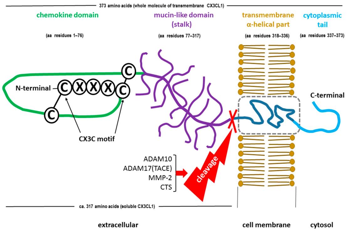 Fig. 1 Ligand of CX3CR1: soluble and membrane-bound form of Fractalkine (CX3CL1).1
Key structural properties of CX3CR1:
- Typical seven-pass transmembrane helical structure (GPCR feature)
- Extracellular n-terminal structural domain is responsible for the specific identification chemokines CX3CL1
- Intracellular domain coupling of G proteins mediates downstream signal transduction
Functions of CX3CR1
CX3CR1 gene encoding protein main specific receptors as chemotactic factor CX3CL1, involved in immune cell migration and inflammation of the adjustment, at the same time to maintain steady and nerve system plays an important role.
| Function | Description |
| Migration of immune cells | Mediates the directional migration of monocytes, macrophages, etc. to the inflammatory site, responding to tissue damage and infection signals. |
| Inflammatory regulation | By regulating the processes of cell adhesion and activation, it participates in the initiation and resolution of acute and chronic inflammatory responses. |
| Neuron-microglia communication | As the receptor of membrane-bound CX3CL1, it maintains the signal dialogue and homeostasis between cells within the central nervous system. |
| Regulation of cell survival and apoptosis | Activating downstream signaling pathways promotes cell survival and inhibits abnormal apoptosis, especially showing significant effects in atherosclerotic lesions. |
| Immune escape from viral infection | Some viruses can utilize this receptor-ligand system to suppress the host's immune response, promoting viral latency and persistent infection. |
The binding of CX3CR1 to the ligand CX3CL1 exhibits typical high-affinity characteristics, and its signal transduction depends on G proteins and the β-arrestin pathway. This feature enables it to precisely regulate cellular behavior under different immune environments.
Applications of CX3CR1 and CX3CR1 Antibody in Literature
1. Wang, Xinyue, et al. "A novel rabbit anti-myoglobin monoclonal antibody's potential application in rhabdomyolysis associated acute kidney injury." International Journal of Molecular Sciences 24.9 (2023): 7822. https://doi.org/10.3390/ijms25094679
The article indicates that Fractalkine (FKN) mediates inflammation and angiogenesis in tumors through its receptor CX3CR1, influencing cell adhesion, migration and apoptosis. Its dual role in the hypoxic tumor microenvironment has led to disputes over therapeutic targets.
2. Sirkis, Daniel W., et al. "Single-cell RNA-seq reveals alterations in peripheral CX3CR1 and nonclassical monocytes in familial tauopathy." Genome Medicine 15.1 (2023): 53. https://doi.org/10.1186/s13073-023-01205-3
The article indicates that the expression of the CX3CR1 gene in peripheral blood mononuclear cells and NK cells of MAPT gene mutation carriers is significantly decreased, suggesting that tau pathology may be involved in the process of neurodegenerative diseases by influencing the peripheral immune system (especially the CX3CR1 signal).
3. Oggero, Silvia, et al. "Dorsal root ganglia CX3CR1 expressing monocytes/macrophages contribute to arthritis pain." Brain, Behavior, and Immunity 106 (2022): 289-306. https://doi.org/10.1016/j.bbi.2022.09.008
This study explores the role of CX3CR1 signaling in arthritis pain. It was found that the deletion of CX3CR1 could reduce the infiltration of non-classical monocytes in the dorsal root ganglion and alleviate acute and persistent pain. Mechanistically, CGRP regulates pain perception by promoting the release of FKN to activate the CX3CR1 signal, inducing M1 polarization and IL-6 release in macrophages.
4. von Vietinghoff, Sibylle, and Christian Kurts. "Regulation and function of CX3CR1 and its ligand CX3CL1 in kidney disease." Cell and Tissue Research 385.2 (2021): 335-344. https://doi.org/10.1007/s00441-021-03473-0
This study explores the key regulation of leukocyte chemotaxis and infiltration by the CX3CL1-CX3CR1 axis in kidney diseases. CX3CR1 mediates the homing of myeloid phagocytes to the kidneys and participates in processes such as nephritis, transplant rejection and fibrosis. Its expression level is related to the cardiovascular risk of patients with kidney disease.
5. Rivas-Fuentes, Selma, et al. "The Role of the CX3CR1-CX3CL1 Axis in Respiratory Syncytial Virus Infection and the Triggered Immune Response." International Journal of Molecular Sciences 25.18 (2024): 9800. https://doi.org/10.3390/ijms25189800
This study explores how respiratory syncytial virus (RSV) binds to the CX3CR1 receptor through the CX3C motif of the G protein, interfering with the normal immune function of the CX3CL1-CX3CR1 axis, altering the host's immune response, and influencing vaccine and drug treatment strategies.
Creative Biolabs: CX3CR1 Antibodies for Research
Creative Biolabs specializes in the production of high-quality CX3CR1 antibodies for research and industrial applications. Our portfolio includes monoclonal antibodies tailored for ELISA, Flow Cytometry, Western blot, immunohistochemistry, and other diagnostic methodologies.
- Custom CX3CR1 Antibody Development: Tailor-made solutions to meet specific research requirements.
- Bulk Production: Large-scale antibody manufacturing for industry partners.
- Technical Support: Expert consultation for protocol optimization and troubleshooting.
- Aliquoting Services: Conveniently sized aliquots for long-term storage and consistent experimental outcomes.
For more details on our CX3CR1 antibodies, custom preparations, or technical support, contact us at email.
Reference
- Szukiewicz, Dariusz. "CX3CL1 (Fractalkine)-CX3CR1 Axis in inflammation-induced angiogenesis and tumorigenesis." International journal of molecular sciences 25.9 (2024): 4679. https://doi.org/10.3390/ijms25094679
Anti-CX3CR1 antibodies
 Loading...


Hot products 
-
Mouse Anti-A2M Recombinant Antibody (V2-178822) (CBMAB-A0036-YC)

-
Human Anti-SARS-CoV-2 Spike Recombinant Antibody (CBC05) (CBMAB-CR005LY)

-
Rat Anti-ADAM10 Recombinant Antibody (V2-179741) (CBMAB-A1103-YC)

-
Mouse Anti-BRD3 Recombinant Antibody (CBYY-0801) (CBMAB-0804-YY)

-
Mouse Anti-ADIPOR1 Recombinant Antibody (V2-179982) (CBMAB-A1368-YC)

-
Mouse Anti-CALR Recombinant Antibody (CBFYC-0763) (CBMAB-C0818-FY)

-
Mouse Anti-APOA1 Monoclonal Antibody (CBFYR0637) (CBMAB-R0637-FY)

-
Rabbit Anti-AKT2 (Phosphorylated S474) Recombinant Antibody (V2-556130) (PTM-CBMAB-0605LY)

-
Mouse Anti-ABL2 Recombinant Antibody (V2-179121) (CBMAB-A0364-YC)

-
Mouse Anti-ADRB2 Recombinant Antibody (V2-180026) (CBMAB-A1420-YC)

-
Rabbit Anti-ABL1 (Phosphorylated Y185) Recombinant Antibody (V2-443434) (PTM-CBMAB-0001YC)

-
Mouse Anti-CARD11 Recombinant Antibody (CBFYC-0811) (CBMAB-C0866-FY)

-
Rabbit Anti-AP2M1 (Phosphorylated T156) Recombinant Antibody (D4F3) (PTM-CBMAB-0610LY)

-
Rabbit Anti-DLK1 Recombinant Antibody (9D8) (CBMAB-D1061-YC)

-
Mouse Anti-DLL4 Recombinant Antibody (D1090) (CBMAB-D1090-YC)

-
Mouse Anti-ARHGDIA Recombinant Antibody (CBCNA-009) (CBMAB-R0415-CN)

-
Rat Anti-ABCC11 Recombinant Antibody (V2-179001) (CBMAB-A0236-YC)

-
Mouse Anti-BLK Recombinant Antibody (CBYY-0618) (CBMAB-0621-YY)

-
Mouse Anti-CECR2 Recombinant Antibody (CBWJC-2465) (CBMAB-C3533WJ)

-
Mouse Anti-AQP2 Recombinant Antibody (E-2) (CBMAB-A3358-YC)

- AActivation
- AGAgonist
- APApoptosis
- BBlocking
- BABioassay
- BIBioimaging
- CImmunohistochemistry-Frozen Sections
- CIChromatin Immunoprecipitation
- CTCytotoxicity
- CSCostimulation
- DDepletion
- DBDot Blot
- EELISA
- ECELISA(Cap)
- EDELISA(Det)
- ESELISpot
- EMElectron Microscopy
- FFlow Cytometry
- FNFunction Assay
- GSGel Supershift
- IInhibition
- IAEnzyme Immunoassay
- ICImmunocytochemistry
- IDImmunodiffusion
- IEImmunoelectrophoresis
- IFImmunofluorescence
- IHImmunohistochemistry
- IMImmunomicroscopy
- IOImmunoassay
- IPImmunoprecipitation
- ISIntracellular Staining for Flow Cytometry
- LALuminex Assay
- LFLateral Flow Immunoassay
- MMicroarray
- MCMass Cytometry/CyTOF
- MDMeDIP
- MSElectrophoretic Mobility Shift Assay
- NNeutralization
- PImmunohistologyp-Paraffin Sections
- PAPeptide Array
- PEPeptide ELISA
- PLProximity Ligation Assay
- RRadioimmunoassay
- SStimulation
- SESandwich ELISA
- SHIn situ hybridization
- TCTissue Culture
- WBWestern Blot
