HLA-DRB1 Antibodies
Background
The HLA-DRB1 gene, as a key member of the human major histocompatibility complex (MHC) II region, is mainly expressed on the surface of antigen-presenting cells. The protein encoded by this gene interacts with foreign peptide segments and presents them to T-cell receptors, playing a core regulatory role in immune recognition and response. Its high polymorphism directly affects an individual's susceptibility to infections, autoimmune diseases, and transplant rejection, making it an important target in immunogenetics research. This gene was jointly discovered along with the MHC system in the 1970s. Its three-dimensional structure and peptide binding mechanism were gradually resolved through crystallographic techniques, leading to breakthroughs in immunological theories. The continuous research on HLA-DRB1 has deepened our understanding of immune regulation, disease associations, and individualized medical care, and has practical applications in clinical diagnosis and drug development.
Structure of HLA-DRB1
The HLA-DRB1 gene is a key gene located in the MHC II region on human chromosome 6. The protein encoded by this gene typically has a molecular weight ranging from 25 to 33 kDa. The specific value depends on different allele types and their post-translational modifications.
| Species | Human | Mouse | Rhesus monkey | Chimpanzee |
| Molecular Weight (kDa) | ~25-33 | ~27-31 | ~26-32 | ~25-33 |
| Primary Structural Differences | High polymorphism determines the antigen binding specificity | Homologous to human, but differing in key antigen-binding sites | Extremely similar to humans, it serves as an important experimental model | Sequence and human are the most close to, the structure is highly conservative |
The protein encoded by this gene consists of approximately 266 amino acids. Its three-dimensional structure features an open antigen-binding groove composed of α helices and β sheets. The core functional region of this protein is the antigen-binding site formed by its secondary structure (the "bottom" composed of β sheets and the "side wall" composed of α helices), which can specifically bind and present exogenous peptide segments. The high polymorphism on the protein surface is mainly concentrated on the amino acid residues of the antigen-binding groove, and these differences directly determine the specificity of immune recognition and disease susceptibility in different individuals.
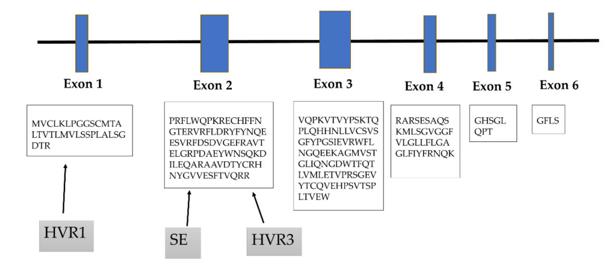 Fig. 1 HLA-DRB1 gene is encoded by six exons.1
Key structural properties of HLA-DRB1:
- Composed of alpha helix with beta fold open the antigen slots
- Highly polymorphic amino acid residues line the antigen-binding groove surface
- The peptide binding domain realizes the specific recognition and stable binding of foreign antigen through the hydrogen bond network
Functions of HLA-DRB1
The core function of the HLA-DRB1 gene is to present antigenic peptide segments during the adaptive immune response. However, it is also deeply involved in various immune regulatory processes, including the establishment of self-immune tolerance and the regulation of the intensity of immune responses.
| Function | Description |
| Antigen Presentation | The genetically encoded proteins form MHC II molecules on the surface of antigen-presenting cells, bind and display exogenous peptide segments, which are recognized by CD4+ T cells. |
| Immune Recognition Initiation | By specifically binding to the T-cell receptor, it initiates the adaptive immune response against pathogens, serving as the first "specific alarm" in the immune defense mechanism. |
| Autoimmune Regulation | Its high polymorphism affects the presentation of self-peptide segments. Certain specific genotypes are significantly associated with the risks of various autoimmune diseases such as ankylosing spondylitis and rheumatoid arthritis. |
| Decision on Organ Rejection | The degree of HLA-DRB1 matching between the donor and the recipient is a crucial factor determining the severity of organ transplant rejection, and it directly affects the success rate of the transplant. |
| Disease Susceptibility Association | Different allele genotypes result in genetic differences in an individual's resistance or susceptibility to specific infectious diseases (such as tuberculosis, leprosy). |
The HLA-DRB1 protein has a highly selective but relatively low affinity for antigenic peptide segments. This is in line with the biological requirement of presenting multiple different antigens, ensuring that the immune system can extensively monitor and respond flexibly to constantly changing pathogen threats.
Applications of HLA-DRB1 and HLA-DRB1 Antibody in Literature
1. Brynedal, Boel, et al. "HLA-A confers an HLA-DRB1 independent influence on the risk of multiple sclerosis." PloS one 2.7 (2007): e664. https://doi.org/10.1371/journal.pone.0000664
This study found that the HLA-A gene has an independent effect on the risk of multiple sclerosis (MS) that is separate from the HLA-DRB1 gene. Among the 1084 MS patients and 1347 control subjects, the HLA-A*02 allele significantly reduced the risk. The results confirmed that the genes in the HLA class I region have an additional effect on the onset of MS, suggesting that both the regulation and effect mechanisms of the immune system are involved in the occurrence of MS.
2. Wysocki, Tomasz, Marzena Olesińska, and Agnieszka Paradowska-Gorycka. "Current understanding of an emerging role of HLA-DRB1 gene in rheumatoid arthritis–from research to clinical practice." Cells 9.5 (2020): 1127. https://doi.org/10.3390/cells9051127
The article indicates that the onset of rheumatoid arthritis is closely related to the HLA-DRB1 gene. This gene affects the structure of immune molecules, antigen binding, and non-joint symptoms, and is also related to the treatment response. A deeper understanding of its mechanism of action is conducive to promoting the development of personalized diagnosis and treatment for RA.
3. Danquah, Bright D., et al. "Mass Spectrometric analysis of antibody—Epitope peptide complex dissociation: Theoretical concept and practical procedure of binding strength characterization." Molecules 25.20 (2020): 4776. https://doi.org/10.1186/ar1837
This study compared the HLA-DRB1/DQB1 genes of 159 RA patients with severe extra-articular manifestations and 178 RA patients without such manifestations. The results showed that only Felty syndrome was significantly associated with HLA-DRB1*0401; other extra-articular manifestations were mainly related to the DRB1*04 genotype carrying double-dose shared epitopes.
4. Alcina, Antonio, et al. "Multiple sclerosis risk variant HLA-DRB1* 1501 associates with high expression of DRB1 gene in different human populations." PloS one 7.1 (2012): e29819. https://doi.org/10.1371/journal.pone.0029819
Studies have shown that the risk of multiple sclerosis is closely related to HLA-DRB1*1501. The expression levels of genes such as DRB1 and DQB1 in carriers of the risk genotype are significantly increased. Research indicates that the high expression of specific alleles, together with their structural characteristics, jointly drive the susceptibility to the disease, rather than being solely determined by antigen presentation.
5. Liu, Mohan, et al. "Identification of HLA-DRB1 association to adalimumab immunogenicity." PLoS One 13.4 (2018): e0195325. https://doi.org/10.1371/journal.pone.0195325
This study, through HLA typing, found that HLA-DRB1*03 and HLA-DRB1*011 could increase the risk of adalimumab-resistant antibodies (AAA) formation, while HLA-DRB1*01 and HLA-DRB1*07 had protective effects. This indicates that specific HLA alleles play a crucial role in antibody production.
Creative Biolabs: HLA-DRB1 Antibodies for Research
Creative Biolabs specializes in the production of high-quality HLA-DRB1 antibodies for research and industrial applications. Our portfolio includes monoclonal antibodies tailored for ELISA, Flow Cytometry, Western blot, immunohistochemistry, and other diagnostic methodologies.
- Custom HLA-DRB1 Antibody Development: Tailor-made solutions to meet specific research requirements.
- Bulk Production: Large-scale antibody manufacturing for industry partners.
- Technical Support: Expert consultation for protocol optimization and troubleshooting.
- Aliquoting Services: Conveniently sized aliquots for long-term storage and consistent experimental outcomes.
For more details on our HLA-DRB1 antibodies, custom preparations, or technical support, contact us at email.
Reference
- Wysocki, Tomasz, Marzena Olesińska, and Agnieszka Paradowska-Gorycka. "Current understanding of an emerging role of HLA-DRB1 gene in rheumatoid arthritis–from research to clinical practice." Cells 9.5 (2020): 1127. https://doi.org/10.3390/cells9051127
Anti-HLA-DRB1 antibodies
 Loading...


Hot products 
-
Rabbit Anti-CBL Recombinant Antibody (D4E10) (CBMAB-CP0149-LY)

-
Mouse Anti-BLNK Recombinant Antibody (CBYY-0623) (CBMAB-0626-YY)

-
Mouse Anti-CD24 Recombinant Antibody (HIS50) (CBMAB-C10123-LY)

-
Mouse Anti-CTNND1 Recombinant Antibody (CBFYC-2414) (CBMAB-C2487-FY)

-
Mouse Anti-BPGM Recombinant Antibody (CBYY-1806) (CBMAB-2155-YY)

-
Rabbit Anti-ENO2 Recombinant Antibody (BA0013) (CBMAB-0272CQ)

-
Mouse Anti-CORO1A Recombinant Antibody (4G10) (V2LY-1206-LY806)

-
Mouse Anti-ACO2 Recombinant Antibody (V2-179329) (CBMAB-A0627-YC)

-
Rat Anti-EPO Recombinant Antibody (16) (CBMAB-E1578-FY)

-
Mouse Anti-AAV-5 Recombinant Antibody (V2-503417) (CBMAB-V208-1369-FY)

-
Mouse Anti-BIRC3 Recombinant Antibody (315304) (CBMAB-1214-CN)

-
Mouse Anti-CGAS Recombinant Antibody (CBFYM-0995) (CBMAB-M1146-FY)

-
Mouse Anti-C1QC Recombinant Antibody (CBFYC-0600) (CBMAB-C0654-FY)

-
Mouse Anti-ASB9 Recombinant Antibody (1D8) (CBMAB-A0529-LY)

-
Mouse Anti-DLC1 Recombinant Antibody (D1009) (CBMAB-D1009-YC)

-
Mouse Anti-ENO2 Recombinant Antibody (85F11) (CBMAB-0276CQ)

-
Mouse Anti-CD1C Recombinant Antibody (L161) (CBMAB-C2173-CQ)

-
Mouse Anti-CFL1 (Phospho-Ser3) Recombinant Antibody (CBFYC-1770) (CBMAB-C1832-FY)

-
Mouse Anti-FN1 Monoclonal Antibody (D6) (CBMAB-1240CQ)

-
Mouse Anti-ARG1 Recombinant Antibody (CBYCL-103) (CBMAB-L0004-YC)

- AActivation
- AGAgonist
- APApoptosis
- BBlocking
- BABioassay
- BIBioimaging
- CImmunohistochemistry-Frozen Sections
- CIChromatin Immunoprecipitation
- CTCytotoxicity
- CSCostimulation
- DDepletion
- DBDot Blot
- EELISA
- ECELISA(Cap)
- EDELISA(Det)
- ESELISpot
- EMElectron Microscopy
- FFlow Cytometry
- FNFunction Assay
- GSGel Supershift
- IInhibition
- IAEnzyme Immunoassay
- ICImmunocytochemistry
- IDImmunodiffusion
- IEImmunoelectrophoresis
- IFImmunofluorescence
- IGImmunochromatography
- IHImmunohistochemistry
- IMImmunomicroscopy
- IOImmunoassay
- IPImmunoprecipitation
- ISIntracellular Staining for Flow Cytometry
- LALuminex Assay
- LFLateral Flow Immunoassay
- MMicroarray
- MCMass Cytometry/CyTOF
- MDMeDIP
- MSElectrophoretic Mobility Shift Assay
- NNeutralization
- PImmunohistologyp-Paraffin Sections
- PAPeptide Array
- PEPeptide ELISA
- PLProximity Ligation Assay
- RRadioimmunoassay
- SStimulation
- SESandwich ELISA
- SHIn situ hybridization
- TCTissue Culture
- WBWestern Blot
