HSF1 Antibodies
Background
HSF1 (heat shock factor 1) exists in the form of a transcription factor and is mainly found in the cytoplasm and nucleus of vertebrates. This factor is activated and transported into the nucleus under cellular stress conditions, and regulates the expression of molecular chaperone by binding to the heat shock element in the promoter region of the heat shock protein gene, thereby maintaining intracellular protein homeostasis. Organisms highly rely on HSF1 to restore protein folding balance under stress conditions such as high temperature, oxidation or infection, as its activation state directly determines the cell's survival ability in response to stress. This factor was first discovered in fruit flies by Giardina and Lis in 1984. Subsequently, its mammalian homolog HSF1 was confirmed to be the core regulator of the heat shock response, and related research won several international Cell stress Society awards in the early 21st century. Its multi-level regulatory mechanism - including trimerization, phosphorylation modification and chaperone protein feedback regulation - has become a research paradigm for protein homeostasis, greatly promoting the development of fields such as cellular stress response, aging mechanism and cancer treatment.
Structure of HSF1
HSF1 is a transcription factor with a molecular weight of approximately 55 kDa. Its molecular weight is relatively conserved among different species, but it will change under stress due to post-translational modifications such as phosphorylation.
| Species | Human | Mouse | Fruit fly | Nematode | Yeast |
| Molecular Weight (kDa) | ~55 | ~55 | ~82 | ~40 | ~40 |
| Primary Structural Differences | Contains DNA binding domain, three polymerization domain, regulatory domain | Structure and function are highly conserved | Homologous proteins have similar domains | Homologous proteins are functionally conserved | For the most basic HSF homologous proteins |
The HSF1 protein typically contains about 500 amino acids, and its three-dimensional structure features a modular arrangement of functional domains. The core of this protein is its N-terminal DNA-binding domain, which performs transcriptional regulatory functions by recognizing and binding to a specific sequence (heat shock element, HSE) in the promoter region of the heat shock protein gene. The secondary structure of HSF1 is mainly composed of α -helix and β -folding, and these structural elements constitute a stable DNA binding domain framework. Among them, the hydrophobic core in the DNA binding domain is crucial for structural stability, while a β -hairpin structure known as the "wing" penetrates deep into the small grooves of the DNA double helix for recognition and binding, thereby achieving precise regulation of gene expression.
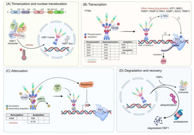 Fig. 1 The activation mechanism of HSF1.1
Key structural properties of HSF1:
- Modular functional domain structure
- N end formed spiral Angle - spiral motif with specific recognition of DNA sequences
- C-terminal zipper structure rich in leucine mediates trimerization activation under stress
- Conservative dna-binding domain through the hydrophobic core
- Regulatory domain contains multiple serine phosphorylation sites, as cell stress signal integration hub
Functions of HSF1
The core function of the HSF1 gene is to act as a transcription factor to activate the expression of heat shock proteins to maintain cellular homeostasis. However, it also plays a regulatory role in a wide range of physiological and pathological processes, including development, metabolism, aging and tumorigenesis.
| Function | Description |
| Heat shock reaction | Activated under stress conditions such as heat and oxidation, it induces the expression of molecular chaperones (such as HSP70, HSP90), helping misfolded proteins to fold correctly or degrade. |
| Developmental Regulation | It is needed at specific stages during embryonic development and is crucial for the normal formation of organs such as the nervous system and reproductive system. |
| Metabolic regulation | It participates in regulating genes related to energy metabolism and affects fat production and insulin sensitivity. |
| Regulation of Aging | Its activity declines with age, leading to the functional deterioration of the protein homeostasis network, and it is one of the key factors contributing to the aging of cells and the body. |
| Tumor occurrence | It is continuously activated in many cancer cells, promoting tumor growth and drug resistance by maintaining the protein homeostasis of cancer cells and inhibiting apoptosis. |
The activation kinetics of HSF1 is characterized by a strict "on-off" mode, which contrasts sharply with the smooth binding curve of myoglobin. In its basic state, HSF1 exists in the form of an inert monomer. Once the stress signal exceeds the threshold, it will rapidly undergo trimerization, nuclear translocation and strongly bind to DNA. This "all or nothing" characteristic ensures that the cell can make a rapid and unified protective response in critical moments.
Applications of HSF1 and HSF1 Antibody in Literature
1. Chin, Yeh, et al. "Targeting HSF1 for cancer treatment: mechanisms and inhibitor development." Theranostics 13.7 (2023): 2281. https://doi.org/10.7150/thno.82431
Research has found that HSF1 is a key factor in the heat shock response, regulating the extensive stress network in cancer cells and promoting tumor development. This article reviews its latest mechanism of action and the progress of targeted inhibitors, providing new ideas for cancer treatment.
2. Zhang, Bingwei, Yumei Fan, and Ke Tan. "HSF1 Activation Mechanisms, Disease Roles, and Small Molecule Therapeutics." International Journal of Biological Sciences 21.8 (2025): 3351. https://doi.org/10.7150/ijbs.110447
Research has found that HSF1 is a key regulatory factor for cellular stress, and its dysfunction is associated with various diseases such as cancer. This article systematically expounds its activation mechanism and pathogenic effects, and summarizes the small molecule inhibitors and activators that have been discovered so far, providing a new direction for targeted therapy.
3. Reyes, Antonia, et al. "Is there a role for HSF1 in viral infections?." FEBS Open Bio 12.6 (2022): 1112-1124. https://doi.org/10.1002/2211-5463.13419
Research has found that HSF1 is a key transcription factor regulating the heat shock response. This article reviews its role in various viral infections, explores the impact of its activation on host health, and provides new ideas for antiviral therapy regulated by HSF1 drugs.
4. Vourc'h, Claire, et al. "HSF1-activated non-coding stress response: satellite lncRNAs and beyond, an emerging story with a complex scenario." Genes 13.4 (2022): 597. https://doi.org/10.3390/genes13040597
Research has found that HSF1 not only regulates heat shock protein-coding genes but also targets multiple non-coding genomic regions. This article focuses on mammals, summarizing the non-coding sites directly bound to HSF1 and the functions of the long non-coding Rnas induced by them, providing a new perspective for exploring stress responses.
5. Prince, Thomas L., et al. "HSF1: primary factor in molecular chaperone expression and a major contributor to cancer morbidity." Cells 9.4 (2020): 1046. https://doi.org/10.3390/cells9041046
Research has found that HSF1 is a core factor for cells to respond to protein toxic stress, abnormally activating and regulating gene expression in various tumors to drive tumor occurrence and metastasis. This article reviews its tumor-promoting mechanism and expression regulation, and explores anti-cancer treatment strategies targeting HSF1.
Creative Biolabs: HSF1 Antibodies for Research
Creative Biolabs specializes in the production of high-quality HSF1 antibodies for research and industrial applications. Our portfolio includes monoclonal antibodies tailored for ELISA, Flow Cytometry, Western blot, immunohistochemistry, and other diagnostic methodologies.
- Custom HSF1 Antibody Development: Tailor-made solutions to meet specific research requirements.
- Bulk Production: Large-scale antibody manufacturing for industry partners.
- Technical Support: Expert consultation for protocol optimization and troubleshooting.
- Aliquoting Services: Conveniently sized aliquots for long-term storage and consistent experimental outcomes.
For more details on our HSF1 antibodies, custom preparations, or technical support, contact us at email.
Reference
- Zhang, Bingwei, Yumei Fan, and Ke Tan. "HSF1 Activation Mechanisms, Disease Roles, and Small Molecule Therapeutics." International Journal of Biological Sciences 21.8 (2025): 3351. https://doi.org/10.7150/ijbs.110447
Anti-HSF1 antibodies
 Loading...


Hot products 
-
Mouse Anti-CD247 Recombinant Antibody (6B10.2) (CBMAB-C1583-YY)

-
Mouse Anti-AAV9 Recombinant Antibody (V2-634029) (CBMAB-AP023LY)

-
Mouse Anti-DLC1 Recombinant Antibody (D1009) (CBMAB-D1009-YC)

-
Mouse Anti-ALB Recombinant Antibody (V2-363290) (CBMAB-S0173-CQ)

-
Mouse Anti-ESR1 Recombinant Antibody (Y31) (CBMAB-1208-YC)

-
Rat Anti-C5AR1 Recombinant Antibody (8D6) (CBMAB-C9139-LY)

-
Mouse Anti-dsDNA Recombinant Antibody (22) (CBMAB-AP1954LY)

-
Mouse Anti-AKR1C3 Recombinant Antibody (V2-12560) (CBMAB-1050-CN)

-
Mouse Anti-CRYAB Recombinant Antibody (A4345) (CBMAB-A4345-YC)

-
Mouse Anti-AMACR Recombinant Antibody (CB34A) (CBMAB-CA034LY)

-
Mouse Anti-CA9 Recombinant Antibody (CBXC-2079) (CBMAB-C0131-CQ)

-
Rat Anti-4-1BB Recombinant Antibody (V2-1558) (CBMAB-0953-LY)

-
Human Anti-SARS-CoV-2 S1 Monoclonal Antibody (CBFYR-0120) (CBMAB-R0120-FY)

-
Rabbit Anti-BRCA2 Recombinant Antibody (D9S6V) (CBMAB-CP0017-LY)

-
Mouse Anti-ADAM29 Recombinant Antibody (V2-179787) (CBMAB-A1149-YC)

-
Mouse Anti-A2M Recombinant Antibody (V2-178822) (CBMAB-A0036-YC)

-
Mouse Anti-CD24 Recombinant Antibody (HIS50) (CBMAB-C10123-LY)

-
Mouse Anti-CD33 Recombinant Antibody (6C5/2) (CBMAB-C8126-LY)

-
Mouse Anti-AKT1 (Phosphorylated S473) Recombinant Antibody (V2-505430) (PTM-CBMAB-0067LY)

-
Mouse Anti-BIRC5 Recombinant Antibody (6E4) (CBMAB-CP2646-LY)

- AActivation
- AGAgonist
- APApoptosis
- BBlocking
- BABioassay
- BIBioimaging
- CImmunohistochemistry-Frozen Sections
- CIChromatin Immunoprecipitation
- CTCytotoxicity
- CSCostimulation
- DDepletion
- DBDot Blot
- EELISA
- ECELISA(Cap)
- EDELISA(Det)
- ESELISpot
- EMElectron Microscopy
- FFlow Cytometry
- FNFunction Assay
- GSGel Supershift
- IInhibition
- IAEnzyme Immunoassay
- ICImmunocytochemistry
- IDImmunodiffusion
- IEImmunoelectrophoresis
- IFImmunofluorescence
- IGImmunochromatography
- IHImmunohistochemistry
- IMImmunomicroscopy
- IOImmunoassay
- IPImmunoprecipitation
- ISIntracellular Staining for Flow Cytometry
- LALuminex Assay
- LFLateral Flow Immunoassay
- MMicroarray
- MCMass Cytometry/CyTOF
- MDMeDIP
- MSElectrophoretic Mobility Shift Assay
- NNeutralization
- PImmunohistologyp-Paraffin Sections
- PAPeptide Array
- PEPeptide ELISA
- PLProximity Ligation Assay
- RRadioimmunoassay
- SStimulation
- SESandwich ELISA
- SHIn situ hybridization
- TCTissue Culture
- WBWestern Blot
