
LIG1 Antibodies
Background
DNA ligase 1 encoded by the LIG1 gene is a key enzyme protein existing in the cell nucleus, mainly involved in the DNA replication and repair processes of eukaryotes. This enzyme maintains genomic stability by catalyzing the formation of phosphodiester bonds between DNA strands, achieving the connection of Okazaki fragments and base excection repair. During the S phase of the cell cycle, the activity of LIG1 is significantly enhanced to support efficient DNA replication. The loss of its function can lead to genetic diseases such as LIG1 syndrome. This enzyme was jointly discovered by multiple laboratories in 1967 and is the first purified eukaryotic DNA ligase. The analysis of its three-dimensional structure has revealed the classic conformation of the three major domains and the NAD+ -dependent catalytic mechanism. As a model molecule for DNA metabolism research, LIG1 provides an important model for understanding the mechanism of nuclease action, genomic maintenance networks, and the development of anti-cancer drugs.
Structure of LIG1
The DNA ligase I encoded by the LIG1 gene is a large nucleoprotein with a molecular weight of approximately 102 kDa. This protein has a highly conserved catalytic core among different species, but there are significant differences in the N-terminal regulatory domain sequence.
| Species | Human | Mouse | Bovine | Yeast |
| Molecular Weight (kDa) | 102 | 100 | 101 | 90 |
| Primary Structural Differences | Classic copy connection function | Deletion of the N-terminal proliferating cell nuclear antigen binding domain | Retain the core catalytic domain | Simplified version of ligase |
This enzyme is composed of 919 amino acids and presents a classic three-domain structure. Its N-terminal domain is responsible for the antigen-anchoring and localization signals of proliferating cell nuclei, the central domain forms the adenylation catalytic core, and the C-terminal OB folding domain mediates DNA binding. The conserved Lys568 residue in the active center covalently binds with ATP to form an enzyme-AMP intermediate, which then catalyzes the formation of a phosphodiester bond between the 5' -phosphate and 3' -hydroxyl groups of the DNA strand, completing the covalent connection of the DNA fragment.
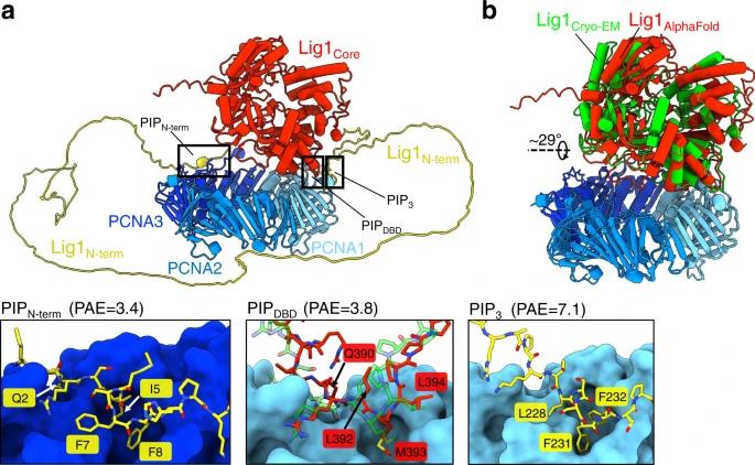 Fig. 1 AlphaFold prediction of the Lig1–PCNA complex.1
Key structural properties of LIG1:
- Classic three-domain architecture
- Active center is responsible for the catalytic adenosine
- OB folding structure specific to the DNA binding domain
Functions of LIG1
The core function of the LIG1 gene is to complete the connection of DNA strands to maintain genomic integrity. It mainly participates in the following key biological processes:
| Function | Description |
| DNA replication | Link Okazaki fragments in lag chain synthesis to ensure high fidelity and continuity of DNA replication. |
| Base excision repair | Sealing the single-strand break gap is a key step in repairing common DNA damages such as oxidative damage and alkylation damage. |
| Nucleotide excision repair | Participate in the final connection step of large-scale DNA damage repair pathways and jointly remove helical twist damage caused by ultraviolet rays, etc. |
| Cell cycle regulation | The active peak in S phase, the regulation of cell cycle protein, ensure synchronous replication and cell cycle process. |
| Prevention of genetic diseases | Functional deficiency can lead to LIG1 syndrome, characterized by immune deficiency and developmental delay, highlighting its fundamental role in disease prevention. |
Unlike some DNA ligases that use ATP, human LIG1 specifically utilizes NAD+ as an adenylyl donor, a characteristic that gives it a unique regulatory pathway in eukaryotic DNA metabolism.
Applications of LIG1 and LIG1 Antibody in Literature
1. Tang, Qun, et al. "Structures of LIG1 that engage with mutagenic mismatches inserted by polβ in base excision repair." Nature communications 13.1 (2022): 3860. https://doi.org/10.1038/s41467-022-31585-w
This paper reveals the mechanism by which LIG1 selectively links G:T mismatch and hinders A:C mismatch by analyzing the structure of the complex of LIG1 and mismatched DNA, and finds that APE1 can act as a correction enzyme to work in coordination with polβ and LIG1 to maintain the accuracy of base excision repair.
2. Xu, Lei, et al. "hsa_circ_0007919 induces LIG1 transcription by binding to FOXA1/TET1 to enhance the DNA damage response and promote gemcitabine resistance in pancreatic ductal adenocarcinoma." Molecular cancer 22.1 (2023): 195. https://doi.org/10.1186/s12943-023-01887-8
Studies have found that circular RNA hsa_circ_0007919 in pancreatic cancer upregulates LIG1 expression by recruiting the FOXA1/TET1 complex and reducing the methylation of the LIG1 gene promoter. This enhances the DNA damage repair ability, leading to cell resistance to gemcitabine chemotherapy. This mechanism reveals the key role of LIG1 in circrNA-mediated chemotherapy resistance.
3. Song, Ding-ming, et al. "LIG1 is a novel marker for bladder cancer prognosis: evidence based on experimental studies, machine learning and single-cell sequencing." Frontiers in Immunology 15 (2024): 1419126. https://doi.org/10.3389/fimmu.2024.1419126
This study reveals that LIG1 is highly expressed in bladder cancer and is associated with a poor prognosis. LIG1 drives tumor progression by promoting key malignant behaviors such as cancer cell proliferation, invasion, and epithelial-mesenchymal transition, and can serve as a potential risk biomarker.
4. Fracassi, Giulia, et al. "CRISPR/Cas9 screens identify LIG1 as a sensitizer of PARP inhibitors in castration-resistant prostate cancer." The Journal of Clinical Investigation 135.4 (2025). https://doi.org/10.1172/JCI179393
This study reveals that DNA ligase 1 (LIG1) deficiency and PARP inhibitors produce synthetic lethal effects in various cancers, including prostate cancer. LIG1 deletion can exacerbate DNA replication stress and double-strand breaks, leading to apoptosis. Its expression status can serve as a potential biomarker for patient stratification and combination therapy.
5. Blair, Kerry, et al. "Mechanism of human Lig1 regulation by PCNA in Okazaki fragment sealing." Nature communications 13.1 (2022): 7833. https://doi.org/10.1038/s41467-022-35475-z
This study, through structural biology, discovered that human DNA ligase 1 cooperates with PCNA via its two PIP motifs to form a double-loop structure anchored to the DNA incision. PIPDBD plays a key role in catalysis. This mechanism enables the substrate transfer of FEN1 to LIG1 and clarifies the molecular model by which PCNA regulates the maturation of lag chains.
Creative Biolabs: LIG1 Antibodies for Research
Creative Biolabs specializes in the production of high-quality LIG1 antibodies for research and industrial applications. Our portfolio includes monoclonal antibodies tailored for ELISA, Flow Cytometry, Western blot, immunohistochemistry, and other diagnostic methodologies.
- Custom LIG1 Antibody Development: Tailor-made solutions to meet specific research requirements.
- Bulk Production: Large-scale antibody manufacturing for industry partners.
- Technical Support: Expert consultation for protocol optimization and troubleshooting.
- Aliquoting Services: Conveniently sized aliquots for long-term storage and consistent experimental outcomes.
For more details on our LIG1 antibodies, custom preparations, or technical support, contact us at email.
Reference
- Blair, Kerry, et al. "Mechanism of human Lig1 regulation by PCNA in Okazaki fragment sealing." Nature communications 13.1 (2022): 7833. https://doi.org/10.1038/s41467-022-35475-z
Anti-LIG1 antibodies
 Loading...
 Compare




Hot products 
-
Mouse Anti-ENPP1 Recombinant Antibody (CBFYE-0159) (CBMAB-E0375-FY)

-
Mouse Anti-AK4 Recombinant Antibody (V2-180419) (CBMAB-A1891-YC)

-
Mouse Anti-CD33 Recombinant Antibody (6C5/2) (CBMAB-C8126-LY)

-
Mouse Anti-ATM Recombinant Antibody (2C1) (CBMAB-A3970-YC)

-
Mouse Anti-ARG1 Recombinant Antibody (CBYCL-103) (CBMAB-L0004-YC)

-
Rabbit Anti-Acetyl-Histone H3 (Lys36) Recombinant Antibody (V2-623395) (CBMAB-CP0994-LY)

-
Mouse Anti-FN1 Monoclonal Antibody (D6) (CBMAB-1240CQ)

-
Mouse Anti-ANXA7 Recombinant Antibody (A-1) (CBMAB-A2941-YC)

-
Mouse Anti-ASH1L Monoclonal Antibody (ASH5H03) (CBMAB-1372-YC)

-
Mouse Anti-BIRC3 Recombinant Antibody (315304) (CBMAB-1214-CN)

-
Mouse Anti-dsRNA Recombinant Antibody (2) (CBMAB-D1807-YC)

-
Mouse Anti-AMIGO2 Recombinant Antibody (CBYY-C0756) (CBMAB-C2192-YY)

-
Mouse Anti-8-oxoguanine Recombinant Antibody (V2-7719) (CBMAB-1898CQ)

-
Mouse Anti-BCL2L1 Recombinant Antibody (H5) (CBMAB-1025CQ)

-
Mouse Anti-8-oxoguanine Recombinant Antibody (V2-7697) (CBMAB-1869CQ)

-
Rat Anti-ADGRE4 Recombinant Antibody (V2-160163) (CBMAB-F0011-CQ)

-
Mouse Anti-CRYAB Recombinant Antibody (A4345) (CBMAB-A4345-YC)

-
Mouse Anti-CD59 Recombinant Antibody (CBXC-2097) (CBMAB-C4421-CQ)

-
Rabbit Anti-CCN1 Recombinant Antibody (CBWJC-3580) (CBMAB-C4816WJ)

-
Mouse Anti-CD8 Recombinant Antibody (C1083) (CBMAB-C1083-LY)

- AActivation
- AGAgonist
- APApoptosis
- BBlocking
- BABioassay
- BIBioimaging
- CImmunohistochemistry-Frozen Sections
- CIChromatin Immunoprecipitation
- CTCytotoxicity
- CSCostimulation
- DDepletion
- DBDot Blot
- EELISA
- ECELISA(Cap)
- EDELISA(Det)
- ESELISpot
- EMElectron Microscopy
- FFlow Cytometry
- FNFunction Assay
- GSGel Supershift
- IInhibition
- IAEnzyme Immunoassay
- ICImmunocytochemistry
- IDImmunodiffusion
- IEImmunoelectrophoresis
- IFImmunofluorescence
- IGImmunochromatography
- IHImmunohistochemistry
- IMImmunomicroscopy
- IOImmunoassay
- IPImmunoprecipitation
- ISIntracellular Staining for Flow Cytometry
- LALuminex Assay
- LFLateral Flow Immunoassay
- MMicroarray
- MCMass Cytometry/CyTOF
- MDMeDIP
- MSElectrophoretic Mobility Shift Assay
- NNeutralization
- PImmunohistologyp-Paraffin Sections
- PAPeptide Array
- PEPeptide ELISA
- PLProximity Ligation Assay
- RRadioimmunoassay
- SStimulation
- SESandwich ELISA
- SHIn situ hybridization
- TCTissue Culture
- WBWestern Blot
