MAP4 Antibodies
Background
MAP4 is a microtubule-binding protein widely present in eukaryotic cells, mainly involved in the assembly, stability and spatial organization of microtubule networks. This protein supports key biological processes such as cell morphology maintenance, intracellular material transport and mitosis by regulating microtubule dynamics and interacting with the cytoskeleton. During neuronal development, MAP4 plays a crucial regulatory role in the microtubule orientation arrangement of axons and dendrites, and its functional abnormalities are closely related to neurodegenerative diseases and cancer metastasis. Since its discovery in the 1980s, MAP4, as a typical representative of the microtubule regulatory protein family, has seen its phosphorylation regulatory mechanism and subcellular localization characteristics widely studied, providing an important model system for understanding cytoskeleton dynamics, cell polarity establishment, and disease occurrence mechanisms.
Structure of MAP4
MAP4 is a protein with a relatively large molecular weight, typically within the range of approximately 200 to 220 kDa, with specific values varying depending on species and splicing variants.
| Species | Human | Mouse | Bovine | Rat |
| Molecular Weight (kDa) | ~ 200–220 | ~ 200–215 | ~ 205–218 | ~ 198–212 |
| Primary Structural Differences | Contains multiple microtubule-binding repeat domains | The N-terminal sequence is highly homologous | The C-end domain is relatively conservative | Splicing variant types |
This protein contains multiple microtubule-binding domains and interacts with the microtubule surface through its coiled helices and acidic regions. Its secondary structure is mainly composed of α -helix and random curling, forming a flexible molecular structure that can dynamically bind to tubulin. MAP4 regulates its affinity for microtubules through phosphorylation at specific serine/threonine sites, thereby influencing the stability of microtubules and the process of cell division. This protein is widely expressed within cells and plays a particularly crucial role in maintaining microtubule dynamics and spindle assembly.
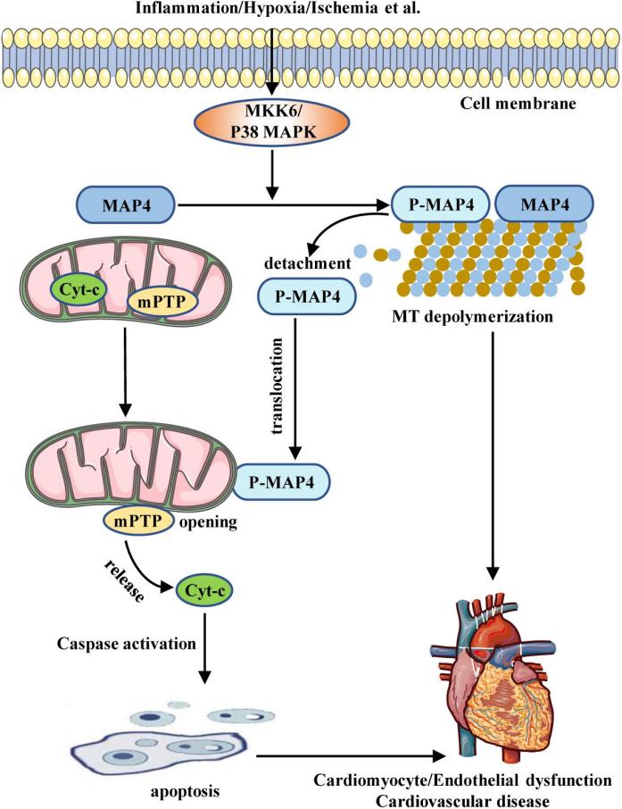 Fig. 1 Schematic illustrating the role of MAP4 in CVD.1
Key structural properties of MAP4:
- Contains multiple microtubule binding domains (MTBD)
- N-terminal acidic region rich in proline
- C-terminal conserved microtubule binding module
- Connect the functional domain through the flexible linker zone
Functions of MAP4
The main function of MAP4 is to regulate the dynamic stability of microtubules and the organization of the cytoskeleton. In addition, it is also involved in a variety of cellular processes, including mitosis, intracellular transport and maintenance of cell morphology.
| Function | Description |
| Microtubule stability | By binding to the surface of microtubules, it inhibits the depolymerization of microtubules and enhances the structural integrity of the cytoskeleton. |
| Mitotic regulation | During the mitotic phase, it participates in spindle assembly and the correct separation of chromosomes, and its phosphorylation state affects the mitotic process. |
| Intracellular substance transport | As a molecular scaffold, it assists motor proteins (such as kinesin and dynamin) in directional transport along microtubules. |
| Establishment of cell polarity | Through region-specific microtubule organization, it promotes the differentiation of neuronal axons and the formation of epithelial cell polarity. |
| Stress response | When cells are under stress, the microtubule network is regulated by altering the phosphorylation state to maintain cell structure and function. |
MAP4 interacts with microtubules in a dynamic manner through its multiple microtubule binding domains. Its regulatory function is highly dependent on phosphorylation modification, especially phosphorylation at the Ser/Thr site significantly reduces its affinity for microtubules, thereby rapidly responding to intracellular signal changes.
Applications of MAP4 and MAP4 Antibody in Literature
1. Xa, Gaochun, et al. "MAP4 acts as an oncogene and prognostic marker and affects radioresistance by mediating epithelial–mesenchymal transition in lung adenocarcinoma." Journal of Cancer Research and Clinical Oncology 150.2 (2024): 88. https://doi.org/10.1007/s00432-024-05614-8
This study explores the high expression of MAP4 in lung adenocarcinoma and its prognostic value, and finds that MAP4 promotes tumor cell migration, invasion and radiation resistance by regulating epithelial-mesenchymal transition (EMT). The nomogram constructed based on MAP4 and pT staging has good predictive performance. MAP4 is expected to become a new target for prognosis assessment and radiosensitization of lung adenocarcinoma.
2. Zhang, Siwei, et al. "Expression of Syk and MAP4 proteins in ovarian cancer." Journal of Cancer Research and Clinical Oncology 145.4 (2019): 909-919. https://doi.org/10.1007/s00432-019-02856-9
This study evaluated the expression of MAP4 in ovarian cancer and found that it was significantly correlated with histological subtypes, stages, grades and residual tumors, but not significantly associated with the overall survival of patients. The expression of MAP4 is correlated with that of Syk and calpain-1, especially in mucinous carcinoma, suggesting that these proteins may jointly participate in the early tumor spread process.
3. Zhang, Huangxian, et al. "miR-103-3p regulates the differentiation and autophagy of myoblasts by targeting MAP4." International Journal of Molecular Sciences 24.4 (2023): 4130. https://doi.org/10.3390/ijms24044130
In this study, through the C2C12 cell model, it was found that miR-103-3p inhibits myoblast differentiation and autophagy by directly targeting MAP4. MAP4 not only promotes myotubule formation, but also interacts with LC3 to regulate the autophagy process, thereby revealing a new mechanism of action of the miR-103-3p/MAP4 pathway in skeletal muscle formation.
4. Pan, Yunzhi, et al. "FBXW7 loss of function promotes esophageal squamous cell carcinoma progression via elevating MAP4 and ERK phosphorylation." Journal of Experimental & Clinical Cancer Research 42.1 (2023): 75. https://doi.org/10.1186/s13046-023-02630-3
This study reveals that the loss of FBXW7 function inhibits MAP4 degradation through CHEK1-mediated phosphorylation at the MAP4 T521 site, thereby activating the MAPK/ERK pathway and promoting the progression of esophageal squamous cell carcinoma. Low expression of FBXW7 and high MAP4 suggest a poor prognosis for patients. Targeting ERK and VEGFA can inhibit the growth of FBXW7-deficient tumors.
5. Zeng, Wenchao, et al. "Overexpression of BRINP3 predicts poor prognosis and promotes cancer cell proliferation and migration via MAP4 in osteosarcoma." Disease Markers 2022.1 (2022): 2698869. https://doi.org/10.1155/2022/2698869
This study found that BRINP3, which is highly expressed in osteosarcoma, stabilizes MAP4 protein through interaction, thereby promoting the proliferation and invasion of tumor cells. The BRINP3/MAP4 axis can serve as a potential diagnostic marker and therapeutic target for osteosarcoma.
Creative Biolabs: MAP4 Antibodies for Research
Creative Biolabs specializes in the production of high-quality MAP4 antibodies for research and industrial applications. Our portfolio includes monoclonal antibodies tailored for ELISA, Flow Cytometry, Western blot, immunohistochemistry, and other diagnostic methodologies.
- Custom MAP4 Antibody Development: Tailor-made solutions to meet specific research requirements.
- Bulk Production: Large-scale antibody manufacturing for industry partners.
- Technical Support: Expert consultation for protocol optimization and troubleshooting.
- Aliquoting Services: Conveniently sized aliquots for long-term storage and consistent experimental outcomes.
For more details on our MAP4 antibodies, custom preparations, or technical support, contact us at email.
Reference
- Li, Lingfei, et al. "MAP4 as a new candidate in cardiovascular disease." Frontiers in physiology 11 (2020): 1044. https://doi.org/10.3389/fphys.2020.01044
Anti-MAP4 antibodies
 Loading...


Hot products 
-
Mouse Anti-AMIGO2 Recombinant Antibody (CBYY-C0756) (CBMAB-C2192-YY)

-
Mouse Anti-ADGRE2 Recombinant Antibody (V2-261270) (CBMAB-C0813-LY)

-
Mouse Anti-APC Recombinant Antibody (CBYC-A661) (CBMAB-A3036-YC)

-
Mouse Anti-EMP3 Recombinant Antibody (CBFYE-0100) (CBMAB-E0207-FY)

-
Armenian hamster Anti-CD40 Recombinant Antibody (HM40-3) (CBMAB-C10365-LY)

-
Mouse Anti-CRYAB Recombinant Antibody (A4345) (CBMAB-A4345-YC)

-
Mouse Anti-BRD3 Recombinant Antibody (CBYY-0801) (CBMAB-0804-YY)

-
Mouse Anti-CAT Recombinant Antibody (724810) (CBMAB-C8431-LY)

-
Mouse Anti-BIRC3 Recombinant Antibody (16E63) (CBMAB-C3367-LY)

-
Mouse Anti-CD46 Recombinant Antibody (CBFYC-0076) (CBMAB-C0085-FY)

-
Mouse Anti-ACVR1C Recombinant Antibody (V2-179685) (CBMAB-A1041-YC)

-
Mouse Anti-AP4E1 Recombinant Antibody (32) (CBMAB-A2996-YC)

-
Mouse Anti-APP Recombinant Antibody (DE2B4) (CBMAB-1122-CN)

-
Mouse Anti-ATP1A2 Recombinant Antibody (M7-PB-E9) (CBMAB-A4013-YC)

-
Rat Anti-FABP3 Recombinant Antibody (CBXF-2299) (CBMAB-F1612-CQ)

-
Mouse Anti-BSN Recombinant Antibody (219E1) (CBMAB-1228-CN)

-
Mouse Anti-ENPP1 Recombinant Antibody (CBFYE-0159) (CBMAB-E0375-FY)

-
Mouse Anti-CD247 Recombinant Antibody (6B10.2) (CBMAB-C1583-YY)

-
Mouse Anti-ELAVL4 Recombinant Antibody (6B9) (CBMAB-1132-YC)

-
Mouse Anti-APOH Recombinant Antibody (4D9A4) (CBMAB-A3249-YC)

- AActivation
- AGAgonist
- APApoptosis
- BBlocking
- BABioassay
- BIBioimaging
- CImmunohistochemistry-Frozen Sections
- CIChromatin Immunoprecipitation
- CTCytotoxicity
- CSCostimulation
- DDepletion
- DBDot Blot
- EELISA
- ECELISA(Cap)
- EDELISA(Det)
- ESELISpot
- EMElectron Microscopy
- FFlow Cytometry
- FNFunction Assay
- GSGel Supershift
- IInhibition
- IAEnzyme Immunoassay
- ICImmunocytochemistry
- IDImmunodiffusion
- IEImmunoelectrophoresis
- IFImmunofluorescence
- IGImmunochromatography
- IHImmunohistochemistry
- IMImmunomicroscopy
- IOImmunoassay
- IPImmunoprecipitation
- ISIntracellular Staining for Flow Cytometry
- LALuminex Assay
- LFLateral Flow Immunoassay
- MMicroarray
- MCMass Cytometry/CyTOF
- MDMeDIP
- MSElectrophoretic Mobility Shift Assay
- NNeutralization
- PImmunohistologyp-Paraffin Sections
- PAPeptide Array
- PEPeptide ELISA
- PLProximity Ligation Assay
- RRadioimmunoassay
- SStimulation
- SESandwich ELISA
- SHIn situ hybridization
- TCTissue Culture
- WBWestern Blot
