SDC1 Antibodies
Background
The protein encoded by the SDC1 gene is a transmembrane glycosaminoglycan located on the cell surface, mainly expressed in epithelial cells and plasma cells. This protein binds to various extracellular matrix components, growth factors, and cytokines through its heparan sulfate chains, playing a crucial role in cell adhesion, migration, and signal transduction. SDC1 not only participates in regulating wound healing and tissue regeneration processes, but also plays an important role in inflammatory responses and tumor progression. Changes in its expression level are closely related to the invasion and metastasis of various cancers. As an important member of the Syndecan family, SDC1 was first cloned and identified in 1989. Subsequent extensive research has revealed its bridging role between the cell microenvironment and intracellular signaling pathways. The unique structural characteristics of this molecule enable it to simultaneously bind to multiple ligands, and this versatility makes it an important regulatory node in maintaining tissue homeostasis and the occurrence and development of diseases.
Structure of SDC1
The core protein encoded by the SDC1 gene has a molecular weight of approximately 33 kDa, but after glycosylation modification, the mature protein can reach 100-200 kDa. There are certain differences in structure and function among SDC1 proteins from different species. .
| Species | Human | Mouse | Rat | Cow |
| Molecular Weight (kDa) | 33 | 32.8 | 32.9 | 33.1 |
| Primary Structural Differences | The extracellular domain contains 3 heparan sulfate binding sites | The extracellular domain is shorter and has a different glycosylation pattern | Highly homologous to rats | There are slight differences in the extracellular domain cleavage sites |
The SDC1 protein is composed of approximately 310 amino acids. Its core protein has a linear structure, and the extracellular segment is rich in serine-glycine repeat sequences as attachment sites for the heparan sulfate chains. The protein structure includes a short intracellular domain, a single transmembrane region, and a large extracellular domain. The intracellular segment contains a conserved domain that can interact with the cytoskeleton. The extracellular segment of SDC1 usually connects to 3-5 heparan sulfate glycan chains, which are charged polysaccharides and give the protein the ability to bind to various ligands. The transmembrane region is composed of 25 hydrophobic amino acids, and the four conserved tyrosine residues in the intracellular segment play a regulatory role in signal transduction.
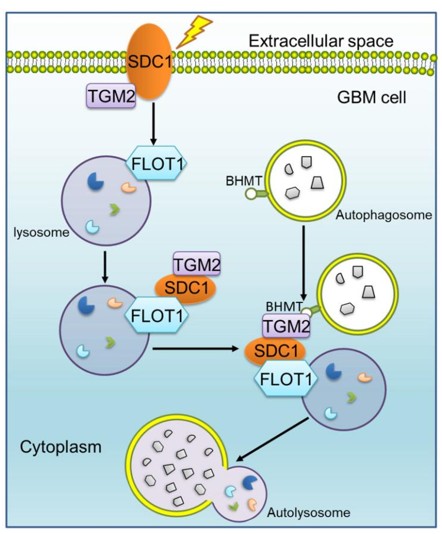 Fig. 1 SDC1 molecules mediated GBM cells after radiotherapy autophagy flow integration mechanism.1
Key structural properties of SDC1:
- Transmembrane core protein with extracellular heparan sulfate chains
- The negatively charged region of the extracellular segment mediates ligand binding
- The conserved intracellular domain connects the cytoskeleton
- Three heparan sulfate modification sites enhance functional diversity
Functions of SDC1
The main function of SDC1 is to mediate the interaction between cells and the microenvironment. However, it is also involved in various physiological processes, such as tissue repair, inflammation regulation, and tumor progression.
| Function | Description |
| Cell adhesion | SDC1 binds to matrix components through heparan sulfate chains, mediating the adhesion of epithelial cells to the basement membrane. |
| Signal transduction | As a co-receptor, it binds to growth factors (such as FGF), activating downstream signaling pathways to regulate cell proliferation and migration. |
| Tissue repair | It promotes the migration of keratinocytes and the formation of granulation tissue during wound healing. |
| Inflammation regulation | It regulates the recruitment and activation of immune cells at the inflammatory site by binding to chemokines and cytokines. |
| Tumor progression | The expression changes of SDC1 affect the invasiveness of tumor cells, and its shedding form can promote the remodeling of the tumor microenvironment. |
The expression regulation curve of SDC1 shows a tissue-specific distribution, contrasting with the widespread expression pattern of integrins, indicating its unique functional positioning in maintaining epithelial homeostasis.
Applications of SDC1 and SDC1 Antibody in Literature
1. Zeng, Liang, et al. "SDC1-TGM2-FLOT1-BHMT complex determines radiosensitivity of glioblastoma by influencing the fusion of autophagosomes with lysosomes." Theranostics 13.11 (2023): 3725. https://doi.org/10.7150/thno.81999
The study found that SDC1 and TGM2 are highly expressed in glioblastoma with radiotherapy resistance, indicating a poor prognosis. The mechanism is that SDC1 carries TGM2, which is transported to the lysosome via FLOT1 and binds to BHMT on autophagosomes, promoting the autophagic flux and leading to radiotherapy resistance.
2. Zhang, Chuan-long, et al. "SDC1 and ITGA2 as novel prognostic biomarkers for PDAC related to IPMN." Scientific Reports 13.1 (2023): 18727. https://doi.org/10.1038/s41598-023-44646-x
The research found that through the analysis of sequencing data from pancreatic ductal adenocarcinoma and precancerous lesions, it was discovered that SDC1 and ITGA2 are key prognostic markers. The high expression of SDC1 involves the interferon response, while ITGA2 is enriched in the epithelial-mesenchymal transition pathway, providing new targets for the treatment of pancreatic cancer.
3. Lu, Lihong, et al. "Sirt7/HIC1 complex participates in hyperglycaemia‐mediated EndMT via modulation of SDC1 expression in diabetic kidney disease and metabolic memory." Journal of Cellular and Molecular Medicine 28.9 (2024): e18336. https://doi.org/10.1111/jcmm.18336
The research has found that in diabetic nephropathy, even when blood sugar levels are under control, the binding of high glucose-induced Sirt7 and HIC1 to SDC1 still inhibits the transcription of SDC1, continuously driving endothelial-mesenchymal transformation, forming metabolic memory, and exacerbating renal damage.
4. Li, Kaizhi, et al. "Loss of SDC1 expression is associated with poor prognosis of colorectal cancer patients in Northern China." Disease Markers 2019.1 (2019): 3768708. https://doi.org/10.1155/2019/3768708
The study found that syndecan-1 was significantly downregulated in colorectal cancer and metastatic lymph nodes. Its absence was closely associated with poor tumor differentiation, advanced stage, lymph node metastasis, and poor prognosis of patients, and it was a key indicator of malignant progression.
5. Li, Gen, et al. "MZ1, a BRD4 inhibitor, exerted its anti-cancer effects by suppressing SDC1 in glioblastoma." BMC cancer 24.1 (2024): 220. https://doi.org/10.1186/s12885-024-11966-8
The research found that the BRD4 degrader MZ1 targeting super enhancers can effectively inhibit the growth of glioblastoma. Mechanistically, MZ1 downregulates cell cycle and EMT-related genes by degrading BRD4, and it was discovered that SDC1 is a new oncogenic gene.
Creative Biolabs: SDC1 Antibodies for Research
Creative Biolabs specializes in the production of high-quality SDC1 antibodies for research and industrial applications. Our portfolio includes monoclonal and polyclonal antibodies tailored for ELISA, Flow Cytometry, Western blot, immunohistochemistry, and other diagnostic methodologies.
- Custom SDC1 Antibody Development: Tailor-made solutions to meet specific research requirements.
- Bulk Production: Large-scale antibody manufacturing for industry partners.
- Technical Support: Expert consultation for protocol optimization and troubleshooting.
- Aliquoting Services: Conveniently sized aliquots for long-term storage and consistent experimental outcomes.
For more details on our SDC1 antibodies, custom preparations, or technical support, contact us at email.
Reference
- Zeng, Liang, et al. "SDC1-TGM2-FLOT1-BHMT complex determines radiosensitivity of glioblastoma by influencing the fusion of autophagosomes with lysosomes." Theranostics 13.11 (2023): 3725. Distributed under Open Access license CC BY 4.0, without modification. https://doi.org/10.7150/thno.81999
Anti-SDC1 antibodies
 Loading...


Hot products 
-
Mouse Anti-CD247 Recombinant Antibody (6B10.2) (CBMAB-C1583-YY)

-
Mouse Anti-GLP1R Recombinant Antibody (4F3) (CBMAB-G0521-LY)

-
Rat Anti-CCR2 Recombinant Antibody (475301) (CBMAB-C1338-LY)

-
Mouse Anti-FPR2 Recombinant Antibody (1D6) (CBMAB-F2628-CQ)

-
Mouse Anti-CD63 Recombinant Antibody (CBXC-1200) (CBMAB-C1467-CQ)

-
Mouse Anti-CD46 Recombinant Antibody (CBFYC-0076) (CBMAB-C0085-FY)

-
Rabbit Anti-CAMK2A Recombinant Antibody (BA0032) (CBMAB-0137CQ)

-
Mouse Anti-ESR1 Recombinant Antibody (Y31) (CBMAB-1208-YC)

-
Mouse Anti-COL1A2 Recombinant Antibody (CF108) (V2LY-1206-LY626)

-
Mouse Anti-APCS Recombinant Antibody (CBYC-A663) (CBMAB-A3054-YC)

-
Mouse Anti-BrdU Recombinant Antibody (IIB5) (CBMAB-1038CQ)

-
Mouse Anti-EPO Recombinant Antibody (CBFYR0196) (CBMAB-R0196-FY)

-
Mouse Anti-ARHGDIA Recombinant Antibody (CBCNA-009) (CBMAB-R0415-CN)

-
Mouse Anti-DDC Recombinant Antibody (8E8) (CBMAB-0992-YC)

-
Mouse Anti-AFDN Recombinant Antibody (V2-58751) (CBMAB-L0408-YJ)

-
Mouse Anti-BLNK Recombinant Antibody (CBYY-0623) (CBMAB-0626-YY)

-
Mouse Anti-BIRC3 Recombinant Antibody (16E63) (CBMAB-C3367-LY)

-
Mouse Anti-CD24 Recombinant Antibody (ALB9) (CBMAB-0176CQ)

-
Mouse Anti-ASTN1 Recombinant Antibody (H-9) (CBMAB-1154-CN)

-
Mouse Anti-ADGRE2 Recombinant Antibody (V2-261270) (CBMAB-C0813-LY)

- AActivation
- AGAgonist
- APApoptosis
- BBlocking
- BABioassay
- BIBioimaging
- CImmunohistochemistry-Frozen Sections
- CIChromatin Immunoprecipitation
- CTCytotoxicity
- CSCostimulation
- DDepletion
- DBDot Blot
- EELISA
- ECELISA(Cap)
- EDELISA(Det)
- ESELISpot
- EMElectron Microscopy
- FFlow Cytometry
- FNFunction Assay
- GSGel Supershift
- IInhibition
- IAEnzyme Immunoassay
- ICImmunocytochemistry
- IDImmunodiffusion
- IEImmunoelectrophoresis
- IFImmunofluorescence
- IGImmunochromatography
- IHImmunohistochemistry
- IMImmunomicroscopy
- IOImmunoassay
- IPImmunoprecipitation
- ISIntracellular Staining for Flow Cytometry
- LALuminex Assay
- LFLateral Flow Immunoassay
- MMicroarray
- MCMass Cytometry/CyTOF
- MDMeDIP
- MSElectrophoretic Mobility Shift Assay
- NNeutralization
- PImmunohistologyp-Paraffin Sections
- PAPeptide Array
- PEPeptide ELISA
- PLProximity Ligation Assay
- RRadioimmunoassay
- SStimulation
- SESandwich ELISA
- SHIn situ hybridization
- TCTissue Culture
- WBWestern Blot
