STEAP1 Antibodies
Background
The STEAP1 gene encodes a six-transmembrane protein, which is mainly located on the cell membrane and endosomal membranes. It is expressed in various tissues (especially the prostate, bladder, and certain cancer cells). This protein participates in the intracellular transport and reduction of iron and copper ions as a metal reductase, playing a crucial role in maintaining the homeostasis of metal ions, regulating cellular metabolism, and balancing redox equilibrium. STEAP1 was first identified in 2001. The study of its structural characteristics and functional mechanisms has revealed its metal ion processing ability in normal physiological processes, and it is often highly expressed in malignant tumors such as prostate cancer, closely related to tumor growth, invasion, and treatment response. As a diagnostic marker and therapeutic target for cancer, the structural and functional research of STEAP1 continuously promotes a deeper understanding of tumor metabolism and targeted treatment strategies.
Structure of STEAP1
STEAP1 is a six-transmembrane protein with a molecular weight of approximately 54 kDa. Its molecular weight may vary slightly among different species due to differences in amino acid sequences.
| Species | Human | Mouse | Rat |
| Molecular Weight (kDa) | About 54 | About 53.8 | About 53.9 |
| Primary Structural Differences | Containing 483 amino acids and having a conserved NADPH binding domain | Highly homologous amino acid sequences, conservative transmembrane region structure | Across the membrane structure and reduction is similar to human highly active center |
This protein is composed of approximately 483 amino acids and its primary structure folds into a transmembrane topology dominated by α-helices. The protein core contains a transmembrane channel composed of hydrophobic amino acid residues, which is used for the recognition and transport of metal ions. The key functional structures include an NADPH binding site located on the cytoplasmic side, and a metal reduction center composed of conserved histidine residues in the transmembrane region, which work together to complete the electron transfer and reduction of metal ions.
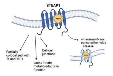 Fig. 1 Structural and physiologic properties of STEAP1.1
Key structural properties of STEAP1:
- Six transmembrane α-helical topological structures
- Hydrophobic transmembrane channels surround the metal ion binding sites.
- Conservative NADPH combined with domain provides electron transfer ability
- Histidine residues are involved in metal ion coordination and reduction catalysis
Functions of STEAP1
The core function of the STEAP1 protein is to participate in the reduction and transport of metal ions within the cell. Additionally, it is involved in several key physiological and pathological processes, including cellular metabolic regulation and tumor progression.
| Function | Description |
| Metal ion reduction | As a metal-reducing enzyme, it mainly reduces trivalent iron (Fe³⁺) and divalent copper (Cu²⁺) to their more readily usable forms (Fe²⁺, Cu⁺). |
| Ion Transport | Promotes the transmembrane transport of reduced metal ions, providing essential cofactors for cellular metabolism (such as the mitochondrial respiratory chain). |
| Cellular Proliferation Regulation | By influencing the intracellular metal ion homeostasis and redox balance, it participates in regulating the cell cycle and proliferation process. |
| Cancer-related functions | Highly expressed in various cancer cells such as prostate cancer, promoting tumor growth, invasion, and resistance to treatments (such as radiotherapy). |
| Oxidative Stress Regulation | Its reducing activity is correlated with the level of reactive oxygen species (ROS) within the cells, thereby indirectly influencing the oxidative stress state of the cells. |
Unlike the cooperative allosteric regulation of hemoglobin, the metal reduction activity of STEAP1 mainly relies on its conserved NADPH binding domain for unidirectional electron transfer. This determines its fundamental "guardian" role in maintaining the homeostasis of metal ions and is abnormally exploited in the tumor microenvironment to support the rapid proliferation of cancer cells.
Applications of STEAP1 and STEAP1 Antibody in Literature
1. Xu, Michael, et al. "STEAP1–4 (six-transmembrane epithelial antigen of the prostate 1–4) and their clinical implications for prostate cancer." Cancers 14.16 (2022): 4034. https://doi.org/10.3390/cancers14164034
The article indicates that the STEAP protein family, which is highly expressed in prostate cancer, especially STEAP1, is a key target for driving tumor progression. This article reviews its structure and function, and explores its potential as a biomarker and a new target for immunotherapy in clinical applications.
2. Bhatia, Vipul, et al. "Targeting advanced prostate cancer with STEAP1 chimeric antigen receptor T cell and tumor-localized IL-12 immunotherapy." Nature Communications 14.1 (2023): 2041. https://doi.org/10.1038/s41467-023-37874-2
The article indicates that STEAP1 is a new target for prostate cancer and that targeted CAR-T therapy is effective. The study found that antigen escape is the main cause of drug resistance. Combined with local IL-12 treatment, it can reshape the tumor microenvironment, enhance efficacy and trigger a broad-spectrum immune response.
3. Nakamura, Hajime, Yohei Arihara, and Kohichi Takada. "Targeting STEAP1 as an anticancer strategy." Frontiers in Oncology 13 (2023): 1285661. https://doi.org/10.3389/fonc.2023.1285661
The article indicates that STEAP1, as a transmembrane protein, is highly expressed in various cancers and is associated with poor prognosis. It is a highly promising therapeutic target. Antibody-drug conjugates and CAR-T cell therapies developed based on it have shown promising application prospects in clinical trials.
4. Rocha, Sandra M., et al. "STEAP1 knockdown decreases the sensitivity of prostate cancer cells to paclitaxel, docetaxel and cabazitaxel." International Journal of Molecular Sciences 24.7 (2023): 6643. https://doi.org/10.3390/ijms24076643
This study shows that the expression level of the STEAP1 protein in prostate cancer cells affects their sensitivity to taxane-based chemotherapy drugs. After knocking down the STEAP1 gene, the original anti-cancer effect of the drug may be reversed, and even a reverse effect of promoting cell growth may occur.
5. Jiang, Jun-nan, et al. "EIF4E regulates STEAP1 expression in peritoneal metastasis." Journal of Cancer 11.4 (2020): 990. https://doi.org/10.7150/jca.29105
The research has found that phosphorylated eIF4E initiates the translation of STEAP1 protein, thereby driving peritoneal metastasis of gastric cancer. Inhibiting the phosphorylation of eIF4E or its binding to the translation complex can effectively block this process, providing a new target for the treatment of metastasis.
Creative Biolabs: STEAP1 Antibodies for Research
Creative Biolabs specializes in the production of high-quality STEAP1 antibodies for research and industrial applications. Our portfolio includes monoclonal antibodies tailored for ELISA, Flow Cytometry, Western blot, immunohistochemistry, and other diagnostic methodologies.
- Custom STEAP1 Antibody Development: Tailor-made solutions to meet specific research requirements.
- Bulk Production: Large-scale antibody manufacturing for industry partners.
- Technical Support: Expert consultation for protocol optimization and troubleshooting.
- Aliquoting Services: Conveniently sized aliquots for long-term storage and consistent experimental outcomes.
For more details on our STEAP1 antibodies, custom preparations, or technical support, contact us at email.
Reference
- Xu, Michael, et al. "STEAP1–4 (six-transmembrane epithelial antigen of the prostate 1–4) and their clinical implications for prostate cancer." Cancers 14.16 (2022): 4034. https://doi.org/10.3390/cancers14164034
Anti-STEAP1 antibodies
 Loading...


Hot products 
-
Rat Anti-(1-5)-α-L-Arabinan Recombinant Antibody (V2-501861) (CBMAB-XB0003-YC)

-
Mouse Anti-ADAM12 Recombinant Antibody (V2-179752) (CBMAB-A1114-YC)

-
Mouse Anti-8-oxoguanine Recombinant Antibody (V2-7719) (CBMAB-1898CQ)

-
Mouse Anti-BACE1 Recombinant Antibody (61-3E7) (CBMAB-1183-CN)

-
Mouse Anti-GFAP Recombinant Antibody (24) (CBMAB-G2927-LY)

-
Mouse Anti-DMD Recombinant Antibody (D1190) (CBMAB-D1190-YC)

-
Rabbit Anti-ENO2 Recombinant Antibody (BA0013) (CBMAB-0272CQ)

-
Mouse Anti-ACKR3 Recombinant Antibody (V2-261265) (CBMAB-C1023-LY)

-
Mouse Anti-DDC Recombinant Antibody (8E8) (CBMAB-0992-YC)

-
Mouse Anti-FPR2 Recombinant Antibody (1D6) (CBMAB-F2628-CQ)

-
Mouse Anti-BrdU Recombinant Antibody (IIB5) (CBMAB-1038CQ)

-
Mouse Anti-CFL1 Recombinant Antibody (CBFYC-1771) (CBMAB-C1833-FY)

-
Mouse Anti-BAX Recombinant Antibody (CBYY-0216) (CBMAB-0217-YY)

-
Mouse Anti-AQP2 Recombinant Antibody (G-3) (CBMAB-A3359-YC)

-
Mouse Anti-BIRC3 Recombinant Antibody (315304) (CBMAB-1214-CN)

-
Mouse Anti-COL12A1 Recombinant Antibody (CBYY-C3117) (CBMAB-C4560-YY)

-
Human Anti-SARS-CoV-2 Spike Recombinant Antibody (CR3022) (CBMAB-CR014LY)

-
Mouse Anti-APP Recombinant Antibody (DE2B4) (CBMAB-1122-CN)

-
Mouse Anti-ARSA Recombinant Antibody (CBYC-A799) (CBMAB-A3679-YC)

-
Mouse Anti-C1QC Recombinant Antibody (CBFYC-0600) (CBMAB-C0654-FY)

- AActivation
- AGAgonist
- APApoptosis
- BBlocking
- BABioassay
- BIBioimaging
- CImmunohistochemistry-Frozen Sections
- CIChromatin Immunoprecipitation
- CTCytotoxicity
- CSCostimulation
- DDepletion
- DBDot Blot
- EELISA
- ECELISA(Cap)
- EDELISA(Det)
- ESELISpot
- EMElectron Microscopy
- FFlow Cytometry
- FNFunction Assay
- GSGel Supershift
- IInhibition
- IAEnzyme Immunoassay
- ICImmunocytochemistry
- IDImmunodiffusion
- IEImmunoelectrophoresis
- IFImmunofluorescence
- IGImmunochromatography
- IHImmunohistochemistry
- IMImmunomicroscopy
- IOImmunoassay
- IPImmunoprecipitation
- ISIntracellular Staining for Flow Cytometry
- LALuminex Assay
- LFLateral Flow Immunoassay
- MMicroarray
- MCMass Cytometry/CyTOF
- MDMeDIP
- MSElectrophoretic Mobility Shift Assay
- NNeutralization
- PImmunohistologyp-Paraffin Sections
- PAPeptide Array
- PEPeptide ELISA
- PLProximity Ligation Assay
- RRadioimmunoassay
- SStimulation
- SESandwich ELISA
- SHIn situ hybridization
- TCTissue Culture
- WBWestern Blot
