
TNKS Antibodies
Background
The TNKS gene encodes a protein called end-anchored polymerase, which functions as a scaffold protein in the cell nucleus and is mainly involved in the regulation of signaling pathways such as Wnt/β-catenin. It plays an important role in genomic stability by regulating telomere length maintenance and DNA damage repair mechanisms. Research has found that TNKS play a key role in the self-renewal of stem cells and tissue regeneration, and their abnormal expression is closely related to the occurrence of various tumors and fibrotic diseases. This gene was first identified through telome-binding protein screening in 1998. Its unique PARP domain characteristics provide a new direction for tumor-targeted therapy, and the related inhibitors have currently entered the clinical trial stage. In-depth research on TNKS continuously drives the development of understanding of the dynamic regulation of chromatin and the mechanisms of cell fate determination.
Structure of TNKS
TNKS is a multi-domain scaffold protein with a molecular weight of approximately 142 kDa. This protein shows significant differences among different subtypes (TNKS1/TNKS2), mainly due to the changes in the number of ANK repeat domains and SAM domains.
| Species | Human | Mouse | Zebrafish | Fruit fly |
| Molecular Weight (kDa) | 142 (TNKS1) | 139 | 135 | 128 |
| Primary Structural Differences | Contains five ANK repeat domains with the C-terminal PARP catalytic domain | ANK repeat number domain highly conservative | The aggregation capability of the SAM domain is relatively weak | Only the core PARP homologous domain is retained |
This protein is composed of 1,327 amino acids, and its tertiary structure presents a typical "rod-like" shape, formed by the linear arrangement of the ANK repeat domain at the N-terminal. The protein achieves self-polymerization internally through multiple homologous oligomerization domains (Sams), among which the HPS domain is responsible for recognizing substrates and mediating the regulation of the Wnt/β-catenin signaling pathway. The conserved PARP catalytic domain is located at the C-terminal, and its unique "helical - turning - helical" motif can specifically catalyze the transfer reaction of nicotinamide adenine dinucleotide, thereby achieving polyADP-ribonylation modification of the target protein.
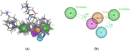 Fig. 1 The defined pharmacophore model of TNKS protein.1
Key structural properties of TNKS:
- Multi-domain series architecture
- Fork head-related domain
- C-end PARP catalytic core
Functions of TNKS
The end-anchor polymerase encoded by the TNKS gene mainly functions as a regulatory hub for signaling pathways such as Wnt/β-catenin, and also participates in maintaining genomic stability.
| Function | Description |
| Wnt signal regulation | The degradation of Axin through PARsylation modification promotes the nuclear entry of β-catenin to activate the transcription of target genes. |
| Telomere length is maintained | Interact with telomere binding protein TRF1, regulate telomerase independence in telomeres extension mechanism. |
| DNA damage repair | Recruit the BRCA1/BARD1 complex to DNA damage sites to regulate the process of homologous recombination repair. |
| Regulation of metabolic homeostasis | Regulation of glucose uptake through GLUT4 vesicle transport affects insulin signaling pathway sensitivity. |
| Cell fate determination | Controlling the balance between self-renewal and differentiation of stem cells, its abnormal expression leads to abnormal tissue regeneration. |
This protein achieves functional transformation through allosteric regulation mechanisms: after recognizing different substrates, its N-terminal ANK repeat domain can induce conformational rearrangement in the C-terminal PARP catalytic domain, thereby achieving specific functional switching in different biological processes such as chromatin remodeling, DNA repair, and metabolic regulation. This structural plasticity makes it a key molecular node connecting epigenetic regulation and signal transduction.
Applications of TNKS and TNKS Antibody in Literature
1. Chang, Chun-Chun, et al. "Combinatorial Virtual Screening Revealed a Novel Scaffold for TNKS Inhibition to Combat Colorectal Cancer." Biomedicines 10.1 (2022): 143. https://doi.org/10.3390/biomedicines10010143
This study discovered a novel Tankyrase inhibitor through virtual screening. Enzyme activity and cell experiments have verified that compound NSC319963 can effectively inhibit TNKS activity, block the Wnt signaling pathway, and significantly inhibit the growth of colorectal cancer cells, indicating that it is a potential TNKS inhibitor.
2. Ma, Yanmei, et al. "Dihydroartemisinin suppresses proliferation, migration, the Wnt/β-catenin pathway and EMT via TNKS in gastric cancer." Oncology Letters 22.4 (2021): 688. https://doi.org/10.3892/ol.2021.12949
This study found that Tankyrase (TNKS) expression was upregulated in gastric cancer. Knockdown of TNKS or treatment with artesunate (DHA) can effectively inhibit the proliferation and migration ability of gastric cancer cells by suppressing the Wnt/β-catenin signaling pathway and the epithelial-mesenchymal transition process.
3. Zhifei, Ma, et al. "circ5615 functions as a ceRNA to promote colorectal cancer progression by upregulating TNKS." Cell Death and Disease 11.5 (2020). https://doi.org/10.1038/s41419-020-2514-0
This study found that the circular RNA circ5615, which is highly expressed in colorectal cancer, adsorbs miR-149-5p to release its inhibitory effect on the target gene Tankyrase (TNKS), thereby activating the Wnt/β-catenin signaling pathway and ultimately promoting the malignant progression of tumors.
4. Solberg, Nina Therese, et al. "MEK inhibition induces canonical WNT signaling through YAP in KRAS mutated HCT-15 cells, and a cancer preventive FOXO3/FOXM1 ratio in combination with TNKS inhibition." Cancers 11.2 (2019): 164. https://doi.org/10.3390/cancers11020164
This study reveals that in colorectal cancer cells with co-mutations of APC and KRAS, MEK-dependent YAP signaling can activate the WNT pathway. The combined use of Tankyrase (TNKS) and MEK inhibitors can effectively block this induction circuit and enhance the anti-cancer effect by triggering metabolic stress and regulating the FOXO3/FOXM1 ratio.
5. Lee, Ji Hae, Youngjoo Kwon, and Kyungsil Yoon. "CREB Regulates Cisplatin Resistance by Targeting TNKS and KDM6A in NSCLC cell-Derived Tumor Spheroid." International Journal of Biological Sciences 21.11 (2025): 4851. https://doi.org/10.7150/ijbs.109419
This study reveals that the transcription factor CREB is a key regulatory factor mediating cisplatin resistance in non-small cell lung cancer. In drug-resistant cells, CREB continuously binds to target genes such as TNKS and maintains their expression; Inhibiting CREB can reduce the level of TNKS and restore the sensitivity of cancer cells to cisplatin, providing a new target for overcoming drug resistance.
Creative Biolabs: TNKS Antibodies for Research
Creative Biolabs specializes in the production of high-quality TNKS antibodies for research and industrial applications. Our portfolio includes monoclonal antibodies tailored for ELISA, Flow Cytometry, Western blot, immunohistochemistry, and other diagnostic methodologies.
- Custom TNKS Antibody Development: Tailor-made solutions to meet specific research requirements.
- Bulk Production: Large-scale antibody manufacturing for industry partners.
- Technical Support: Expert consultation for protocol optimization and troubleshooting.
- Aliquoting Services: Conveniently sized aliquots for long-term storage and consistent experimental outcomes.
For more details on our TNKS antibodies, custom preparations, or technical support, contact us at email.
Reference
- Chang, Chun-Chun, et al. "Combinatorial Virtual Screening Revealed a Novel Scaffold for TNKS Inhibition to Combat Colorectal Cancer." Biomedicines 10.1 (2022): 143. https://doi.org/10.3390/biomedicines10010143
Anti-TNKS antibodies
 Loading...
 Compare




Hot products 
-
Mouse Anti-FPR2 Recombinant Antibody (1D6) (CBMAB-F2628-CQ)

-
Mouse Anti-CD164 Recombinant Antibody (CBFYC-0077) (CBMAB-C0086-FY)

-
Mouse Anti-DLG1 Monolconal Antibody (4F3) (CBMAB-0225-CN)

-
Rat Anti-CD63 Recombinant Antibody (7G4.2E8) (CBMAB-C8725-LY)

-
Rat Anti-CCR2 Recombinant Antibody (475301) (CBMAB-C1338-LY)

-
Mouse Anti-AKR1B1 Antibody (V2-2449) (CBMAB-1001CQ)

-
Mouse Anti-BANF1 Recombinant Antibody (3F10-4G12) (CBMAB-A0707-LY)

-
Mouse Anti-ALB Recombinant Antibody (V2-180650) (CBMAB-A2186-YC)

-
Mouse Anti-BPGM Recombinant Antibody (CBYY-1806) (CBMAB-2155-YY)

-
Mouse Anti-DMD Recombinant Antibody (D1190) (CBMAB-D1190-YC)

-
Mouse Anti-CCDC25 Recombinant Antibody (CBLC132-LY) (CBMAB-C9786-LY)

-
Mouse Anti-CTCF Recombinant Antibody (CBFYC-2371) (CBMAB-C2443-FY)

-
Mouse Anti-ENO2 Recombinant Antibody (85F11) (CBMAB-0276CQ)

-
Mouse Anti-AQP2 Recombinant Antibody (G-3) (CBMAB-A3359-YC)

-
Mouse Anti-GLP1R Recombinant Antibody (4F3) (CBMAB-G0521-LY)

-
Mouse Anti-BSN Recombinant Antibody (219E1) (CBMAB-1228-CN)

-
Mouse Anti-B2M Recombinant Antibody (CBYY-0050) (CBMAB-0050-YY)

-
Mouse Anti-ENO2 Recombinant Antibody (H14) (CBMAB-E1341-FY)

-
Mouse Anti-ABIN2 Recombinant Antibody (V2-179106) (CBMAB-A0349-YC)

-
Rat Anti-EPO Recombinant Antibody (16) (CBMAB-E1578-FY)

- AActivation
- AGAgonist
- APApoptosis
- BBlocking
- BABioassay
- BIBioimaging
- CImmunohistochemistry-Frozen Sections
- CIChromatin Immunoprecipitation
- CTCytotoxicity
- CSCostimulation
- DDepletion
- DBDot Blot
- EELISA
- ECELISA(Cap)
- EDELISA(Det)
- ESELISpot
- EMElectron Microscopy
- FFlow Cytometry
- FNFunction Assay
- GSGel Supershift
- IInhibition
- IAEnzyme Immunoassay
- ICImmunocytochemistry
- IDImmunodiffusion
- IEImmunoelectrophoresis
- IFImmunofluorescence
- IGImmunochromatography
- IHImmunohistochemistry
- IMImmunomicroscopy
- IOImmunoassay
- IPImmunoprecipitation
- ISIntracellular Staining for Flow Cytometry
- LALuminex Assay
- LFLateral Flow Immunoassay
- MMicroarray
- MCMass Cytometry/CyTOF
- MDMeDIP
- MSElectrophoretic Mobility Shift Assay
- NNeutralization
- PImmunohistologyp-Paraffin Sections
- PAPeptide Array
- PEPeptide ELISA
- PLProximity Ligation Assay
- RRadioimmunoassay
- SStimulation
- SESandwich ELISA
- SHIn situ hybridization
- TCTissue Culture
- WBWestern Blot
