WT1 Antibodies
Background
The WT1 gene is a zinc finger transcription factor protein located on the short arm of human chromosome 11, which mainly plays a core regulatory role in the development of embryonic kidneys, gonads and mesodermal tissues. This gene generates multiple isomers through selective splicing, which bind to DNA targets in a sequence-specific manner. It can both activate and inhibit the expression of downstream genes, thereby precisely regulating cell differentiation and organ formation. In 1990, researchers first discovered that its mutation was closely related to the occurrence of childhood kidney tumors - Wilms tumors, which provided a key breakthrough for understanding the developmental mechanism defects in tumor occurrence. The molecular structure of the WT1 gene consists of four zinc finger domains and functionally variable regulatory regions. Its mutation lineage covers a variety of genetic diseases (such as Denys-Drash syndrome and Frasier syndrome), and it remains an important molecular model for studying developmental biology, tumorigenesis, and epigenetic regulation to this day.
Structure of WT1
The molecular weight of the protein encoded by the WT1 gene is approximately 47-52 kDa, and the specific value varies due to different splicing isomers (such as ±KTS variants). This protein contains four highly conserved C2H2-type zinc finger domains, which form its DNA binding core module.
| Species | Human | Mouse | Rat |
| Molecular Weight (kDa) | 49-52 | 48-51 | 48-51 |
| Primary Structural Differences | Contains four zinc-finger domains with ±KTS splice variants | Zinc finger structure is highly homologous to human | Conservative arrangement of zinc finger domains |
The N-terminal of the WT1 protein is rich in proline/glutamine and has transcriptional regulatory functions, while the C-terminal binds to specific DNA sequences (such as the EGR1 binding site) through a zinc finger structure. The third and fourth zinc fingers form the crucial DNA recognition helix, while the second zinc finger stabilizes the protein-DNA complex. This structural feature enables it to precisely regulate gene expression through conformational changes, playing a key role in the development of the kidneys and gonads.
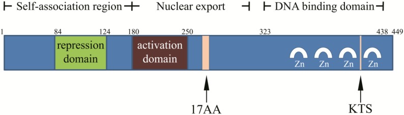 Fig. 1 Structure of WT1 protein.1
Key structural properties of WT1:
- Four C2H2-type zinc finger domains constitute the DNA binding core
- N-terminal proline-/ glutamine-rich transcriptional regulatory region
- Zinc interfinger junction region determines DNA binding specificity of ±KTS splicing variants
Functions of WT1
The core function of the WT1 gene is to act as a transcriptional regulatory factor in embryonic development and tumor suppression. This gene achieves diverse biological functions through different splicing variants.
| Function | Description |
| Transcriptional regulation | By binding the GC-rich sequence through the zinc finger domain, the expression of the target gene can be activated or inhibited. |
| Development of the urinary system | Regulate the differentiation of renal mesenchymal tissue into epithelium and guide the normal formation of nephrons. |
| Gonadal development | Participate in testicular cord formation and miao le tube degradation, affect the sex determination. |
| Tumor suppression | By regulating the expression of cyclin, the occurrence of nephroblastoma is inhibited. |
| RNA metabolism | The KTS splicing variant is involved in the regulation of mRNA splicing and affects the post-processing of gene expression. |
The functional state of the WT1 protein is highly dependent on the type of its splicing isomer and the cellular environment. This characteristic makes it a key molecule in developmental biology and oncology research.
Applications of WT1 and WT1 Antibody in Literature
1. Hong, Xizhen, et al. "WT1+ glomerular parietal epithelial progenitors promote renal proximal tubule regeneration after severe acute kidney injury." Theranostics 13.4 (2023): 1311. https://doi.org/10.7150/thno.79326
The article indicates that after acute kidney injury, the regeneration mechanism of the proximal renal tubules remains unclear. This study reveals that glomerular parietal epithelial cells expressing Wilms' tumor 1 (WT1) promote regeneration by differentiating into renal tubular epithelial cells, and the deletion of the WT1 gene aggravates the damage, confirming that WT1+ cells possess stem cell-like characteristics.
2. Zhang, Ye, et al. "The role of WT1 in breast cancer: clinical implications, biological effects and molecular mechanism." International journal of biological sciences 16.8 (2020): 1474. https://doi.org/10.7150/ijbs.39958
The article indicates that the WT1 gene plays a complex role in breast cancer. It is not only related to the prognosis of patients and can serve as a therapeutic target, but also drives the progression of breast cancer by influencing biological processes such as the proliferation, apoptosis and invasion of cancer cells.
3. Oka, Yoshihiro, et al. "WT1 peptide cancer vaccine for patients with hematopoietic malignancies and solid cancers." The Scientific World Journal 7.1 (2007): 649-665. https://doi.org/10.1100/tsw.2007.119
The article indicates that the WT1 gene is highly expressed in various hematological malignancies and solid tumors and is an important tumor antigen. Peptide vaccines based on WT1 can stimulate specific immune responses and have shown anti-tumor efficacy in clinical trials, providing a new strategy for cancer immunotherapy.
4. Jing, Y. J., et al. "WT1 Inhibits Human Renal Carcinoma Cell Proliferation and Induces G2/M Arrest by Upregulating IL‐24 Expression." BioMed Research International 2022.1 (2022): 1093945. https://doi.org/10.1155/2022/1093945
Studies have shown that the WT1 gene in renal cancer cells inhibits tumor proliferation by regulating JUN to upregulate the expression of IL-24 and induce G2/M phase arrest of the cell cycle. The WT1-IL-24 pathway reveals a potential new therapeutic target for renal cancer.
5. Katuri, Varalakshmi, et al. "WT1 regulates angiogenesis in Ewing Sarcoma." Oncotarget 5.9 (2014): 2436. https://doi.org/10.18632/oncotarget.1610
This study confirmed that the WT1 gene is a key regulatory factor for tumor angiogenesis in Ewing's sarcoma. It can directly affect the morphology and density of blood vessels by regulating the expression of various pro-angiogenic molecules such as VEGF, thereby driving tumor growth.
Creative Biolabs: WT1 Antibodies for Research
Creative Biolabs specializes in the production of high-quality WT1 antibodies for research and industrial applications. Our portfolio includes monoclonal antibodies tailored for ELISA, Flow Cytometry, Western blot, immunohistochemistry, and other diagnostic methodologies.
- Custom WT1 Antibody Development: Tailor-made solutions to meet specific research requirements.
- Bulk Production: Large-scale antibody manufacturing for industry partners.
- Technical Support: Expert consultation for protocol optimization and troubleshooting.
- Aliquoting Services: Conveniently sized aliquots for long-term storage and consistent experimental outcomes.
For more details on our WT1 antibodies, custom preparations, or technical support, contact us at email.
Reference
- Zhang, Ye, et al. "The role of WT1 in breast cancer: clinical implications, biological effects and molecular mechanism." International journal of biological sciences 16.8 (2020): 1474. https://doi.org/10.7150/ijbs.39958
Anti-WT1 antibodies
 Loading...


Hot products 
-
Mouse Anti-GP1BA Recombinant Antibody (6D1) (CBMAB-0271-CN)

-
Mouse Anti-BIRC7 Recombinant Antibody (88C570) (CBMAB-L0261-YJ)

-
Mouse Anti-GPRC5C Recombinant Antibody (CBFYH-0464) (CBMAB-H0521-FY)

-
Mouse Anti-ADAM29 Recombinant Antibody (V2-179787) (CBMAB-A1149-YC)

-
Mouse Anti-ACKR3 Recombinant Antibody (V2-261265) (CBMAB-C1023-LY)

-
Mouse Anti-BCL2L1 Recombinant Antibody (H5) (CBMAB-1025CQ)

-
Mouse Anti-CCT6A/B Recombinant Antibody (CBXC-0168) (CBMAB-C5570-CQ)

-
Mouse Anti-FOXA3 Recombinant Antibody (2A9) (CBMAB-0377-YC)

-
Mouse Anti-FLI1 Recombinant Antibody (CBXF-0733) (CBMAB-F0435-CQ)

-
Mouse Anti-CCS Recombinant Antibody (CBFYC-1093) (CBMAB-C1150-FY)

-
Mouse Anti-ATP1A2 Recombinant Antibody (M7-PB-E9) (CBMAB-A4013-YC)

-
Mouse Anti-AMACR Recombinant Antibody (CB34A) (CBMAB-CA034LY)

-
Mouse Anti-BHMT Recombinant Antibody (CBYY-0547) (CBMAB-0550-YY)

-
Armenian hamster Anti-CD40 Recombinant Antibody (HM40-3) (CBMAB-C10365-LY)

-
Mouse Anti-GFAP Recombinant Antibody (5) (CBMAB-G0346-LY)

-
Mouse Anti-CCND2 Recombinant Antibody (DCS-3) (CBMAB-G1318-LY)

-
Mouse Anti-DISP2 Monoclonal Antibody (F66A4B1) (CBMAB-1112CQ)

-
Mouse Anti-14-3-3 Pan Recombinant Antibody (V2-9272) (CBMAB-1181-LY)

-
Mouse Anti-ARID3A Antibody (A4) (CBMAB-0128-YC)

-
Mouse Anti-CD33 Recombinant Antibody (P67.6) (CBMAB-C10189-LY)

- AActivation
- AGAgonist
- APApoptosis
- BBlocking
- BABioassay
- BIBioimaging
- CImmunohistochemistry-Frozen Sections
- CIChromatin Immunoprecipitation
- CTCytotoxicity
- CSCostimulation
- DDepletion
- DBDot Blot
- EELISA
- ECELISA(Cap)
- EDELISA(Det)
- ESELISpot
- EMElectron Microscopy
- FFlow Cytometry
- FNFunction Assay
- GSGel Supershift
- IInhibition
- IAEnzyme Immunoassay
- ICImmunocytochemistry
- IDImmunodiffusion
- IEImmunoelectrophoresis
- IFImmunofluorescence
- IGImmunochromatography
- IHImmunohistochemistry
- IMImmunomicroscopy
- IOImmunoassay
- IPImmunoprecipitation
- ISIntracellular Staining for Flow Cytometry
- LALuminex Assay
- LFLateral Flow Immunoassay
- MMicroarray
- MCMass Cytometry/CyTOF
- MDMeDIP
- MSElectrophoretic Mobility Shift Assay
- NNeutralization
- PImmunohistologyp-Paraffin Sections
- PAPeptide Array
- PEPeptide ELISA
- PLProximity Ligation Assay
- RRadioimmunoassay
- SStimulation
- SESandwich ELISA
- SHIn situ hybridization
- TCTissue Culture
- WBWestern Blot
