CD7 Antibodies
Background
CD7 encodes a type I transmembrane glycoprotein that is located on the cell surface and is mainly expressed in the early stages of T lymphocyte and natural killer (NK) cell development. The protein expressed by this gene plays a key role in immune cell interactions by mediating intercellular adhesion and signal transduction, especially in the regulation of T cell activation processes. Due to the high expression characteristics of CD7 in T-cell leukemia and some lymphomas, it has become an important target in the field of immunotherapy. The CAR-T therapy targeting this gene is currently in the clinical trial stage. This gene was first identified in 1980. The research on its function has deepened people's understanding of the development and activation mechanisms of immune cells, providing a new molecular basis for tumor immunotherapy.
Structure of CD7
CD7 is a type I transmembrane glycoprotein with a molecular weight of approximately 40 kDa. The molecular weight of this protein varies among different species, mainly due to the degree of glycosylation modification and the subtle changes in amino acid sequences.
| Species | Human | Mouse | Rhesus monkey | Rat |
| Molecular Weight (kDa) | 40 | 42-45 | 40-42 | 43 |
| Primary Structural Differences | Contains multiple extracellular immunoglobulin-like domains | CD7 has higher homology with people | Highly conserved to the human sequence | There are interspecific differences in glycosylation sites |
CD7 is composed of 240 amino acids, and its extracellular region contains three immunoglobulin superfamily domains, forming an extended conformation. This protein is anchored to the cell membrane through the transmembrane region and participates in signal transduction through a shorter intracellular tail. Its structure is closely related to its function: the N-terminal immunoglobulin-like domain mediates the binding of homologous or heterologous ligands, and this interaction is crucial for the activation and adhesion processes of T cells and NK cells.
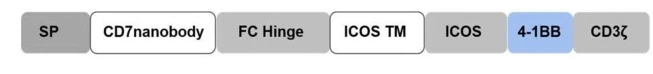 Fig. 1 Schematic structure of the CD7 CAR T-cells.1
Key structural properties of CD7:
- Extracellular section contains three immunoglobulin superfamily (IgSF) domain structure
- Anchored to the cell membrane by glycosylphosphatidylinositol (GPI)
- A functional domain that mediates homophilic binding
Functions of CD7
The main function of CD7 is to act as a co-stimulatory molecule of immune synapses, participating in lymphocyte activation and signal transduction. In addition, it also intervenes in various physiological processes such as immune regulation and cell adhesion.
| Function | Description |
| T cell activation | By binding to ligands, it provides co-stimulatory signals to promote the proliferation of T cells and the production of cytokines. |
| Cell adhesion | Mediate the interaction between immune cells, influencing the binding of T cells to antigen-presenting cells and synaptic formation. |
| Signal transduction | Its intracellular segment can recruit various signaling molecules and participate in regulating the activation threshold and response intensity of immune cells. |
| Immune regulation | In autoimmune and inflammatory response plays a regulatory role, its abnormal expression is associated with a variety of immune diseases. |
| Tumor immune escape | In some lymphomas and leukemias, the expression changes of CD7 may affect the immune surveillance and escape of tumor cells. |
CD7 specifically binds to ligands through its extracellular immunoglobulin-like domain. Its signal has the characteristics of moderate intensity and long duration, which helps maintain the stability of immune responses, especially playing a key role in early immune activation.
Applications of CD7 and CD7 Antibody in Literature
1. Liu, Jile, et al. "Targeted CD7 CAR T-cells for treatment of T-Lymphocyte leukemia and lymphoma and acute myeloid leukemia: recent advances." Frontiers in Immunology 14 (2023): 1170968. https://doi.org/10.3389/fimmu.2023.1170968
The article indicates that CD7 is highly expressed in T-cell leukemia and lymphoma and is an ideal target for CAR-T therapy. However, CD7 CAR-T cells are prone to auto-killing and exhaustion, which affects the therapeutic effect. In recent years, through technologies such as gene editing and protein blocking, significant breakthroughs have been made in CD7 CAR-T therapy, significantly overcoming the limitations of traditional methods.
2. Liu, Xuan, et al. "Naturally selected CD7-directed CAR-T bridging allo-HSCT in refractory acute myeloid leukemia: a case report and review." Frontiers in Immunology 15 (2024): 1461908. https://doi.org/10.3389/fimmu.2024.1461908
The article indicates that CD7 is expressed in approximately 30% of refractory/relapsed acute myeloid leukemia (R/R-AML) cases and is a potential immunotherapy target. A CD7-positive R/R-AML patient achieved remission after CD7 CAR-T treatment through natural screening and was promptly connected to allogeneic hematopoietic stem cell transplantation, achieving disease-free survival for 12 months. This confirmed that the combined strategy has clinical potential.
3. Kim, Miriam Y., et al. "CD7-deleted hematopoietic stem cells can restore immunity after CAR T cell therapy." JCI insight 6.16 (2021): e149819. https://doi.org/10.1172/jci.insight.149819
The article indicates that the treatment of T-cell malignancies with universal CD7 CAR-T (UCART7) will lead to the loss of normal T/NK cells, causing immune deficiency. Research shows that the transplantation of hematopoietic stem cells with CD7 knockout (CD7-KO HSCs) can effectively reconstruct T/NK cells after UCART7 treatment, maintain immune function, and provide potential protective strategies for clinical practice.
4. Wang, Shiqi, et al. "An Anti-CD7 Antibody–Drug Conjugate Target Showing Potent Antitumor Activity for T-Lymphoblastic Leukemia (T-ALL)." Biomolecules 14.1 (2024): 106. https://doi.org/10.3390/biom14010106
In this study, a novel anti-CD7 antibody-drug conjugate (ADC, J87-Dxd) was successfully developed for the treatment of T-cell acute lymphoblastic leukemia (T-ALL). J87-Dxd can be efficiently internalized and induce apoptosis of CD7-positive T-ALL cells, with an IC50 of 6.3 nM in vitro. In the tumor-bearing mouse model, the survival rate reached 80%, and no obvious organ toxicity occurred, demonstrating good therapeutic potential.
5. Bai, Yanliang, et al. "CD7-positive leukemic blasts with DNMT3A mutations predict poor prognosis in patients with acute myeloid leukemia." Frontiers in Oncology 14 (2024): 1342998. https://doi.org/10.3389/fonc.2024.1342998
This study explores the combined prognostic value of DNMT3A mutation and CD7 expression in acute myeloid leukemia (AML). The results show that patients with DNMT3A mutation and CD7 positive (CD7+) have a lower complete response rate, a higher recurrence rate, and significantly shorter overall survival and recurrence-free survival, which are independent poor prognostic factors for this type of AML.
Creative Biolabs: CD7 Antibodies for Research
Creative Biolabs specializes in the production of high-quality CD7 antibodies for research and industrial applications. Our portfolio includes monoclonal antibodies tailored for ELISA, Flow Cytometry, Western blot, immunohistochemistry, and other diagnostic methodologies.
- Custom CD7 Antibody Development: Tailor-made solutions to meet specific research requirements.
- Bulk Production: Large-scale antibody manufacturing for industry partners.
- Technical Support: Expert consultation for protocol optimization and troubleshooting.
- Aliquoting Services: Conveniently sized aliquots for long-term storage and consistent experimental outcomes.
For more details on our CD7 antibodies, custom preparations, or technical support, contact us at email.
Reference
- Dai, Hai-ping, et al. "Haploidentical CD7 CAR T-cells induced remission in a patient with TP53 mutated relapsed and refractory early T-cell precursor lymphoblastic leukemia/lymphoma." Biomarker Research 10.1 (2022): 6. https://doi.org/10.1186/s40364-022-00352-w
Anti-CD7 antibodies
 Loading...


Hot products 
-
Rat Anti-ABCC11 Recombinant Antibody (V2-179001) (CBMAB-A0236-YC)

-
Mouse Anti-A2M Recombinant Antibody (V2-178822) (CBMAB-A0036-YC)

-
Mouse Anti-FOSB Recombinant Antibody (CBXF-3593) (CBMAB-F2522-CQ)

-
Mouse Anti-CD1C Recombinant Antibody (L161) (CBMAB-C2173-CQ)

-
Mouse Anti-BAD (Phospho-Ser136) Recombinant Antibody (CBYY-0138) (CBMAB-0139-YY)

-
Mouse Anti-ACLY Recombinant Antibody (V2-179314) (CBMAB-A0610-YC)

-
Mouse Anti-AKT1/AKT2/AKT3 (Phosphorylated T308, T309, T305) Recombinant Antibody (V2-443454) (PTM-CBMAB-0030YC)

-
Mouse Anti-BLK Recombinant Antibody (CBYY-0618) (CBMAB-0621-YY)

-
Mouse Anti-EPO Recombinant Antibody (CBFYR0196) (CBMAB-R0196-FY)

-
Mouse Anti-BACE1 Recombinant Antibody (CBLNB-121) (CBMAB-1180-CN)

-
Mouse Anti-ATP1A2 Recombinant Antibody (M7-PB-E9) (CBMAB-A4013-YC)

-
Mouse Anti-ADAM12 Recombinant Antibody (V2-179752) (CBMAB-A1114-YC)

-
Mouse Anti-BRCA2 Recombinant Antibody (CBYY-0790) (CBMAB-0793-YY)

-
Mouse Anti-CCND2 Recombinant Antibody (DCS-3) (CBMAB-G1318-LY)

-
Rabbit Anti-AKT3 Recombinant Antibody (V2-12567) (CBMAB-1057-CN)

-
Mouse Anti-CFL1 (Phospho-Ser3) Recombinant Antibody (CBFYC-1770) (CBMAB-C1832-FY)

-
Mouse Anti-AAV9 Recombinant Antibody (V2-634029) (CBMAB-AP023LY)

-
Mouse Anti-ADIPOR2 Recombinant Antibody (V2-179983) (CBMAB-A1369-YC)

-
Mouse Anti-AMACR Recombinant Antibody (CB34A) (CBMAB-CA034LY)

-
Mouse Anti-CD46 Recombinant Antibody (CBFYC-0076) (CBMAB-C0085-FY)

- AActivation
- AGAgonist
- APApoptosis
- BBlocking
- BABioassay
- BIBioimaging
- CImmunohistochemistry-Frozen Sections
- CIChromatin Immunoprecipitation
- CTCytotoxicity
- CSCostimulation
- DDepletion
- DBDot Blot
- EELISA
- ECELISA(Cap)
- EDELISA(Det)
- ESELISpot
- EMElectron Microscopy
- FFlow Cytometry
- FNFunction Assay
- GSGel Supershift
- IInhibition
- IAEnzyme Immunoassay
- ICImmunocytochemistry
- IDImmunodiffusion
- IEImmunoelectrophoresis
- IFImmunofluorescence
- IGImmunochromatography
- IHImmunohistochemistry
- IMImmunomicroscopy
- IOImmunoassay
- IPImmunoprecipitation
- ISIntracellular Staining for Flow Cytometry
- LALuminex Assay
- LFLateral Flow Immunoassay
- MMicroarray
- MCMass Cytometry/CyTOF
- MDMeDIP
- MSElectrophoretic Mobility Shift Assay
- NNeutralization
- PImmunohistologyp-Paraffin Sections
- PAPeptide Array
- PEPeptide ELISA
- PLProximity Ligation Assay
- RRadioimmunoassay
- SStimulation
- SESandwich ELISA
- SHIn situ hybridization
- TCTissue Culture
- WBWestern Blot
