CD81 Antibodies
Background
The CD81 gene encodes a quadruplex transmembrane protein, which serves as a cell surface receptor and is widely distributed in various tissues such as lymphocytes and nerve cells. Its encoded protein participates in cell adhesion, movement and signal transduction processes by forming a tetramer supramolecular complex, and is particularly important in immune regulation - it, together with CD19 and CD21, constitutes a B-cell co-receptor complex, regulating the activation threshold of B lymphocytes. This gene was first identified in 1990 and is the earliest core member of the four-transmembrane protein family to be confirmed to have immune regulatory functions. The study of its three-dimensional structure has revealed a unique intermolecular interaction pattern. As a key co-receptor for hepatitis C virus invading host cells, CD81 has become an important target for the study of viral pathogenic mechanisms and immunotherapy. Its unique molecular conformation provides a classic model for transmembrane signal transduction research.
Structure of CD81
CD81 is a transmembrane protein with a molecular weight of approximately 25-26 kDa. Its molecular weight varies slightly among different cell types due to the degree of glycosylation modification.
| Species | Human | Mouse | Rat | Macaque |
| Molecular Weight (kDa) | 25.8 | 25.9 | 26.1 | 25.7 |
| Primary Structural Differences | Conservative quadruple transmembrane domain | There are differences in N-glycosylation sites | Extracellular loop area sequence is highly similar | More than 90% homology to human CD81 |
This protein is composed of 228 amino acids, forming four highly conserved transmembrane domains. Its molecular structure consists of two extracellular rings (EC1 and EC2), among which the larger EC2 ring forms a stable spherical domain through disulfide bonds. The EC2 loop has specific hydrophobic regions and glycosylation sites that directly participate in protein-protein interactions. Palmitoylation modification in the near-membrane region enhanced the stability of CD81 on the plasma membrane, while the interaction in the transmembrane region promoted the formation of tetramer complexes. These structural features jointly determined its functions in cell signal transduction and viral invasion.
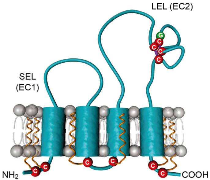 Fig. 1 Schematic representation of the tetraspanin CD81.1
Key structural properties of CD81:
- Four times across the membrane structure field of typical stents
- Large extracellular two-rings form molecular interaction interfaces
- Conserved palmitoylation sites regulate membrane segregation and signal transduction
Functions of CD81
The core function of the CD81 gene-encoded protein is to act as a cell membrane organizer, regulating cell signaling and adhesion by forming a tetramer network. Its main physiological and pathological functions are as follows:
| Function | Description |
| Immune regulation | In the B cell surface receptor complexes with CD19, CD21, regulating lymphocyte activation threshold and signal transduction. |
| Virus invasion | As a key co-receptor for hepatitis C virus (HCV) invading host cells, it specifically binds to the viral envelope glycoprotein E2. |
| Cellular movement | Through the synergistic effect of integrins, it affects cell morphology and migration, and participates in tumor cell metastasis and immune cell chemotaxis. |
| Membrane domain organization | As a core member of the four-transmembrane protein superfamily, it forms specific microdomains on the plasma membrane to regulate the distribution and interaction of membrane proteins. |
| Developmental regulation | Involved in oocyte fertilization, synapse formation and the development of liver cell morphogenesis process. |
The interaction between CD81 and ligands shows spatial specificity, and the conformational changes of its extracellular ring directly determine the binding activity. This characteristic not only explains its regulatory role in the immune response but also reveals the molecular basis of the virus's utilization of host receptors.
Applications of CD81 and CD81 Antibody in Literature
1. Fan, Yé, et al. "Differential proteomics argues against a general role for CD9, CD81 or CD63 in the sorting of proteins into extracellular vesicles." Journal of Extracellular Vesicles 12.8 (2023): 12352. https://doi.org/10.1002/jev2.12352
The article indicates that in MCF7 cells, CD81 and CD9 are mainly co-localized in the plasma membrane, and CD81 has a higher enrichment degree in extracellular vesicles (EVs). Studies have found that the absence of CD9 and CD81 has little effect on the protein composition of EVs, but they jointly regulate the contents of their direct binding proteins CD9P-1 and EWI-2 in EVs. The latter is mainly caused by the decrease in cell expression levels.
2. Gurrieri, Elena, et al. "CD81-guided heterologous EVs present heterogeneous interactions with breast cancer cells." Journal of Biomedical Science 31.1 (2024): 92. https://doi.org/10.1186/s12929-024-01084-9
The study aimed to construct engineered extracellular vesicles (EVs) targeting HER2 using CD81 as a scaffold. This EV can specifically bind to HER2-positive cancer cells and effectively deliver therapeutic cargo such as doxorubicin or long-chain mRNA. It has shown enhanced targeted killing effects in both in vitro and in mouse models, providing a basis for the CD81-based EV targeted therapy platform.
3. Cone, Allaura S., et al. "CD81 fusion alters SARS-CoV-2 Spike trafficking." Mbio 15.9 (2024): e01922-24. https://doi.org/10.1128/mbio.01922-24
The study fused the S protein of the novel coronavirus with the transmembrane protein CD81, successfully redirecting and enriching the degradation pathway of the S protein in extracellular vesicles (EVs). This CD81-S fusion EV can produce in large quantities, bind to ACE2, and effectively induce anti-S antibodies in mice, providing a path for the design of new vaccines.
4. Fénéant, Lucie, Shoshana Levy, and Laurence Cocquerel. "CD81 and hepatitis C virus (HCV) infection." Viruses 6.2 (2014): 535-572. https://doi.org/10.3390/v6020535
The article indicates that the invasion of hepatitis C virus (HCV) into liver cells is a complex multi-step process, among which the four-transmembrane protein CD81 is one of the most clearly defined key invasion factors. This review elaborates in detail on the core role of CD81 in the HCV life cycle and the antiviral immune response of the body.
5. Zona, Laetitia, et al. "CD81-receptor associations—impact for hepatitis C virus entry and antiviral therapies." Viruses 6.2 (2014): 875-892. https://doi.org/10.3390/v6020875
The article indicates that the four-transmembrane protein CD81 plays a key role in the process of HCV invasion of hepatocytes by forming specific microdomains with other membrane proteins. This invasion process is complex and requires the synergistic action of multiple host factors such as CD81 and scavenger receptor BI. Research on this complex provides new ideas for the prevention and treatment of HCV infection.
Creative Biolabs: CD81 Antibodies for Research
Creative Biolabs specializes in the production of high-quality CD81 antibodies for research and industrial applications. Our portfolio includes monoclonal antibodies tailored for ELISA, Flow Cytometry, Western blot, immunohistochemistry, and other diagnostic methodologies.
- Custom CD81 Antibody Development: Tailor-made solutions to meet specific research requirements.
- Bulk Production: Large-scale antibody manufacturing for industry partners.
- Technical Support: Expert consultation for protocol optimization and troubleshooting.
- Aliquoting Services: Conveniently sized aliquots for long-term storage and consistent experimental outcomes.
For more details on our CD81 antibodies, custom preparations, or technical support, contact us at email.
Reference
- Fénéant, Lucie, Shoshana Levy, and Laurence Cocquerel. "CD81 and hepatitis C virus (HCV) infection." Viruses 6.2 (2014): 535-572. https://doi.org/10.3390/v6020535
Anti-CD81 antibodies
 Loading...


Hot products 
-
Mouse Anti-CAPZB Recombinant Antibody (CBYY-C0944) (CBMAB-C2381-YY)

-
Mouse Anti-BCL2L1 Recombinant Antibody (H5) (CBMAB-1025CQ)

-
Mouse Anti-ALDOA Recombinant Antibody (A2) (CBMAB-A2316-YC)

-
Mouse Anti-CDK7 Recombinant Antibody (CBYY-C1783) (CBMAB-C3221-YY)

-
Mouse Anti-APOA1 Monoclonal Antibody (CBFYR0637) (CBMAB-R0637-FY)

-
Mouse Anti-CHRNA9 Recombinant Antibody (8E4) (CBMAB-C9161-LY)

-
Mouse Anti-DES Monoclonal Antibody (440) (CBMAB-AP1857LY)

-
Mouse Anti-ALPL Antibody (B4-78) (CBMAB-1009CQ)

-
Mouse Anti-ANXA7 Recombinant Antibody (A-1) (CBMAB-A2941-YC)

-
Mouse Anti-ADGRE5 Recombinant Antibody (V2-360335) (CBMAB-C2088-CQ)

-
Mouse Anti-DLC1 Recombinant Antibody (D1009) (CBMAB-D1009-YC)

-
Mouse Anti-NSUN6 Recombinant Antibody (D-5) (CBMAB-N3674-WJ)

-
Mouse Anti-G6PD Recombinant Antibody (13B331) (CBMAB-G1553-LY)

-
Mouse Anti-ADAM29 Recombinant Antibody (V2-179787) (CBMAB-A1149-YC)

-
Rat Anti-CD34 Recombinant Antibody (MEC 14.7) (CBMAB-C10196-LY)

-
Mouse Anti-CSPG4 Recombinant Antibody (CBFYM-1050) (CBMAB-M1203-FY)

-
Mouse Anti-AMH Recombinant Antibody (5/6) (CBMAB-A2527-YC)

-
Mouse Anti-AAV-5 Recombinant Antibody (V2-503417) (CBMAB-V208-1369-FY)

-
Mouse Anti-AAV-5 Recombinant Antibody (V2-503416) (CBMAB-V208-1402-FY)

-
Mouse Anti-ACLY Recombinant Antibody (V2-179314) (CBMAB-A0610-YC)

- AActivation
- AGAgonist
- APApoptosis
- BBlocking
- BABioassay
- BIBioimaging
- CImmunohistochemistry-Frozen Sections
- CIChromatin Immunoprecipitation
- CTCytotoxicity
- CSCostimulation
- DDepletion
- DBDot Blot
- EELISA
- ECELISA(Cap)
- EDELISA(Det)
- ESELISpot
- EMElectron Microscopy
- FFlow Cytometry
- FNFunction Assay
- GSGel Supershift
- IInhibition
- IAEnzyme Immunoassay
- ICImmunocytochemistry
- IDImmunodiffusion
- IEImmunoelectrophoresis
- IFImmunofluorescence
- IGImmunochromatography
- IHImmunohistochemistry
- IMImmunomicroscopy
- IOImmunoassay
- IPImmunoprecipitation
- ISIntracellular Staining for Flow Cytometry
- LALuminex Assay
- LFLateral Flow Immunoassay
- MMicroarray
- MCMass Cytometry/CyTOF
- MDMeDIP
- MSElectrophoretic Mobility Shift Assay
- NNeutralization
- PImmunohistologyp-Paraffin Sections
- PAPeptide Array
- PEPeptide ELISA
- PLProximity Ligation Assay
- RRadioimmunoassay
- SStimulation
- SESandwich ELISA
- SHIn situ hybridization
- TCTissue Culture
- WBWestern Blot
