CD83 Antibodies
Background
CD83 is a type I transmembrane glycoprotein mainly expressed on the surface of mature dendritic cells, which plays a key role in the formation of immune synapses and the activation of T cells. This protein participates in the regulation of antigen presentation by mediating intercellular signal transduction and can be released into the extracellular environment in a soluble form to regulate the intensity of the immune response. Research has found that CD83 has an important negative regulatory function in autoimmune diseases and transplant rejection reactions, and its expression level changes are closely related to various immunopathological states. This gene was first identified in 1992. Its unique immunomodulatory properties make it an important target for tumor immunotherapy and intervention of autoimmune diseases. Related research continues to drive in-depth exploration of the regulatory mechanism of immune checkpoints.
Structure of CD83
CD83 is a cell surface glycoprotein with a molecular weight of approximately 45-50 kDa. The specific molecular weight varies depending on the degree of glycosylation modification.
| Species | Human | Mouse | Rat |
| Molecular Weight (kDa) | 45-50 | 44-49 | 45-50 |
| Primary Structural Differences | Containing 244 amino acids, extracellular region contains two immune globulin structure domain | With the human CD83 about 62% homology | Approximately 65% homology to human CD83 |
The CD83 protein is composed of 244 amino acids, and its extracellular region contains two immunoglobulin-like domains (type V and type C2), which form a stable spatial conformation through disulfide bonds. The transmembrane region of this protein anchors it to the cell membrane, while the shorter intracellular region is involved in signal transduction. The glycosylation modifications of CD83 mainly occur on the asparagine residues in the extracellular region, and these glycosylation modifications affect the stability and function of the protein.
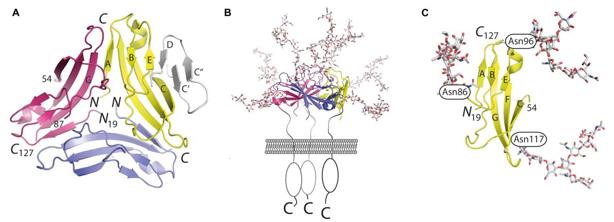 Fig. 1 Atomic structure of CD83.1
Key structural properties of CD83:
- Extracellular region contains two immune globulin structure domain (V and C2)
- Multiple conserved cysteine residues form intramolecular disulfide bonds
- Transmembrane regions anchor proteins to cell membranes
- Intracellular segments contain potential phosphorylation sites involved in signal transduction
- Glycosylation modification sites were mainly located in the extracellular region
Functions of CD83
The main function of CD83 is to participate in the regulation of immune responses, especially playing a key role in the maturation of dendritic cells and the activation of T cells. In addition, this molecule is also involved in the establishment and maintenance of immune tolerance.
| Function | Description |
| Immune regulation | Through the membrane type and the soluble form two-way regulating function of dendritic cells, affect threshold T cell activation. |
| Costimulatory signal | Interacting with ligands provides the second signal required for T cell activation and enhances the formation of immune synapses. |
| Tolerance induction | Involved in the formation of the central tolerance in thymic stromal cells, outer weeks, to maintain the immune balance. |
| Inflammatory regulation | Limit excessive inflammatory responses through negative feedback mechanisms to prevent autoimmune damage. |
| Disease association | The changes in expression levels are closely related to autoimmune diseases, transplant rejection and tumor immune escape. |
The immunomodulatory properties of CD83 exhibit dose-dependent characteristics. Its membrane form and soluble form respectively exert positive and negative regulatory functions. This dual mechanism makes it an important target for immune checkpoint research.
Applications of CD83 and CD83 Antibody in Literature
1. Riaz, Bushra, et al. "CD83 regulates the immune responses in inflammatory disorders." International Journal of Molecular Sciences 24.3 (2023): 2831. https://doi.org/10.3390/ijms24032831
Research has found that CD83 is a key molecule for the maturation of dendritic cells and participates in immune responses by regulating T cell differentiation. Its role in autoimmune diseases and pathogen immune escape remains unclear. This article reviews the function of CD83 in inflammatory diseases.
2. Grosche, Linda, et al. "The CD83 molecule–an important immune checkpoint." Frontiers in Immunology 11 (2020): 721. https://doi.org/10.3389/fimmu.2020.00721
Research has found that CD83 is expressed in a variety of immune cells. It is not only a co-stimulatory molecule but also plays a key role in controlling immune responses, maintaining immune tolerance and inflammatory balance. Its immunomodulatory function shows great therapeutic potential.
3. Li, Ziduo, et al. "CD83: activation marker for antigen presenting cells and its therapeutic potential." Frontiers in immunology 10 (2019): 1312. https://doi.org/10.3389/fimmu.2019.01312
Research has found that CD83 is a key immunomodulatory molecule. Membrane-type CD83 promotes immune activation, while soluble CD83 has immunosuppressive functions. Therapies based on this characteristic (such as sCD83 or anti-CD83 antibodies) have shown great potential in the treatment of autoimmune diseases, transplant rejection and other diseases.
4. Wu, Zhiwen, et al. "CD83 expression characterizes precursor exhausted T cell population." Communications biology 6.1 (2023): 258. https://doi.org/10.1038/s42003-023-04631-6
Research has found that CD83 can serve as a specific surface marker for precursor exhausted T cells (TPEX) in tumor-infiltrating T cells, effectively distinguishing between terminal exhausted T cells and bystander T cells, and has stronger proliferation and anti-tumor capabilities.
5. Akauliya, Madhav, et al. "CD83 expression regulates antibody production in response to influenza A virus infection." Virology Journal 17.1 (2020): 194. https://doi.org/10.1186/s12985-020-01465-0
Research has found that CD83 is crucial in influenza A virus infection. CD83 gene knockout mice exhibited dysregulation of B cell and T cell homeostasis and were unable to produce effective virus-specific IgG antibodies, revealing the crucial role of CD83 in antiviral immunity.
Creative Biolabs: CD83 Antibodies for Research
Creative Biolabs specializes in the production of high-quality CD83 antibodies for research and industrial applications. Our portfolio includes monoclonal antibodies tailored for ELISA, Flow Cytometry, Western blot, immunohistochemistry, and other diagnostic methodologies.
- Custom CD83 Antibody Development: Tailor-made solutions to meet specific research requirements.
- Bulk Production: Large-scale antibody manufacturing for industry partners.
- Technical Support: Expert consultation for protocol optimization and troubleshooting.
- Aliquoting Services: Conveniently sized aliquots for long-term storage and consistent experimental outcomes.
For more details on our CD83 antibodies, custom preparations, or technical support, contact us at email.
Reference
- Grosche, Linda, et al. "The CD83 molecule–an important immune checkpoint." Frontiers in Immunology 11 (2020): 721. https://doi.org/10.3389/fimmu.2020.00721
Anti-CD83 antibodies
 Loading...


Hot products 
-
Rabbit Anti-ALDOA Recombinant Antibody (D73H4) (CBMAB-A2314-YC)

-
Mouse Anti-8-oxoguanine Recombinant Antibody (V2-7719) (CBMAB-1898CQ)

-
Mouse Anti-BCL6 Recombinant Antibody (CBYY-0442) (CBMAB-0445-YY)

-
Mouse Anti-AGO2 Recombinant Antibody (V2-634169) (CBMAB-AP203LY)

-
Mouse Anti-ALOX5 Recombinant Antibody (33) (CBMAB-1890CQ)

-
Mouse Anti-ACTG1 Recombinant Antibody (V2-179597) (CBMAB-A0916-YC)

-
Mouse Anti-APOH Recombinant Antibody (4D9A4) (CBMAB-A3249-YC)

-
Mouse Anti-FN1 Monoclonal Antibody (D6) (CBMAB-1240CQ)

-
Mouse Anti-AKT1/AKT2/AKT3 (Phosphorylated T308, T309, T305) Recombinant Antibody (V2-443454) (PTM-CBMAB-0030YC)

-
Mouse Anti-AOC3 Recombinant Antibody (CBYY-0014) (CBMAB-0014-YY)

-
Mouse Anti-CARTPT Recombinant Antibody (113612) (CBMAB-C2450-LY)

-
Mouse Anti-BIRC3 Recombinant Antibody (315304) (CBMAB-1214-CN)

-
Rat Anti-4-1BB Recombinant Antibody (V2-1558) (CBMAB-0953-LY)

-
Mouse Anti-BPGM Recombinant Antibody (CBYY-1806) (CBMAB-2155-YY)

-
Mouse Anti-COL12A1 Recombinant Antibody (CBYY-C3117) (CBMAB-C4560-YY)

-
Mouse Anti-CD19 Recombinant Antibody (CBXC-1224) (CBMAB-C1491-CQ)

-
Rabbit Anti-ALOX5AP Recombinant Antibody (CBXF-1219) (CBMAB-F0750-CQ)

-
Mouse Anti-ARHGAP5 Recombinant Antibody (54/P190-B) (CBMAB-P0070-YC)

-
Mouse Anti-GFAP Recombinant Antibody (20) (CBMAB-G2914-LY)

-
Mouse Anti-GFAP Recombinant Antibody (5) (CBMAB-G0346-LY)

- AActivation
- AGAgonist
- APApoptosis
- BBlocking
- BABioassay
- BIBioimaging
- CImmunohistochemistry-Frozen Sections
- CIChromatin Immunoprecipitation
- CTCytotoxicity
- CSCostimulation
- DDepletion
- DBDot Blot
- EELISA
- ECELISA(Cap)
- EDELISA(Det)
- ESELISpot
- EMElectron Microscopy
- FFlow Cytometry
- FNFunction Assay
- GSGel Supershift
- IInhibition
- IAEnzyme Immunoassay
- ICImmunocytochemistry
- IDImmunodiffusion
- IEImmunoelectrophoresis
- IFImmunofluorescence
- IGImmunochromatography
- IHImmunohistochemistry
- IMImmunomicroscopy
- IOImmunoassay
- IPImmunoprecipitation
- ISIntracellular Staining for Flow Cytometry
- LALuminex Assay
- LFLateral Flow Immunoassay
- MMicroarray
- MCMass Cytometry/CyTOF
- MDMeDIP
- MSElectrophoretic Mobility Shift Assay
- NNeutralization
- PImmunohistologyp-Paraffin Sections
- PAPeptide Array
- PEPeptide ELISA
- PLProximity Ligation Assay
- RRadioimmunoassay
- SStimulation
- SESandwich ELISA
- SHIn situ hybridization
- TCTissue Culture
- WBWestern Blot
