CDH5 Antibodies
Background
The CDH5 gene encodes a transmembrane protein called VE-cadherin, which is mainly present at the adhesion junctions between vascular endothelial cells. This protein binds to the same molecules on adjacent cells through its extracellular region, forming a homophilic interaction, thereby maintaining the integrity and permeability barrier function of the endothelial layer. During embryonic development, CDH5 is crucial for the correct assembly and remodeling of the vascular network, and continues to participate in the maintenance of vascular homeostasis and the regulation of inflammatory responses in adulthood. This gene was initially cloned and identified in the 1990s, and its encoded protein is the first member of the calcium-binding protein family specifically expressed in endothelial cells. Subsequent studies have shown that CDH5 not only acts as a structural adhesion molecule but also interacts with various signaling molecules through its intracellular segment, participating in the regulation of key biological processes such as cell survival, proliferation, and angiogenesis. These findings provide an important foundation for understanding vascular biology and related disease mechanisms.
Structure of CDH5
The CDH5 gene encodes the VE-cadherin protein with a molecular weight of approximately 88 kDa. Its extracellular segment contains five cadherin repeat domains, which mediate adhesion between endothelial cells through homophilic interactions.
| Species | Human | Mouse | Rat | Bovine | Pig |
| Molecular Weight (kDa) | 88.2 | 88.0 | 88.1 | 88.3 | 88.2 |
| Primary Structural Differences | Five CAD repeats in the extracellular region, with calcium-binding sites | The degree of genetic similarity with others is approximately 92% | The degree of genetic similarity with others is approximately 91% | The extracellular segment structure is highly conserved | The domain composition is consistent |
The intracellular segment of this protein is connected to the actin cytoskeleton through p120-catenin and β-catenin. The first and second extracellular CAD domains directly participate in the formation of intercellular adhesion. The transmembrane region maintains the correct localization of the protein on the membrane, while the conserved sequences in the extracellular near-membrane region are crucial for maintaining the function of the endothelial barrier.
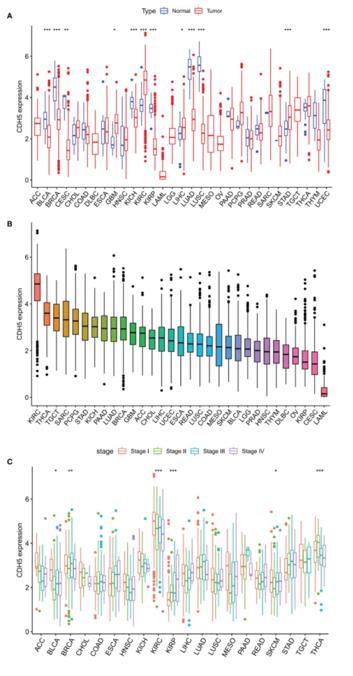 Fig. 1 Differential expression analysis of CDH5 in TCGA pan-cancer.1
Key structural properties of CDH5:
- Five extracellular cadherin repeat domains (CAD1-5)
- The intracellular region is connected to the actin cytoskeleton through integrins
- The transmembrane region maintains the correct localization on the membrane
Functions of CDH5
The VE-cadherin protein encoded by the CDH5 gene mainly functions to maintain the integrity of vascular endothelium and participates in various physiological processes, including angiogenesis and regulation of inflammatory responses.
| Function | Description |
| Cell adhesion | It binds to adjacent cells through the extracellular CAD1-2 domains, forming adhesion junctions. |
| Barrier function | It regulates the permeability between endothelial cells, controlling the exchange of substances between the inside and outside of the blood vessels. |
| Angiogenesis | It mediates the formation of connections between new vascular endothelial cells and participates in the remodeling of the vascular network. |
| Inflammatory regulation | Inflammatory factors can downregulate its expression, leading to increased endothelial permeability, facilitating the extravasation of white blood cells. |
| Mechanical conduction | It senses changes in blood flow shear stress and regulates the function of endothelial cells through intracellular signaling pathways. |
The intracellular segment of VE-cadherin protein is anchored to the actin cytoskeleton by binding to p120-catenin and β-catenin. Its expression downregulation or structural abnormalities can lead to vascular leakage, playing a key role in various vascular-related diseases.
Applications of CDH5 and CDH5 Antibody in Literature
1. Li, Yuantao, et al. "A comprehensive pan-cancer analysis of CDH5 in immunological response." Frontiers in Immunology 14 (2023): 1239875. https://doi.org/10.3389/fimmu.2023.1239875
The research has found that CDH5 is abnormally expressed in various tumors and is significantly correlated with prognosis, immune infiltration, etc. Especially in bladder cancer, the low expression of CDH5 can promote the function of CD8+ T cells, suggesting that it may serve as a potential new tumor marker and immune checkpoint.
2. He, Qi, et al. "The Cdh5-CreERT2 transgene causes conditional Shb gene deletion in hematopoietic cells with consequences for immune cell responses to tumors." Scientific Reports 9.1 (2019): 7548. https://doi.org/10.1038/s41598-019-44039-z
The study found that the Cdh5-CreERT2 tamoxifen-induced approach not only targets endothelial cells but also leads to genetic recombination in hematopoietic cells (such as bone marrow and spleen cells), thereby affecting the results of tumor immunology experiments. It is recommended to adopt a bone marrow transplantation strategy to obtain a pure endothelial cell-specific phenotype.
3. Zheng, Huiwen, et al. "Cdh5-mediated Fpn1 deletion exerts neuroprotective effects during the acute phase and inhibitory effects during the recovery phase of ischemic stroke." Cell Death & Disease 14.2 (2023): 161. https://doi.org/10.1038/s41419-023-05688-1
The study found that knocking out the iron export protein Fpn1 in endothelial cells (mediated by VE-cadherin-Cre) has a dual effect on ischemic stroke: in the acute phase, it reduces brain iron to alleviate damage, but in the later stage, due to iron deficiency, it hinders neural repair, providing new ideas for stroke treatment.
4. Wang, Yishuai, et al. "The rs7404339 AA genotype in CDH5 contributes to increased risks of Kawasaki disease and coronary artery lesions in a Southern Chinese child population." Frontiers in Cardiovascular Medicine 9 (2022): 760982. https://doi.org/10.3389/fcvm.2022.760982
The study found that the rs7404339 polymorphism of the CDH5 gene is associated with the susceptibility to Kawasaki disease in children in southern China. The AA genotype is a risk factor. Children carrying the AA genotype, especially those under 5 years old and male, have a higher risk of developing the disease and are more prone to coronary artery damage.
5. Liu, Yinping, et al. "RETRACTED ARTICLE: ANGPTL4 functions as an oncogene through regulation of the ETV5/CDH5/AKT/MMP9 axis to promote angiogenesis in ovarian cancer." Journal of Ovarian Research 15.1 (2022): 131. https://doi.org/10.1186/s13048-022-01060-7
The study found that ANGPTL4 is highly expressed in ovarian cancer. It activates the CDH5/AKT/MMP9 signaling pathway by upregulating the transcription factor ETV5, thereby promoting tumor angiogenesis and metastasis. This suggests that it could be a potential therapeutic target.
Creative Biolabs: CDH5 Antibodies for Research
Creative Biolabs specializes in the production of high-quality CDH5 antibodies for research and industrial applications. Our portfolio includes monoclonal and polyclonal antibodies tailored for ELISA, Flow Cytometry, Western blot, immunohistochemistry, and other diagnostic methodologies.
- Custom CDH5 Antibody Development: Tailor-made solutions to meet specific research requirements.
- Bulk Production: Large-scale antibody manufacturing for industry partners.
- Technical Support: Expert consultation for protocol optimization and troubleshooting.
- Aliquoting Services: Conveniently sized aliquots for long-term storage and consistent experimental outcomes.
For more details on our CDH5 antibodies, custom preparations, or technical support, contact us at email.
Reference
- Li, Yuantao, et al. "A comprehensive pan-cancer analysis of CDH5 in immunological response." Frontiers in Immunology 14 (2023): 1239875. Distributed under Open Access license CC BY 4.0, without modification. https://doi.org/10.3389/fimmu.2023.1239875
Anti-CDH5 antibodies
 Loading...


Hot products 
-
Mouse Anti-ALX1 Recombinant Antibody (96k) (CBMAB-C0616-FY)

-
Mouse Anti-ENPP1 Recombinant Antibody (CBFYE-0159) (CBMAB-E0375-FY)

-
Mouse Anti-CD24 Recombinant Antibody (ALB9) (CBMAB-0176CQ)

-
Mouse Anti-ATP1A2 Recombinant Antibody (M7-PB-E9) (CBMAB-A4013-YC)

-
Mouse Anti-BRCA2 Recombinant Antibody (CBYY-1728) (CBMAB-2077-YY)

-
Mouse Anti-AMH Recombinant Antibody (5/6) (CBMAB-A2527-YC)

-
Mouse Anti-ELAVL4 Recombinant Antibody (6B9) (CBMAB-1132-YC)

-
Mouse Anti-CDKL5 Recombinant Antibody (CBFYC-1629) (CBMAB-C1689-FY)

-
Rat Anti-EMCN Recombinant Antibody (28) (CBMAB-E0280-FY)

-
Mouse Anti-ARHGDIA Recombinant Antibody (CBCNA-009) (CBMAB-R0415-CN)

-
Mouse Anti-CORO1A Recombinant Antibody (4G10) (V2LY-1206-LY806)

-
Rat Anti-EPO Recombinant Antibody (16) (CBMAB-E1578-FY)

-
Mouse Anti-CASQ1 Recombinant Antibody (CBFYC-0863) (CBMAB-C0918-FY)

-
Mouse Anti-AKR1B1 Antibody (V2-2449) (CBMAB-1001CQ)

-
Mouse Anti-C5B-9 Recombinant Antibody (CBFYA-0216) (CBMAB-X0304-FY)

-
Mouse Anti-ADIPOR2 Recombinant Antibody (V2-179983) (CBMAB-A1369-YC)

-
Mouse Anti-BCL6 Recombinant Antibody (CBYY-0435) (CBMAB-0437-YY)

-
Mouse Anti-AAV-5 Recombinant Antibody (V2-503417) (CBMAB-V208-1369-FY)

-
Mouse Anti-ACTN4 Recombinant Antibody (V2-6075) (CBMAB-0020CQ)

-
Mouse Anti-Acetyl SMC3 (K105/K106) Recombinant Antibody (V2-634053) (CBMAB-AP052LY)

- AActivation
- AGAgonist
- APApoptosis
- BBlocking
- BABioassay
- BIBioimaging
- CImmunohistochemistry-Frozen Sections
- CIChromatin Immunoprecipitation
- CTCytotoxicity
- CSCostimulation
- DDepletion
- DBDot Blot
- EELISA
- ECELISA(Cap)
- EDELISA(Det)
- ESELISpot
- EMElectron Microscopy
- FFlow Cytometry
- FNFunction Assay
- GSGel Supershift
- IInhibition
- IAEnzyme Immunoassay
- ICImmunocytochemistry
- IDImmunodiffusion
- IEImmunoelectrophoresis
- IFImmunofluorescence
- IGImmunochromatography
- IHImmunohistochemistry
- IMImmunomicroscopy
- IOImmunoassay
- IPImmunoprecipitation
- ISIntracellular Staining for Flow Cytometry
- LALuminex Assay
- LFLateral Flow Immunoassay
- MMicroarray
- MCMass Cytometry/CyTOF
- MDMeDIP
- MSElectrophoretic Mobility Shift Assay
- NNeutralization
- PImmunohistologyp-Paraffin Sections
- PAPeptide Array
- PEPeptide ELISA
- PLProximity Ligation Assay
- RRadioimmunoassay
- SStimulation
- SESandwich ELISA
- SHIn situ hybridization
- TCTissue Culture
- WBWestern Blot
