CISH Antibodies
Background
The CISH gene encodes a signal transduction inhibitory protein present in the cytoplasm, which mainly regulates the immune response process by participating in the JAK-STAT pathway. Its protein structure contains the SH2 domain, which can bind to cytokine receptors and target STAT proteins for ubiquitination and degradation, thereby negatively regulating T cell activation and proliferation. This gene plays a key role in immune balance, and its functional deficiency may trigger an excessive immune response. Studies have shown that the polymorphism of the CISH gene is associated with the susceptibility to various infectious diseases, especially showing a significant correlation in the malaria and tuberculosis populations. Due to its pivotal position in immune regulation, this gene has become an important research object in the fields of autoimmune diseases and cancer treatment, providing a molecular basis for the development of targeted therapeutic strategies.
Structure of CISH
CISH is a cytoplasmic protein with a molecular weight of approximately 28 kDa. This protein is composed of approximately 258 amino acids, and its core structure contains a SH2 domain, which mediates protein-protein interactions by recognizing phosphorylated tyrosine residues. CISH protein, as a key negative regulatory factor of the JAK-STAT signaling pathway, inhibits signal transduction by binding to activated cytokine receptors and promoting the ubiquitination and degradation of STAT5. This protein plays a significant role in maintaining immune homeostasis, and its abnormal expression is closely related to the occurrence and development of various immune diseases and tumors.
| Species | Human | Mouse | Zebrafish |
| Molecular Weight (kDa) | 28 | 27.8 | 26.5 |
| Primary Structural Differences | Inhibit cytokine signaling pathways such as IL-2 | The CISH function of humans is highly conserved | Basic regulation role in innate immunity |
The tertiary structure of CISH forms a typical SH2 domain fold, which consists of a central β sheet and bilateral α -helixes and can specifically recognize phosphorylated tyrosine sites. Its C-terminal extension region participates in the assembly of the E3 ubiquitin ligase complex, jointly completing the targeted degradation of signal proteins.
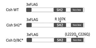 Fig. 1 Schematic diagram of Cish constructs used in our experiments.1
Key structural properties of CISH:
- Typical SH2 domain core conformation
- Phosphorylated tyrosine recognizes pockets
- Highly conserved FLVRES sequence motifs
Functions of CISH
The core function of the CISH protein is to negatively regulate cytokine signaling pathways. In addition, it is also involved in various cellular physiological processes, including the maintenance of immune homeostasis and the occurrence and development of tumors.
| Function | Description |
| Inhibition of the JAK-STAT pathway | By binding to activated cytokine receptors through the SH2 domain, it competitively blocks the phosphorylation and activation of STAT5. |
| Degradation of signal proteins | Recruit the E3 ubiquitin ligase complex to mediate the ubiquitination and degradation of signaling molecules such as STAT5. |
| Regulation of T cell function | During the activation of T cells, excessive proliferation is restricted to maintain the balance of immune tolerance. |
| Tumor suppressive effect | By inhibiting growth-promoting signaling pathways, the occurrence and development of various solid tumors can be prevented. |
| Participation in metabolic regulation | Effects of insulin signal transduction, involved in regulating cell metabolic balance. |
The inhibitory effect of CISH on the JAK-STAT pathway shows a rapid response characteristic, and its inhibitory efficiency increases with the enhancement of signal strength. This negative feedback mechanism is of key significance for maintaining the homeostasis of the immune system. The loss of function of this protein can significantly increase the risk of various autoimmune diseases and tumors.
Applications of CISH and CISH Antibody in Literature
1. Lakkavaram, Asha L., et al. "Cish knockout mice exhibit similar outcomes to malaria infection despite altered hematopoietic responses." Frontiers in Microbiology 14 (2023): 1288876.https://doi.org/10.3389/fmicb.2023.1288876
The article indicates that in BALB/c mice, knockout of the CISH gene can improve hematopoietic function during the anemia stage of malaria, but does not affect the course of acute infection or the final outcome of cerebral malaria. The deficiency of this protein has no significant effect on the severity of the disease.
2. Kotas, Maya E., et al. "CISH constrains the tuft–ILC2 circuit to set epithelial and immune tone." Mucosal immunology 14.6 (2021): 1295-1305. https://doi.org/10.1038/s41385-021-00430-6
Studies have shown that CISH protein, as an inhibitory factor, can continuously restrict the expansion and activation of type 2 natural lymphocytes (ILC2). The absence of CISH can enhance anti-worm immunity but weakens the innate control over Salmonella, revealing its key regulatory role in mucosal immunity.
3. Boudin, Laurys, et al. "CISH expression is associated with metastasis-free interval in triple-negative breast cancer and refines the prognostic value of pdl1 expression." Cancers 14.14 (2022): 3356. https://doi.org/10.3390/cancers14143356
Studies have shown that high expression of CISH is associated with a better prognosis in triple-negative breast cancer. The prognosis is best when both CISH and PDL1 are upregulated simultaneously. This dual-gene model suggests that the combined inhibition of CISH and the PD1/PDL1 axis has potential therapeutic value.
4. Acosta, Jasmin C., et al. "Expression of CISH, an Inhibitor of NK Cell Function, Increases in Association with Ovarian Cancer Development and Progression." Biomedicines 11.2 (2023): 299. https://doi.org/10.3390/biomedicines11020299
Studies have shown that in ovarian cancer, the tumor microenvironment induces NK cells to express CISH through high expression of IL-10 and other factors, which may be related to the exhaustion of NK cell function. The level of CISH is positively correlated with tumor progression and the degree of cellular stress.
5. Guittard, Geoffrey, et al. "The Cish SH2 domain is essential for PLC-γ1 regulation in TCR stimulated CD8+ T cells." Scientific Reports 8.1 (2018): 5336. https://doi.org/10.1038/s41598-018-23549-2
Research shows that in CD8+ T cells, CISH specifically binds to and ubiquitinates PLC-γ1 through its SH2 domain, thereby inhibiting the release of calcium ions after the activation of T cell receptors and playing a key immune checkpoint role.
Creative Biolabs: CISH Antibodies for Research
Creative Biolabs specializes in the production of high-quality CISH antibodies for research and industrial applications. Our portfolio includes monoclonal antibodies tailored for ELISA, Flow Cytometry, Western blot, immunohistochemistry, and other diagnostic methodologies.
- Custom CISH Antibody Development: Tailor-made solutions to meet specific research requirements.
- Bulk Production: Large-scale antibody manufacturing for industry partners.
- Technical Support: Expert consultation for protocol optimization and troubleshooting.
- Aliquoting Services: Conveniently sized aliquots for long-term storage and consistent experimental outcomes.
For more details on our CISH antibodies, custom preparations, or technical support, contact us at email.
Reference
- Guittard, Geoffrey, et al. "The Cish SH2 domain is essential for PLC-γ1 regulation in TCR stimulated CD8+ T cells." Scientific Reports 8.1 (2018): 5336. https://doi.org/10.1038/s41598-018-23549-2
Anti-CISH antibodies
 Loading...


Hot products 
-
Rat Anti-AChR Recombinant Antibody (V2-12500) (CBMAB-0990-CN)

-
Mouse Anti-ADGRL2 Recombinant Antibody (V2-58519) (CBMAB-L0166-YJ)

-
Mouse Anti-CCNH Recombinant Antibody (CBFYC-1054) (CBMAB-C1111-FY)

-
Rabbit Anti-B2M Recombinant Antibody (CBYY-0059) (CBMAB-0059-YY)

-
Mouse Anti-GIPC2 Recombinant Antibody (10) (CBMAB-G0476-LY)

-
Mouse Anti-ADIPOR1 Recombinant Antibody (V2-179982) (CBMAB-A1368-YC)

-
Mouse Anti-ALPL Antibody (B4-78) (CBMAB-1009CQ)

-
Mouse Anti-ARHGAP5 Recombinant Antibody (54/P190-B) (CBMAB-P0070-YC)

-
Mouse Anti-ARID1B Recombinant Antibody (KMN1) (CBMAB-A3546-YC)

-
Mouse Anti-ASB9 Recombinant Antibody (1D8) (CBMAB-A0529-LY)

-
Mouse Anti-COL1A2 Recombinant Antibody (CF108) (V2LY-1206-LY626)

-
Mouse Anti-CD59 Recombinant Antibody (CBXC-2097) (CBMAB-C4421-CQ)

-
Mouse Anti-BCL6 Recombinant Antibody (CBYY-0442) (CBMAB-0445-YY)

-
Mouse Anti-dsRNA Recombinant Antibody (2) (CBMAB-D1807-YC)

-
Rabbit Anti-ALDOA Recombinant Antibody (D73H4) (CBMAB-A2314-YC)

-
Mouse Anti-AKT1 Recombinant Antibody (V2-180546) (CBMAB-A2070-YC)

-
Mouse Anti-BACE1 Recombinant Antibody (61-3E7) (CBMAB-1183-CN)

-
Mouse Anti-DISP2 Monoclonal Antibody (F66A4B1) (CBMAB-1112CQ)

-
Mouse Anti-ACLY Recombinant Antibody (V2-179314) (CBMAB-A0610-YC)

-
Mouse Anti-CAT Recombinant Antibody (724810) (CBMAB-C8431-LY)

- AActivation
- AGAgonist
- APApoptosis
- BBlocking
- BABioassay
- BIBioimaging
- CImmunohistochemistry-Frozen Sections
- CIChromatin Immunoprecipitation
- CTCytotoxicity
- CSCostimulation
- DDepletion
- DBDot Blot
- EELISA
- ECELISA(Cap)
- EDELISA(Det)
- ESELISpot
- EMElectron Microscopy
- FFlow Cytometry
- FNFunction Assay
- GSGel Supershift
- IInhibition
- IAEnzyme Immunoassay
- ICImmunocytochemistry
- IDImmunodiffusion
- IEImmunoelectrophoresis
- IFImmunofluorescence
- IGImmunochromatography
- IHImmunohistochemistry
- IMImmunomicroscopy
- IOImmunoassay
- IPImmunoprecipitation
- ISIntracellular Staining for Flow Cytometry
- LALuminex Assay
- LFLateral Flow Immunoassay
- MMicroarray
- MCMass Cytometry/CyTOF
- MDMeDIP
- MSElectrophoretic Mobility Shift Assay
- NNeutralization
- PImmunohistologyp-Paraffin Sections
- PAPeptide Array
- PEPeptide ELISA
- PLProximity Ligation Assay
- RRadioimmunoassay
- SStimulation
- SESandwich ELISA
- SHIn situ hybridization
- TCTissue Culture
- WBWestern Blot
