COP1 Antibodies
Background
COP1 is an E3 ubiquitin ligase widely present in eukaryotes, mainly functioning as a key negative regulatory factor for photomorphogenesis. This protein can inhibit the expression of light-response genes by mediating the ubiquitination and degradation of specific transcription factors, thereby playing a core role in plant dark morphogenesis. Since its first identification in 1991, COP1 has become one of the hot models in plant biology research due to its significant role in light signal transduction. Its conserved domain composition and regulatory mechanisms were later found to have significant functions in animal development and human disease-related pathways, providing a key molecular basis for understanding the environmental adaptability and developmental regulation of organisms.
Structure of COP1
COP1 is an E3 ubiquitin ligase with a molecular weight of approximately 63 kDa. Its molecular weight varies slightly among different species due to differences in domain composition. For instance, the predicted molecular weight of Arabidopsis thaliana COP1 protein is approximately 64 kDa, while that of human COP1 (also known as RFWD2) is about 65 kDa. This protein is composed of multiple domains, including the N-terminal ring finger domain, the coiled helical domain, and the C-terminal WD40 repeat domain. These structures together form its functional core. Among them, the WD40 repeat domain mediates specific recognition and binding to downstream substrates (such as transcription factors like HY5), while the ring finger domain is responsible for catalyzing ubiquitin transfer, thereby achieving ubiquitination and degradation of the target protein. This modular structure enables COP1 to precisely regulate the photomorphogenesis of plants and multiple signaling pathways in animal cells.
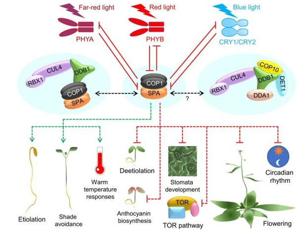 Fig. 1 COP1/SPA acts as a central regulator of plant growth and development.1
Key structural properties of COP1:
- Modular domain organization
- The WD40 repeat structure forms a β-propeller conformation
- Ring finger domain structure has the E3 ubiquitin ligase activity
- Curly spiral structure domain involved in protein polymerization and two positioning control in the cell
Functions of COP1
The main function of the COP1 gene is to serve as a core negative regulatory factor for phomorphogenesis and to exert E3 ubiquitin ligase activity in various biological processes. The specific functions are summarized as follows:
| Function | Description |
| Optical signal transduction | Under dark conditions, the COP1 protein enters the cell nucleus and mediates the ubiquitination and degradation of photomorphogenesis promoters (such as HY5), thereby inhibiting the expression of photoresponsive genes. |
| skotomorphogenesis | By continuously degrading transcription factors that promote photomorphogenesis, it dominates the growth and development patterns of plants in dark environments, such as promoting hypocotyl elongation. |
| Environmental adaptation regulation | By integrating multiple environmental signals such as light, temperature and biological clocks, and targeting different substrates, it regulates the timing and morphology of plant growth and development. |
| Animal cell function | In animals, COP1 homologous proteins (such as RFWD2) are involved in regulating processes like the cell cycle, DNA damage repair, and tumorigenesis. |
| Substrate specific recognition | The WD40 repeat domain at its C-terminal can specifically recognize and bind to target proteins containing specific degradation signals, such as VP or Degron motifs. |
Its mechanism of action is different from the typical signal cascade amplification. Instead, it directly determines the direction of the developmental pathway by precisely controlling the stability of key regulatory proteins in a "switch-like" manner.
Applications of COP1 and COP1 Antibody in Literature
1. Luo, Dakui, et al. "CUL4B-DDB1-COP1-mediated UTX downregulation promotes colorectal cancer progression." Experimental Hematology & Oncology 12.1 (2023): 77. https://doi.org/10.1186/s40164-023-00440-z
This study found that the COP1-CUL4B-DDB1 complex can ubiquitinate and degrade histone demethylase UTX, thereby promoting the progression of colorectal cancer (CRC). UTX has anti-cancer function, and its deficiency can be partially reversed by EZH2 inhibitors.
2. Ponnu, Jathish, and Ute Hoecker. "Illuminating the COP1/SPA ubiquitin ligase: fresh insights into its structure and functions during plant photomorphogenesis." Frontiers in Plant Science 12 (2021): 662793. https://doi.org/10.3389/fpls.2021.662793
COP1 is a Ring-type E3 ubiquitin ligase that is widely present in plants and animals. In plants, the COP1/SPA complex negatively regulates photomorphogenesis by degrading substrates. In humans, COP1 is involved in the regulation of tumorigenesis.
3. Xu, Dongqing, Danmeng Zhu, and Xing Wang Deng. "The role of COP1 in repression of photoperiodic flowering." F1000Research 5 (2016): F1000-Faculty. https://doi.org/10.12688/f1000research.7346.1
The article indicates that COP1 is an E3 ubiquitin ligase. It collaborates with SPA proteins to degrade core regulatory factors such as CO in photoperiodic flowering, thereby integrating light signals with the biological clock and negatively regulating the photoperiodic flowering time of plants.
4. Choi, Hyun Ho, and Mong-Hong Lee. "CSN6-COP1 axis in cancer." Aging (Albany NY) 7.7 (2015): 461. https://doi.org/10.18632/aging.100778
The article indicates that CSN6 enhances stability by interacting with COP1 and inhibiting its autoubiquitination. The two work together as E3 ligases to degrade the tumor suppressor protein 14-3-3σ and activate the Akt pathway, thereby promoting the survival of cancer cells and tumorigenesis.
5. Senapati, Dhirodatta, et al. "COP 1 regulates the stability of CAM 7 to promote photomorphogenic growth." Plant Direct 3.6 (2019): e00144. https://doi.org/10.1002/pld3.144
The article indicates that under low light intensity, calmodulin CAM7 interacts with ubiquitin ligase COP1 in the cell nucleus, synergistically promoting HY5 expression and photomorphogenesis. The function of COP1 is crucial for maintaining the protein level of CAM7.
Creative Biolabs: COP1 Antibodies for Research
Creative Biolabs specializes in the production of high-quality COP1 antibodies for research and industrial applications. Our portfolio includes monoclonal antibodies tailored for ELISA, Flow Cytometry, Western blot, immunohistochemistry, and other diagnostic methodologies.
- Custom COP1 Antibody Development: Tailor-made solutions to meet specific research requirements.
- Bulk Production: Large-scale antibody manufacturing for industry partners.
- Technical Support: Expert consultation for protocol optimization and troubleshooting.
- Aliquoting Services: Conveniently sized aliquots for long-term storage and consistent experimental outcomes.
For more details on our COP1 antibodies, custom preparations, or technical support, contact us at email.
Reference
- Ponnu, Jathish, and Ute Hoecker. "Illuminating the COP1/SPA ubiquitin ligase: fresh insights into its structure and functions during plant photomorphogenesis." Frontiers in Plant Science 12 (2021): 662793. https://doi.org/10.3389/fpls.2021.662793
Anti-COP1 antibodies
 Loading...


Hot products 
-
Mouse Anti-DMPK Recombinant Antibody (CBYCD-324) (CBMAB-D1200-YC)

-
Mouse Anti-8-oxoguanine Recombinant Antibody (V2-7697) (CBMAB-1869CQ)

-
Mouse Anti-ADGRE2 Recombinant Antibody (V2-261270) (CBMAB-C0813-LY)

-
Rat Anti-EMCN Recombinant Antibody (28) (CBMAB-E0280-FY)

-
Mouse Anti-APCS Recombinant Antibody (CBYC-A663) (CBMAB-A3054-YC)

-
Mouse Anti-AGO2 Recombinant Antibody (V2-634169) (CBMAB-AP203LY)

-
Mouse Anti-ELAVL4 Recombinant Antibody (6B9) (CBMAB-1132-YC)

-
Mouse Anti-CD24 Recombinant Antibody (HIS50) (CBMAB-C10123-LY)

-
Mouse Anti-AFM Recombinant Antibody (V2-634159) (CBMAB-AP185LY)

-
Mouse Anti-APC Recombinant Antibody (CBYC-A661) (CBMAB-A3036-YC)

-
Mouse Anti-Acetyl SMC3 (K105/K106) Recombinant Antibody (V2-634053) (CBMAB-AP052LY)

-
Mouse Anti-C5AR1 Recombinant Antibody (R63) (CBMAB-C9553-LY)

-
Mouse Anti-GFAP Recombinant Antibody (24) (CBMAB-G2927-LY)

-
Mouse Anti-G6PD Recombinant Antibody (13B331) (CBMAB-G1553-LY)

-
Mouse Anti-AKT1/AKT2/AKT3 (Phosphorylated T308, T309, T305) Recombinant Antibody (V2-443454) (PTM-CBMAB-0030YC)

-
Mouse Anti-BMI1 Recombinant Antibody (CBYC-P026) (CBMAB-P0108-YC)

-
Mouse Anti-APOH Recombinant Antibody (4D9A4) (CBMAB-A3249-YC)

-
Mouse Anti-ALX1 Recombinant Antibody (96k) (CBMAB-C0616-FY)

-
Rat Anti-C5AR1 Recombinant Antibody (8D6) (CBMAB-C9139-LY)

-
Mouse Anti-CCN2 Recombinant Antibody (CBFYC-2383) (CBMAB-C2456-FY)

- AActivation
- AGAgonist
- APApoptosis
- BBlocking
- BABioassay
- BIBioimaging
- CImmunohistochemistry-Frozen Sections
- CIChromatin Immunoprecipitation
- CTCytotoxicity
- CSCostimulation
- DDepletion
- DBDot Blot
- EELISA
- ECELISA(Cap)
- EDELISA(Det)
- ESELISpot
- EMElectron Microscopy
- FFlow Cytometry
- FNFunction Assay
- GSGel Supershift
- IInhibition
- IAEnzyme Immunoassay
- ICImmunocytochemistry
- IDImmunodiffusion
- IEImmunoelectrophoresis
- IFImmunofluorescence
- IGImmunochromatography
- IHImmunohistochemistry
- IMImmunomicroscopy
- IOImmunoassay
- IPImmunoprecipitation
- ISIntracellular Staining for Flow Cytometry
- LALuminex Assay
- LFLateral Flow Immunoassay
- MMicroarray
- MCMass Cytometry/CyTOF
- MDMeDIP
- MSElectrophoretic Mobility Shift Assay
- NNeutralization
- PImmunohistologyp-Paraffin Sections
- PAPeptide Array
- PEPeptide ELISA
- PLProximity Ligation Assay
- RRadioimmunoassay
- SStimulation
- SESandwich ELISA
- SHIn situ hybridization
- TCTissue Culture
- WBWestern Blot
