DTNB Antibodies
Background
The DTNB gene encodes an enzyme protein named "Disulfide Bond Reductase B", which is mainly located in the cytoplasm and is highly expressed in tissues such as the heart, skeletal muscle, and liver. This protein regulates the oxidation-reduction state of sulfhydryl-disulfide bonds, participating in maintaining the intracellular redox balance and signal transduction processes. It plays a crucial role in protecting cells from oxidative stress and maintaining normal metabolic functions. Abnormalities in its function are associated with various diseases, including cardiomyopathy, neurodegenerative diseases, and metabolic syndrome. Since its discovery, the DTNB gene and its encoded protein have become important research objects in the field of redox biology, providing key clues for understanding the cellular antioxidant mechanism, protein modification, and related pathological and physiological processes.
Structure of DTNB
The protein encoded by the DTNB gene has a molecular weight of approximately 27.8 kDa. This molecular weight remains relatively stable across different species, mainly due to the conservation of its core functional domain.
| Species | Human | Mouse | Rat | Bovine |
| Molecular Weight (kDa) | 27.8 | ~27.5 | ~27.6 | ~27.9 |
| Primary Structural Differences | Regulate glutathione metabolism | Highly similar to humans | Often used in oxidative stress models | Highly expressed in the myocardium |
This protein is composed of 247 amino acids. Its core is a conserved thioredoxin domain, which is the basis for its redox enzyme function. This domain contains a typical CXXC active site motif, in which the two cysteine residues catalyze the exchange reaction of the sulfhydryl groups and disulfide bonds of the substrate protein through reversible formation or reduction of disulfide bonds. This structure enables it to effectively participate in the intracellular antioxidant defense system and maintain redox homeostasis.
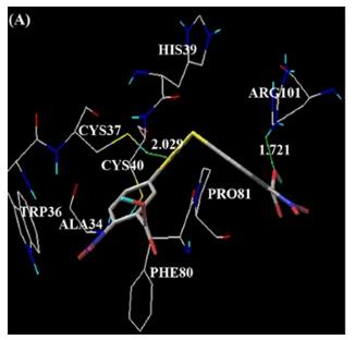 Fig. 1 DTNB.1
Key structural properties of DTNB:
- Core of Thioredoxin (Thioredoxin) folding structure domain
- Active center contains characteristic motif CXXC amino acid sequence
- Electron transfer occurs through the thiol group of the active site cysteine
Functions of DTNB
The protein encoded by the DTNB gene mainly functions to maintain redox homeostasis within cells and participates in various cellular signal transduction and metabolic processes.
| Function | Description |
| Sulfenic-Dithiol Exchange | Through its CXXC motif in the active center, it catalyzes the formation and reduction of disulfide bonds between proteins or between proteins and glutathione, regulating protein functions. |
| Redox Signal Transduction | As a crucial redox sensor, it participates in regulating various signaling pathways such as cell proliferation, differentiation, and apoptosis. |
| Antioxidant Defense | By reducing the oxidized protein sulfhydryl groups, it protects cells from oxidative damage caused by reactive oxygen species (ROS). |
| Maintaining Glutathione Balance | Works in synergy with the glutathione system to maintain an appropriate ratio of reduced glutathione (GSH) to oxidized glutathione (GSSG) within the cells. |
| Metabolic Regulation | By influencing the active state of key metabolic enzymes (such as phosphoglycerate dehydrogenase), it participates in the regulation of processes such as sugar metabolism. |
The active mechanism of this protein makes its action curve more similar to that of an "efficient molecular 'switch'". By rapidly and reversibly altering the redox state of the target protein, it precisely regulates downstream physiological processes. This is fundamentally different from the action mode of proteins with storage or transport functions (such as myoglobin).
Applications of DTNB and DTNB Antibody in Literature
1. Neumann, Alexander, et al. "Rare variants in IFFO1, DTNB, NLRC3 and SLC22A10 associate with Alzheimer's disease CSF profile of neuronal injury and inflammation." Molecular psychiatry 27.4 (2022): 1990-1999. https://doi.org/10.1038/s41380-022-01437-6
Research has found that rare variations in genes such as IFFO1 and DTNB are associated with inflammatory indicators of nerve damage and increase the risk of dementia through this mechanism. The GABBR2 and CASZ1 genes are related to synaptic function, but do not affect the development of Alzheimer's disease.
2. Connellan, JOHN M., and J. E. Folk. "Mechanism of the inactivation of guinea pig liver transglutaminase by 5, 5′-dithiobis-(2-nitrobenzoic acid)." Journal of Biological Chemistry 244.12 (1969): 3173-3181. https://doi.org/10.1016/s0021-9258(18)93110-8
Research has found that in the absence of calcium ions, DTNB reacts with transglutaminase, causing it to lose its transfer and hydrolysis activities. This is attributed to the formation of disulfide bonds within the enzyme, which leads to the loss of substrate binding ability, while esterase activity remains unaffected.
3. Lara, Humberto H., et al. "Antiviral propierties of 5, 5'-dithiobis-2-nitrobenzoic acid and bacitracin against T-tropic human immunodeficiency virus type 1." Virology journal 8.1 (2011): 137. https://doi.org/10.1186/1743-422X-8-137
Studies have found that bacitracin and DTNB can inhibit the entry of HIV-1, especially effective against T-addicted strains. DTNB can act on both the cell membrane and the viral envelope simultaneously and exert a sustained antiviral effect in the late stage of the viral cycle, demonstrating the potential of a local microbicide.
4. Westwood, J. H., and P. Thomas. "Studies on the structure and immunological activity of carcinoembryonic antigen–the role of disulphide bonds." British Journal of Cancer 32.6 (1975): 708-719. https://doi.org/10.1038/bjc.1975.282
Research has found that carcinoembryonic antigen CEA contains six disulfide bonds and has no free thiol groups. After reduction, DTNB can react with it, but the antigenic activity is partially lost after treatment. Complete oxidation would destroy half of the antigenic activity, indicating that the disulfide bond structure is crucial for maintaining its antigenicity.
5. Gostimskaya, Irina S., Gary Cecchini, and Andrei D. Vinogradov. "Topography and chemical reactivity of the active–inactive transition-sensitive SH-group in the mitochondrial NADH: ubiquinone oxidoreductase (complex I)." Biochimica et Biophysica Acta (BBA)-Bioenergetics 1757.9-10 (2006): 1155-1161. https://doi.org/10.1016/j.bbabio.2006.04.016
Research has found that the activity of mitochondrial complex I depends on the thiol group located at the endometrium-stromal interface. In the inactivated state, it can be rapidly inhibited by NEM and DTNB. In intact mitochondria, DTNB can only inhibit the enzyme when the membrane is damaged.
Creative Biolabs: DTNB Antibodies for Research
Creative Biolabs specializes in the production of high-quality DTNB antibodies for research and industrial applications. Our portfolio includes monoclonal antibodies tailored for ELISA, Flow Cytometry, Western blot, immunohistochemistry, and other diagnostic methodologies.
- Custom DTNB Antibody Development: Tailor-made solutions to meet specific research requirements.
- Bulk Production: Large-scale antibody manufacturing for industry partners.
- Technical Support: Expert consultation for protocol optimization and troubleshooting.
- Aliquoting Services: Conveniently sized aliquots for long-term storage and consistent experimental outcomes.
For more details on our DTNB antibodies, custom preparations, or technical support, contact us at email.
Reference
- Gowthaman, Uthaman, Mannu Jayakanthan, and Durai Sundar. "Molecular docking studies of dithionitrobenzoic acid and its related compounds to protein disulfide isomerase: computational screening of inhibitors to HIV-1 entry." BMC bioinformatics 9.Suppl 12 (2008): S14. https://doi.org/10.1186/1471-2105-9-S12-S14
Anti-DTNB antibodies
 Loading...


Hot products 
-
Mouse Anti-APP Recombinant Antibody (5C2A1) (CBMAB-A3314-YC)

-
Mouse Anti-AK4 Recombinant Antibody (V2-180419) (CBMAB-A1891-YC)

-
Mouse Anti-ACO2 Recombinant Antibody (V2-179329) (CBMAB-A0627-YC)

-
Mouse Anti-ALOX5 Recombinant Antibody (33) (CBMAB-1890CQ)

-
Mouse Anti-DLG1 Monolconal Antibody (4F3) (CBMAB-0225-CN)

-
Mouse Anti-2C TCR Recombinant Antibody (V2-1556) (CBMAB-0951-LY)

-
Mouse Anti-CAPZB Recombinant Antibody (CBYY-C0944) (CBMAB-C2381-YY)

-
Mouse Anti-AAV9 Recombinant Antibody (V2-634029) (CBMAB-AP023LY)

-
Mouse Anti-APOA1 Monoclonal Antibody (CBFYR0637) (CBMAB-R0637-FY)

-
Mouse Anti-FTH1 Recombinant Antibody (CBXF-1896) (CBMAB-F3426-CQ)

-
Mouse Anti-CCDC25 Recombinant Antibody (CBLC132-LY) (CBMAB-C9786-LY)

-
Mouse Anti-ENPP1 Recombinant Antibody (CBFYE-0159) (CBMAB-E0375-FY)

-
Armenian hamster Anti-CD40 Recombinant Antibody (HM40-3) (CBMAB-C10365-LY)

-
Mouse Anti-BIRC3 Recombinant Antibody (315304) (CBMAB-1214-CN)

-
Mouse Anti-CFL1 (Phospho-Ser3) Recombinant Antibody (CBFYC-1770) (CBMAB-C1832-FY)

-
Mouse Anti-ARIH1 Recombinant Antibody (C-7) (CBMAB-A3563-YC)

-
Rabbit Anti-CAMK2A Recombinant Antibody (BA0032) (CBMAB-0137CQ)

-
Mouse Anti-CASP7 Recombinant Antibody (10-01-62) (CBMAB-C2005-LY)

-
Mouse Anti-ACE2 Recombinant Antibody (V2-179293) (CBMAB-A0566-YC)

-
Mouse Anti-FLI1 Recombinant Antibody (CBXF-0733) (CBMAB-F0435-CQ)

- AActivation
- AGAgonist
- APApoptosis
- BBlocking
- BABioassay
- BIBioimaging
- CImmunohistochemistry-Frozen Sections
- CIChromatin Immunoprecipitation
- CTCytotoxicity
- CSCostimulation
- DDepletion
- DBDot Blot
- EELISA
- ECELISA(Cap)
- EDELISA(Det)
- ESELISpot
- EMElectron Microscopy
- FFlow Cytometry
- FNFunction Assay
- GSGel Supershift
- IInhibition
- IAEnzyme Immunoassay
- ICImmunocytochemistry
- IDImmunodiffusion
- IEImmunoelectrophoresis
- IFImmunofluorescence
- IGImmunochromatography
- IHImmunohistochemistry
- IMImmunomicroscopy
- IOImmunoassay
- IPImmunoprecipitation
- ISIntracellular Staining for Flow Cytometry
- LALuminex Assay
- LFLateral Flow Immunoassay
- MMicroarray
- MCMass Cytometry/CyTOF
- MDMeDIP
- MSElectrophoretic Mobility Shift Assay
- NNeutralization
- PImmunohistologyp-Paraffin Sections
- PAPeptide Array
- PEPeptide ELISA
- PLProximity Ligation Assay
- RRadioimmunoassay
- SStimulation
- SESandwich ELISA
- SHIn situ hybridization
- TCTissue Culture
- WBWestern Blot
