FBN1 Antibodies
Background
The FBN1 gene encodes a large glycoprotein called fibrin-1, which is the main structural component of microfibers in the extracellular matrix. It provides structural support and elastic function for connective tissue by participating in the assembly of microfibers, and regulates the activity of signaling molecules such as transforming growth factor β (TGF-β). Mutations in this gene can lead to connective tissue diseases such as Marfan syndrome, affecting the normal development and function of the cardiovascular, ocular and skeletal systems. The FBN1 gene was first identified in the late 1980s. The clarification of its function not only revealed the basic mechanism of microfiber assembly but also greatly promoted our understanding of the role of extracellular matrix in tissue homeostasis and genetic diseases, becoming an important basis for related pathological research and clinical diagnosis.
Structure of FBN1
Fibrin-1 encoded by the FBN1 gene is a large glycoprotein with a molecular weight of approximately 350 kDa. This protein is mainly composed of epidermal growth factor-like domains and transforming growth factor - β -binding protein-like domains. The difference in the number of calcium-binding epidermal growth factor-like domains is the main factor leading to the subtle changes in molecular weight among different species.
| Species | Human | Mouse | Bovine |
| Molecular Weight (kDa) | 350 | 347 | 352 |
| Primary Structural Differences | 47 EGF-like domains and 7 TGF-β -binding protein-like domains | 46 EGF-like domains, and the domain composition is highly conserved from human | Domain number consistent with basic human structure, sequence homology of more than 95% |
Fibrin-1 assembles into an extracellular microfiber network through its unique linearly arranged domain. Each monomer contains multiple calcium-binding epidermal growth factor-like domains, which are stably folded through disulfide bonds, providing rigidity for the protein. Transforming growth factor - β -binding protein-like domains mediate protein-protein interactions, and the C-terminal region is crucial for the correct assembly of fibrils. This multi-domain organizational form enables fibrin-1 to simultaneously undertake the dual functions of structural support and signal regulation.
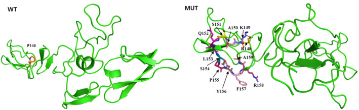 Fig. 1 3D structure prediction of FBN1.1
Key structural properties of FBN1:
- Complex linear arrangement of multiple domains
- Calcium-binding EGF-like domains rich in cysteine
- Transforming growth factor β -binding protein-like domains mediate protein interactions
Functions of FBN1
The main function of the protein encoded by the FBN1 gene is to form the microfiber scaffold of the extracellular matrix and regulate the activity of growth factors. Its specific functions include:
| Function | Description |
| Structural support | As the main component of microfibers, it provides mechanical support and elasticity for elastic tissues and connective tissues. |
| TGF-β regulation | By binding and sustainably releasing TGF-β family cytokines, it regulates tissue development and homeostasis maintenance. |
| Cell signal coordination | Integrate extracellular matrix and intercellular signal transduction, influencing cell adhesion, migration and differentiation processes. |
| Histomorphogenesis | Guide the correct formation of organs such as heart valves, lenses and large blood vessels during embryonic development. |
| Matrix stability | Maintain the integrity of the organizational structure through cross-linked networks to counteract repeated mechanical stresses. |
This protein achieves conformational rigidity through its multiple calcium-binding EGF-like domains. This structural characteristic enables it to not only withstand physical tension but also precisely participate in signal regulation, demonstrating the dual characteristics of structural proteins and regulatory proteins.
Applications of FBN1 and FBN1 Antibody in Literature
1. Sun, Yonghong, et al. "MFAP2 promotes HSCs activation through FBN1/TGF‐β/Smad3 pathway." Journal of Cellular and Molecular Medicine 27.21 (2023): 3235-3246. https://doi.org/10.1111/jcmm.17884
The article indicates that in liver fibrosis, the expression of MFAP2 is upregulated and promotes the activation of hepatic stellate cells and collagen deposition by regulating FBN1. Inhibiting MFAP2 can slow down cell proliferation and induce apoptosis of activated stellate cells, thereby alleviating fibrosis, indicating its clinical therapeutic potential.
2. Åström Malm, Ida, Rachel De Basso, and Peter Blomstrand. "No differences in FBN1 genotype between men with and without abdominal aortic aneurysm." BMC Cardiovascular Disorders 23.1 (2023): 36. https://doi.org/10.1186/s12872-023-03068-3
This study explores the relationship between the FBN1 genotype and abdominal aortic aneurysm (AAA). The results showed that there was no significant difference in the distribution of FBN1 genotypes between the AAA group and the control group. However, among men carrying the FBN1-2/2 genotype, the central artery hardness of AAA patients was significantly higher than that of non-AAA patients.
3. Shimizu, Norihiro, et al. "Novel FBN1 intron variant causes isolated ectopia lentis via in-frame exon skipping." Journal of Human Genetics 70.4 (2025): 199-205. https://doi.org/10.1038/s10038-025-01318-0
The article indicates that in a Japanese IEL family, researchers discovered a novel intron mutation of the FBN1 gene (c.1327+3A>C). This mutation causes exon 11 of FBN1 to be skipped during transcription, resulting in the absence of its protein "hinge region" and ultimately triggering IEL, which provides new insights into the mechanism of FBN1-related diseases.
4. Dougarem, Djouhayna, et al. "A novel heterozygous intronic FBN1 variant contributes to aberrant RNA splicing in marfan syndrome." Molecular Genetics & Genomic Medicine 12.9 (2024): e70004. https://doi.org/10.1002/mgg3.70004
In this study, a novel non-classical splicing site variation c.443-3C>G of the FBN1 gene was identified in a Marfan syndrome family. This variation leads to two abnormal mRNA transcripts, triggering frameshift and in-frame insertions. Experiments have confirmed its pathogenicity, expanding the pathogenic variation spectrum of FBN1.
5. Wang, Tao, et al. "Acromicric dysplasia with stiff skin syndrome‐like severe cutaneous presentation in an 8‐year‐old boy with a missense FBN1 mutation: Case report and literature review." Molecular Genetics & Genomic Medicine 8.7 (2020): e1282. https://doi.org/10.1002/mgg3.1282
In this study, acrofacial dysplasia caused by A heterozygous missense mutation of c.5243G>A in the FBN1 gene was diagnosed in a Chinese child patient, accompanied by severe skin stiffness similar to scleroderma. This case reveals the pleiotropy of the FBN1 gene, where a single hotspot mutation can lead to complex clinical symptoms.
Creative Biolabs: FBN1 Antibodies for Research
Creative Biolabs specializes in the production of high-quality FBN1 antibodies for research and industrial applications. Our portfolio includes monoclonal antibodies tailored for ELISA, Flow Cytometry, Western blot, immunohistochemistry, and other diagnostic methodologies.
- Custom FBN1 Antibody Development: Tailor-made solutions to meet specific research requirements.
- Bulk Production: Large-scale antibody manufacturing for industry partners.
- Technical Support: Expert consultation for protocol optimization and troubleshooting.
- Aliquoting Services: Conveniently sized aliquots for long-term storage and consistent experimental outcomes.
For more details on our FBN1 antibodies, custom preparations, or technical support, contact us at email.
Reference
- Dougarem, Djouhayna, et al. "A novel heterozygous intronic FBN1 variant contributes to aberrant RNA splicing in marfan syndrome." Molecular Genetics & Genomic Medicine 12.9 (2024): e70004. https://doi.org/10.1002/mgg3.70004
Anti-FBN1 antibodies
 Loading...


Hot products 
-
Rat Anti-ABCC11 Recombinant Antibody (V2-179001) (CBMAB-A0236-YC)

-
Mouse Anti-C4B Recombinant Antibody (CBYY-C2996) (CBMAB-C4439-YY)

-
Mouse Anti-CCDC25 Recombinant Antibody (CBLC132-LY) (CBMAB-C9786-LY)

-
Mouse Anti-CTNND1 Recombinant Antibody (CBFYC-2414) (CBMAB-C2487-FY)

-
Mouse Anti-CD8 Recombinant Antibody (C1083) (CBMAB-C1083-LY)

-
Mouse Anti-CFL1 Recombinant Antibody (CBFYC-1771) (CBMAB-C1833-FY)

-
Mouse Anti-AKT1 Recombinant Antibody (V2-180546) (CBMAB-A2070-YC)

-
Mouse Anti-AK4 Recombinant Antibody (V2-180419) (CBMAB-A1891-YC)

-
Mouse Anti-BRCA2 Recombinant Antibody (CBYY-0790) (CBMAB-0793-YY)

-
Rat Anti-ADAM10 Recombinant Antibody (V2-179741) (CBMAB-A1103-YC)

-
Mouse Anti-ARSA Recombinant Antibody (CBYC-A799) (CBMAB-A3679-YC)

-
Rabbit Anti-CCL5 Recombinant Antibody (R0437) (CBMAB-R0437-CN)

-
Mouse Anti-CHRNA9 Recombinant Antibody (8E4) (CBMAB-C9161-LY)

-
Mouse Anti-AAV-5 Recombinant Antibody (V2-503417) (CBMAB-V208-1369-FY)

-
Mouse Anti-CDKL5 Recombinant Antibody (CBFYC-1629) (CBMAB-C1689-FY)

-
Rabbit Anti-ABL1 (Phosphorylated Y185) Recombinant Antibody (V2-443434) (PTM-CBMAB-0001YC)

-
Mouse Anti-CASQ1 Recombinant Antibody (CBFYC-0863) (CBMAB-C0918-FY)

-
Mouse Anti-ARIH1 Recombinant Antibody (C-7) (CBMAB-A3563-YC)

-
Mouse Anti-ACLY Recombinant Antibody (V2-179314) (CBMAB-A0610-YC)

-
Mouse Anti-C5AR1 Recombinant Antibody (R63) (CBMAB-C9553-LY)

- AActivation
- AGAgonist
- APApoptosis
- BBlocking
- BABioassay
- BIBioimaging
- CImmunohistochemistry-Frozen Sections
- CIChromatin Immunoprecipitation
- CTCytotoxicity
- CSCostimulation
- DDepletion
- DBDot Blot
- EELISA
- ECELISA(Cap)
- EDELISA(Det)
- ESELISpot
- EMElectron Microscopy
- FFlow Cytometry
- FNFunction Assay
- GSGel Supershift
- IInhibition
- IAEnzyme Immunoassay
- ICImmunocytochemistry
- IDImmunodiffusion
- IEImmunoelectrophoresis
- IFImmunofluorescence
- IGImmunochromatography
- IHImmunohistochemistry
- IMImmunomicroscopy
- IOImmunoassay
- IPImmunoprecipitation
- ISIntracellular Staining for Flow Cytometry
- LALuminex Assay
- LFLateral Flow Immunoassay
- MMicroarray
- MCMass Cytometry/CyTOF
- MDMeDIP
- MSElectrophoretic Mobility Shift Assay
- NNeutralization
- PImmunohistologyp-Paraffin Sections
- PAPeptide Array
- PEPeptide ELISA
- PLProximity Ligation Assay
- RRadioimmunoassay
- SStimulation
- SESandwich ELISA
- SHIn situ hybridization
- TCTissue Culture
- WBWestern Blot
