GAB3 Antibodies
Background
The GAB3 gene encodes a scaffold protein called GRB2-associated binding protein 3, which is mainly expressed in immune cells such as lymphocytes and myeloid cells. This protein participates in regulating the activation, proliferation and differentiation processes of immune cells by mediating signal transduction downstream of cytokine and growth factor receptors. Research has found that GAB3 plays a key role in innate immune responses and inflammatory pathways, and its abnormal expression is related to the pathogenesis of autoimmune diseases and primary immunodeficiency diseases. This gene was first identified in 2001. The study of its function has provided an important molecular basis for understanding the fine regulation of the immune signal network and has also opened up a new exploration direction for targeted therapy of related immune diseases.
Structure of GAB3
GAB3 is a scaffold protein with a molecular weight of approximately 55-65 kDa, and its specific molecular weight varies among different splicing variants. This protein is composed of multiple domains, including the PH domain and the proline-rich region. Subtle differences in these domains can affect its interaction with signaling partners.
| Species | Human | Mouse | Rat |
| Molecular Weight (kDa) | 58 | 55 | 57 |
| Primary Structural Differences | Conserved sequence, highly similar to other mammals | Minor amino acid variations | Slightly different oxygen affinity |
The GAB3 protein is composed of approximately 540 amino acids, and its primary structure exhibits typical scaffold protein characteristics, forming functional conformations through multiple domains. This protein contains an N-terminal PH domain and multiple protein interaction modules at the C-terminal, which together form a binding platform for signal transduction. Its secondary structure is mainly composed of alternating α -helical and β -folded structures. The PH domain forms a conserved three-dimensional conformation for membrane localization, while the proline-rich region at the C-terminal provides a specific binding interface for the SH3 domain through the helical-rotation-helical motif. Key tyrosine residues undergo phosphorylation during signal transduction, directly regulating the activity of downstream pathways. Meanwhile, multiple protein binding sites maintain the dynamic assembly and dissociation of the signal complex through allosteric effects.
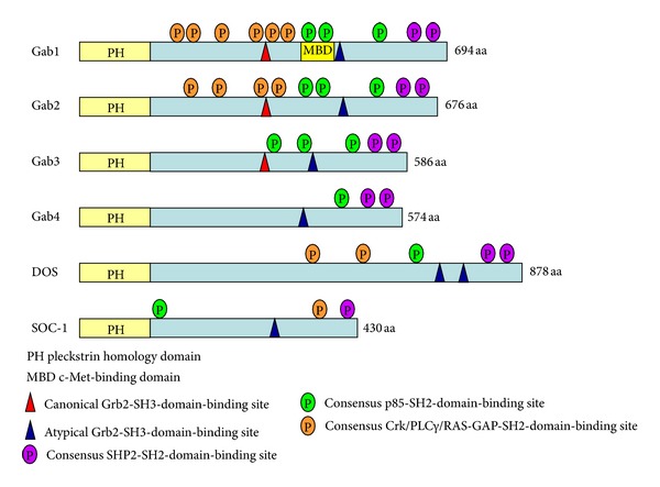 Fig. 1 Schematic structures of Gab family docking proteins.1
Key structural properties of GAB3:
- Multi-domain scaffold protein configuration
- The PH domain mediates cell membrane localization
- Multiple SH3 binding motifs are involved in the assembly of signal complexes
- Tyrosine phosphorylation sites regulate downstream signal transduction
Functions of GAB3
As a scaffold protein, GAB3's main function is to participate in signal transduction and regulation within immune cells. In addition, this protein also plays a role in various immune-related physiological and pathological processes.
| Function | Description |
| Signal transduction | By recruiting downstream signaling molecules, it mediates intracellular signaling triggered by cytokines and growth factors. |
| Activation of immune cells | Regulating the intensity and duration of activation signals in lymphocytes and myeloid cells affects the efficiency of immune responses. |
| Inflammatory regulation | It participates in the fine regulation of inflammatory signaling pathways, and its dysfunction is related to excessive inflammatory responses or autoimmune phenomena. |
| Cell proliferation and survival | By transmitting proliferation-promoting and anti-apoptotic signals, it affects the clonal expansion and survival of immune cells. |
| Maintenance of immune tolerance | Participate in establishing an appropriate signal balance to maintain immune homeostasis and prevent the occurrence of autoimmunity. |
The GAB3-mediated signal response curve exhibits typical saturation kinetics characteristics, in contrast to certain scaffold proteins with synergistic effects, which reflects its functional properties of precisely regulating signal intensity within the physiological range.
Applications of GAB3 and GAB3 Antibody in Literature
1. Jia, Pifeng, et al. "Gab3 overexpression in human glioma mediates Akt activation and tumor cell proliferation." PLoS One 12.3 (2017): e0173473. https://doi.org/10.1371/journal.pone.0173473
This study confirmed that the expression of Gab3 was significantly upregulated in glioma. Functionally, Gab3 promotes the proliferation of glioma cells by activating the Akt signaling pathway, while its knockout inhibits tumor growth. The results show that Gab3 is a key promoting factor for the development of glioma.
2. Wang, Zhengqi, et al. "Gab2 and Gab3 redundantly suppress colitis by modulating macrophage and CD8+ T-cell activation." Frontiers in immunology 10 (2019): 486. https://doi.org/10.3389/fimmu.2019.00486
Research has found that Gab2 and Gab3 are complementary in function. Knockout of the Gab2/3 double gene can induce spontaneous colitis in mice. The mechanism is related to the weakened PI3K-Akt signaling in hematopoietic cells, which leads to excessive activation of macrophages and CD8+ T cells and drives intestinal inflammation.
3. Tao, Meini, et al. "Association analysis of polymorphisms in SLK, ARHGEF9, WWC2, GAB3, and FSHR genes with reproductive traits in different sheep breeds." Frontiers in Genetics 15 (2024): 1371872. https://doi.org/10.3389/fgene.2024.1371872
In this study, genotyping was conducted on breeds such as Dolang sheep using KASP technology. It was found that four loci of the WWC2, SLK, ARHGEF9 and FSHR genes were significantly correlated with the number of lambs born. Although the GAB3 gene locus (G.86134602 G>A) was successfully typed, its association with reproductive traits still needs further verification. Tissue expression profiles show that GAB3 is highly expressed in both reproductive organs and internal organs.
4. Nakaoka, Yoshikazu, and Issei Komuro. "Gab docking proteins in cardiovascular disease, cancer, and inflammation." International journal of inflammation 2013.1 (2013): 141068. https://doi.org/10.1155/2013/141068
The article indicates that Gab3 is an important member of the Gab adaptor protein family. It has no enzymatic activity itself but serves as a signaling hub. When its tyrosine is phosphorylated, it can recruit SH2-domain-containing proteins such as SHP2 and p85, thereby integrating signals from growth factors, cytokines, etc., and regulating physiological processes such as cell proliferation and differentiation. Abnormal signals are closely related to cancer, cardiovascular diseases and inflammation.
5. Cherif, Myriam, et al. "Gab1 is modulated by chronic hypoxia in children with cyanotic congenital heart defect and its overexpression reduces apoptosis in rat neonatal cardiomyocytes." BioMed Research International 2015.1 (2015): 718492. https://doi.org/10.1155/2015/718492
This study focuses on the role of Gab1 under hypoxic conditions. It was found that the expression of Gab1 protein in the myocardium of children with cyanotic congenital heart disease was upregulated. In vitro experiments confirmed that its overexpression could reduce the apoptosis of hypoxic cardiomyocytes. This indicates that Gab1 is a key survival factor for cardiomyocytes in response to hypoxic stress, providing a basis for understanding the role of the Gab family (containing Gab3) in cardiac protection.
Creative Biolabs: GAB3 Antibodies for Research
Creative Biolabs specializes in the production of high-quality GAB3 antibodies for research and industrial applications. Our portfolio includes monoclonal antibodies tailored for ELISA, Flow Cytometry, Western blot, immunohistochemistry, and other diagnostic methodologies.
- Custom GAB3 Antibody Development: Tailor-made solutions to meet specific research requirements.
- Bulk Production: Large-scale antibody manufacturing for industry partners.
- Technical Support: Expert consultation for protocol optimization and troubleshooting.
- Aliquoting Services: Conveniently sized aliquots for long-term storage and consistent experimental outcomes.
For more details on our GAB3 antibodies, custom preparations, or technical support, contact us at email.
Reference
- Nakaoka, Yoshikazu, and Issei Komuro. "Gab docking proteins in cardiovascular disease, cancer, and inflammation." International journal of inflammation 2013.1 (2013): 141068. https://doi.org/10.1155/2013/141068
Anti-GAB3 antibodies
 Loading...


Hot products 
-
Mouse Anti-ENO1 Recombinant Antibody (8G8) (CBMAB-E1329-FY)

-
Mouse Anti-BZLF1 Recombinant Antibody (BZ.1) (CBMAB-AP705LY)

-
Rabbit Anti-B2M Recombinant Antibody (CBYY-0059) (CBMAB-0059-YY)

-
Mouse Anti-CFL1 Recombinant Antibody (CBFYC-1771) (CBMAB-C1833-FY)

-
Mouse Anti-BAD (Phospho-Ser136) Recombinant Antibody (CBYY-0138) (CBMAB-0139-YY)

-
Mouse Anti-ACE2 Recombinant Antibody (V2-179293) (CBMAB-A0566-YC)

-
Mouse Anti-Acetyl-α-Tubulin (Lys40) Recombinant Antibody (V2-623485) (CBMAB-CP2897-LY)

-
Mouse Anti-C5B-9 Recombinant Antibody (CBFYA-0216) (CBMAB-X0304-FY)

-
Rabbit Anti-ALOX5AP Recombinant Antibody (CBXF-1219) (CBMAB-F0750-CQ)

-
Mouse Anti-ATP1B3 Recombinant Antibody (1E9) (CBMAB-A4021-YC)

-
Mouse Anti-FOSB Recombinant Antibody (CBXF-3593) (CBMAB-F2522-CQ)

-
Mouse Anti-ABIN2 Recombinant Antibody (V2-179106) (CBMAB-A0349-YC)

-
Mouse Anti-CD24 Recombinant Antibody (ALB9) (CBMAB-0176CQ)

-
Mouse Anti-FPR2 Recombinant Antibody (1D6) (CBMAB-F2628-CQ)

-
Mouse Anti-CASQ1 Recombinant Antibody (CBFYC-0863) (CBMAB-C0918-FY)

-
Rabbit Anti-CAMK2A Recombinant Antibody (BA0032) (CBMAB-0137CQ)

-
Mouse Anti-CD33 Recombinant Antibody (6C5/2) (CBMAB-C8126-LY)

-
Mouse Anti-BRCA2 Recombinant Antibody (CBYY-1728) (CBMAB-2077-YY)

-
Mouse Anti-ATP5F1A Recombinant Antibody (51) (CBMAB-A4043-YC)

-
Mouse Anti-B2M Recombinant Antibody (CBYY-0050) (CBMAB-0050-YY)

- AActivation
- AGAgonist
- APApoptosis
- BBlocking
- BABioassay
- BIBioimaging
- CImmunohistochemistry-Frozen Sections
- CIChromatin Immunoprecipitation
- CTCytotoxicity
- CSCostimulation
- DDepletion
- DBDot Blot
- EELISA
- ECELISA(Cap)
- EDELISA(Det)
- ESELISpot
- EMElectron Microscopy
- FFlow Cytometry
- FNFunction Assay
- GSGel Supershift
- IInhibition
- IAEnzyme Immunoassay
- ICImmunocytochemistry
- IDImmunodiffusion
- IEImmunoelectrophoresis
- IFImmunofluorescence
- IGImmunochromatography
- IHImmunohistochemistry
- IMImmunomicroscopy
- IOImmunoassay
- IPImmunoprecipitation
- ISIntracellular Staining for Flow Cytometry
- LALuminex Assay
- LFLateral Flow Immunoassay
- MMicroarray
- MCMass Cytometry/CyTOF
- MDMeDIP
- MSElectrophoretic Mobility Shift Assay
- NNeutralization
- PImmunohistologyp-Paraffin Sections
- PAPeptide Array
- PEPeptide ELISA
- PLProximity Ligation Assay
- RRadioimmunoassay
- SStimulation
- SESandwich ELISA
- SHIn situ hybridization
- TCTissue Culture
- WBWestern Blot
