HHATL Antibodies
Background
The HHATL gene (also known as Hhatl) encodes a highly conserved Huntington protein-related acyltransferase-like protein in vertebrates. This protein mainly participates in the development and functional regulation of the nervous system, especially playing a role in cell signal transduction and protein modification processes. Studies have shown that HHATL affects embryonic development and the maintenance of the homeostasis of adult tissues by regulating key pathways such as the Hedgehog signaling pathway. This gene was first identified in the early 21st century, and its structural and functional research has provided important clues for understanding the molecular mechanisms of neurodevelopmental disorders and related diseases. In recent years, in-depth studies on HHATL have not only revealed its regulatory role in cellular metabolism and signal networks, but also promoted the progress in understanding the pathological mechanisms of related genetic diseases.
Structure of HHATL
The protein encoded by the HHATL gene (Hedgehog acyltransferase-like protein) is a transmembrane protein with a molecular weight of approximately 50-55 kDa. Its exact molecular weight may vary slightly among different species and tissues. The core function of this protein is related to its acyltransferase activity, which is responsible for catalyzing the lipid modification (such as palmitoylation) of key ligands in the Hedgehog signaling pathway. This process is crucial for the maturation, secretion, and long-distance signal transmission of the ligands. Its structure contains multiple transmembrane regions and a conserved acyltransferase domain, which together form a hydrophobic catalytic pocket to recognize and bind to specific lipid donors and Hedgehog protein substrates. The active site of the protein usually contains key histidine or aspartic acid residues, which participate in the catalytic reaction and maintain the stability of the enzyme's function. Abnormal regulation of the HHATL protein is closely related to embryonic development defects and the occurrence of certain tumors.
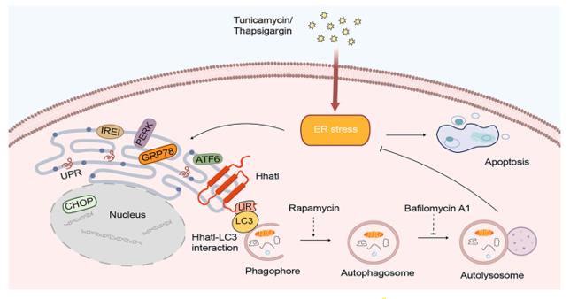 Fig. 1 Model of the role of Hhatl in ER stress.1
Key structural properties of HHATL:
- Conservative transmembrane helical bundle structure
- Catalytic domains with hydrophobic cavities
- Characteristic histidine or aspartic acid residues located in the active center
Functions of HHATL
The main function of the protein encoded by the HHATL gene is to catalyze the lipid modification of ligands in the Hedgehog signaling pathway. However, it is also involved in various physiological and pathological processes, including cell fate determination and maintenance of tissue homeostasis.
| Function | Description |
| Lipid Modification | Catalyzes the palmitoylation of Hedgehog protein, which is a crucial step for its maturation, secretion, and biological activity. |
| Signal Transduction Regulation | By modifying the Hedgehog ligands, the concentration gradients and long-distance signal transmission capabilities of the Hedgehog molecules during the developmental process can be precisely controlled. |
| Embryo Development | It is crucial for the correct formation of the neural tube, limbs and other organs in the embryos of vertebrates. |
| Tissue Homeostasis | It is involved in maintaining the stem cell microenvironment and the regeneration and repair processes of certain tissues in adult organisms. |
| Disease Association | Its dysfunction is closely related to certain developmental defects (such as holoprosencephaly without cranial fissure) and the occurrence and development of tumors (such as basal cell carcinoma). |
Unlike most monomeric enzymes, the activity of HHATL exhibits high substrate specificity. Its catalytic efficiency strictly depends on the synergistic effect of the specific membrane environment and partner proteins, which demonstrates its role as a precise regulatory node in the complex signaling network.
Applications of HHATL and HHATL Antibody in Literature
1. Shi, Xingjuan, et al. "Hhatl ameliorates endoplasmic reticulum stress through autophagy by associating with LC3." Journal of Biological Chemistry 300.6 (2024): 107335. https://doi.org/10.1016/j.jbc.2024.107335
The article indicates that endoplasmic reticulum stress can lead to cell apoptosis. This study found that the endoplasmic reticulum protein HHATL is downregulated during stress, and its overexpression promotes autophagy through LC3 interaction, alleviates stress and apoptosis, suggesting that the HHATL-autophagy pathway may be a potential therapeutic target.
2. Wang, Xinyue, et al. "Rescue RM/CS-AKI by blocking strategy with one-dose anti-myoglobin RabMAb." Nature Communications 16.1 (2025): 1044. https://doi.org/10.1155/ije/6615457
The study found that HHATL is expressed at a low level in diabetic retinopathy. Its overexpression can inhibit the endoplasmic reticulum stress signal of ATF6 and alleviate Müller cell apoptosis under high glucose conditions, suggesting that HHATL may be a potential therapeutic target.
3. Lucas, Cândida, et al. "Yeast Gup1 (2) proteins are homologues of the Hedgehog morphogens acyltransferases HHAT (L): facts and implications." Journal of Developmental Biology 4.4 (2016): 33. https://doi.org/10.3390/jdb4040033
The article indicates that the HHAT and HHATL proteins respectively have homology with the yeast Gup2/Gup1 proteins and are involved in key physiological processes such as Hedgehog signal transduction. This article reviews their functions in eukaryotes and suggests unifying the related nomenclature to facilitate research.
4. Borden, Elizabeth S., et al. "Towards a transcriptomic biomarker for the classification of melanocytic neoplasms." PLoS genetics 21.10 (2025): e1011869. https://doi.org/10.1371/journal.pgen.1011869
This study developed a transcriptomic biomarker based on 23 genes (including HHATL), which can effectively distinguish early melanoma from benign moles, and is expected to improve the accuracy of pathological diagnosis of cutaneous melanoma.
5. Ehrlich, Kenneth C., Carl Baribault, and Melanie Ehrlich. "Epigenetics of muscle-and brain-specific expression of KLHL family genes." International Journal of Molecular Sciences 21.21 (2020): 8394. https://doi.org/10.3390/ijms21218394
This study, through the analysis of the entire genome map, discovered that 17 KLHL/KBTBD genes, including KLHL40/HHATL, have tissue-specific enhancer chromatin in skeletal muscle or the brain. The intron enhancers of these genes can regulate the expression of adjacent genes.
Creative Biolabs: HHATL Antibodies for Research
Creative Biolabs specializes in the production of high-quality HHATL antibodies for research and industrial applications. Our portfolio includes monoclonal antibodies tailored for ELISA, Flow Cytometry, Western blot, immunohistochemistry, and other diagnostic methodologies.
- Custom HHATL Antibody Development: Tailor-made solutions to meet specific research requirements.
- Bulk Production: Large-scale antibody manufacturing for industry partners.
- Technical Support: Expert consultation for protocol optimization and troubleshooting.
- Aliquoting Services: Conveniently sized aliquots for long-term storage and consistent experimental outcomes.
For more details on our HHATL antibodies, custom preparations, or technical support, contact us at email.
Reference
- Shi, Xingjuan, et al. "Hhatl ameliorates endoplasmic reticulum stress through autophagy by associating with LC3." Journal of Biological Chemistry 300.6 (2024): 107335. https://doi.org/10.1016/j.jbc.2024.107335
Anti-HHATL antibodies
 Loading...


Hot products 
-
Mouse Anti-AFM Recombinant Antibody (V2-634159) (CBMAB-AP185LY)

-
Rabbit Anti-CCN1 Recombinant Antibody (CBWJC-3580) (CBMAB-C4816WJ)

-
Mouse Anti-CEMIP Recombinant Antibody (3C12) (CBMAB-K0296-LY)

-
Mouse Anti-CALR Recombinant Antibody (CBFYC-0763) (CBMAB-C0818-FY)

-
Mouse Anti-FYN Recombinant Antibody (10) (CBMAB-S6332-CQ)

-
Mouse Anti-AQP2 Recombinant Antibody (E-2) (CBMAB-A3358-YC)

-
Mouse Anti-CAT Recombinant Antibody (724810) (CBMAB-C8431-LY)

-
Rabbit Anti-DLK1 Recombinant Antibody (9D8) (CBMAB-D1061-YC)

-
Mouse Anti-CARTPT Recombinant Antibody (113612) (CBMAB-C2450-LY)

-
Rat Anti-CD300A Recombinant Antibody (172224) (CBMAB-C0423-LY)

-
Mouse Anti-ACTG1 Recombinant Antibody (V2-179597) (CBMAB-A0916-YC)

-
Mouse Anti-FLT1 Recombinant Antibody (11) (CBMAB-V0154-LY)

-
Mouse Anti-BIRC7 Recombinant Antibody (88C570) (CBMAB-L0261-YJ)

-
Mouse Anti-AP4E1 Recombinant Antibody (32) (CBMAB-A2996-YC)

-
Mouse Anti-CDK7 Recombinant Antibody (CBYY-C1783) (CBMAB-C3221-YY)

-
Mouse Anti-AMH Recombinant Antibody (5/6) (CBMAB-A2527-YC)

-
Mouse Anti-APOA1 Monoclonal Antibody (CBFYR0637) (CBMAB-R0637-FY)

-
Mouse Anti-ALB Recombinant Antibody (V2-55272) (CBMAB-H0819-FY)

-
Mouse Anti-CGAS Recombinant Antibody (CBFYM-0995) (CBMAB-M1146-FY)

-
Mouse Anti-CRTAM Recombinant Antibody (CBFYC-2235) (CBMAB-C2305-FY)

- AActivation
- AGAgonist
- APApoptosis
- BBlocking
- BABioassay
- BIBioimaging
- CImmunohistochemistry-Frozen Sections
- CIChromatin Immunoprecipitation
- CTCytotoxicity
- CSCostimulation
- DDepletion
- DBDot Blot
- EELISA
- ECELISA(Cap)
- EDELISA(Det)
- ESELISpot
- EMElectron Microscopy
- FFlow Cytometry
- FNFunction Assay
- GSGel Supershift
- IInhibition
- IAEnzyme Immunoassay
- ICImmunocytochemistry
- IDImmunodiffusion
- IEImmunoelectrophoresis
- IFImmunofluorescence
- IGImmunochromatography
- IHImmunohistochemistry
- IMImmunomicroscopy
- IOImmunoassay
- IPImmunoprecipitation
- ISIntracellular Staining for Flow Cytometry
- LALuminex Assay
- LFLateral Flow Immunoassay
- MMicroarray
- MCMass Cytometry/CyTOF
- MDMeDIP
- MSElectrophoretic Mobility Shift Assay
- NNeutralization
- PImmunohistologyp-Paraffin Sections
- PAPeptide Array
- PEPeptide ELISA
- PLProximity Ligation Assay
- RRadioimmunoassay
- SStimulation
- SESandwich ELISA
- SHIn situ hybridization
- TCTissue Culture
- WBWestern Blot
