HLA-E Antibodies
Background
The HLA-E gene is located on the short arm of human chromosome 6 and is a member of the non-classical HLA class I gene family. The protein encoded by this gene is widely expressed on the cell surface and participates in the fine regulation of immune responses by interacting with the CD94/NKG2 receptor on the surface of natural killer cells (NK cells). Its structure features conserved peptide-binding grooves, which can present short peptide signals from pathogens or their own proteins, thereby regulating the balance between activation and inhibition of NK cells. Since its functions were revealed in the 1990s, HLA-E has become a research hotspot due to its key role in anti-infection immunity, tumor immunity and pregnancy immune tolerance. The analysis of its molecular mechanism has deepened people's understanding of the immune recognition and regulatory network.
Structure of HLA-E
HLA-E is a non-classical MHC class I protein with a molecular weight of approximately 40 kDa. Its weight is relatively stable among different species and is mainly composed of heavy chains and β 2-microglobulin.
| Species | Human | Mouse | Rhesus monkey |
| Molecular Weight (kDa) | About 40 | About 39 | About 40 |
| Primary Structural Differences | Classic antigen groove narrower | There are species differences in affinity with the CD94/NKG2 receptor | Highly homologous to human HLA-E |
This protein contains approximately 365 amino acids and presents a classic MHC Class I tertiary structure: its α1 and α2 domains form antigenic peptide binding grooves, mainly binding 9-13 peptides from the precursor sequences of other HLA Class I molecules. The α3 domain non-covalently binds to β2 microglobulin. The secondary structure of this protein is mainly composed of β -sheets and α -helices, forming a stable platform to present signal peptides. The arginine residues in its peptide binding groove (such as Arg75) are crucial for anchating the C-terminal of the peptide, while the hydrophobic pocket stabilizes the peptide skeleton. This unique structure enables it to interact extensively with the CD94/NKG2 family receptors on the surface of NK cells, thereby playing a core regulatory role in immune tolerance and response.
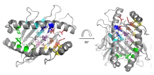 Fig. 1 The peptide‐binding pockets of HLA‐E.1
Key structural properties of HLA-E:
- Typical folded conformation of MHC class I proteins
- Narrow antigenic peptide binding groove structure
- The α3 domain mediates the binding of β2 microglobulin
Functions of HLA-E
The main function of the HLA-E gene is to regulate the activity of natural killer cells (NK cells) and some T cells in the immune system, and its core role is to maintain the balance between immune tolerance and activation.
| Function | Description |
| Induction of immune tolerance | By binding to the CD94/NKG2A inhibitory receptor on the surface of NK cells and transmitting inhibitory signals, it prevents NK cells from attacking normal healthy cells. |
| Anti-infection immunity | In response to viral infection or cell stress, it presents specific peptides that bind to CD94/NKG2C activated receptors and promote NK cell clearance of abnormal cells. |
| Maternal and fetal immune tolerance | The high expression of placental trophoblast cells helps the maternal immune system tolerate the fetus and prevent rejection during pregnancy. |
| Regulation of tumor immune escape | Some tumor cells can inhibit the cytotoxic activity of NK cells and promote tumor immune escape by up-regulating the expression of HLA-E. |
| Transplantation immune regulation | In organ transplantation, the expression level of HLA-E may affect the survival of the graft and the occurrence of rejection reactions. |
The peptide binding characteristics and signal presentation mode of HLA-E distinguish it from the classic MHC class I molecules. Its function is more focused on the regulation of immune checkpoints rather than directly activating the CD8+ T cell response. This characteristic makes it an important research target for immunotherapy and disease intervention.
Applications of HLA-E and HLA-E Antibody in Literature
1. He, Wanlin, et al. "Intracellular trafficking of HLA-E and its regulation." Journal of Experimental Medicine 220.8 (2023): e20221941. https://doi.org/10.1084/jem.20221941
The article indicates that HLA-E is retained in the endoplasmic reticulum due to limited peptide chain supply, and its cytoplasmic tail regulates internal annexation and enrichment in endosomes. This unique transport mode explains its non-classical immune function and provides new ideas for vaccine development.
2. Kim, Se-Jin, and Elham Karamooz. "MR1-and HLA-E-dependent antigen presentation of mycobacterium tuberculosis." International journal of molecular sciences 23.22 (2022): 14412. https://doi.org/10.3390/ijms232214412
The article indicates that MR1 and HLA-E are non-classical antigen-presenting molecules that respectively present microbial metabolites and peptides/glycopeptides in tuberculosis infection, activating specific CD8⁺T cell subsets and providing key targets for the design of novel vaccines.
3. Gillespie, Geraldine M., Max N. Quastel, and Andrew J. McMichael. "HLA‐E: Immune Receptor Functional Mechanisms Revealed by Structural Studies." Immunological Reviews 329.1 (2025): e13434. https://doi.org/10.1111/imr.13434
The article indicates that HLA-E is a non-classical, monomodal molecule that mainly regulates NK cell function by presenting conserved VL9 peptides and can also present pathogen-weakly binding peptides to activate T cells. Its unique structure and immunological function provide a new target for the development of universal immunotherapy.
4. Weitzen, Maya, et al. "Deciphering the HLA-E immunopeptidome with mass spectrometry: an opportunity for universal mRNA vaccines and T-cell-directed immunotherapies." Frontiers in immunology 15 (2024): 1442783. https://doi.org/10.3389/fimmu.2024.1442783
The article indicates that HLA-E can present pathogenic and tumor-associated peptides to activate T cells, and its monomorphism is an advantage of universal immunotherapy. Mass spectrometry and other techniques are being used to analyze its presenting peptide spectra, laying the foundation for the development of broad-spectrum new treatments for cancer and infections.
5. Kraemer, Thomas, Rainer Blasczyk, and Christina Bade-Doeding. "HLA‐E: a novel player for histocompatibility." Journal of immunology research 2014.1 (2014): 352160. https://doi.org/10.1155/2014/352160
The article indicates that HLA-E has only two major functional variants, mainly presenting conserved signaling peptides to regulate NK cell function. The subtle differences in the peptide chain sequences it presents can significantly affect the binding strength with CD94/NKG2 receptors or TCRS and the outcome of immune responses.
Creative Biolabs: HLA-E Antibodies for Research
Creative Biolabs specializes in the production of high-quality HLA-E antibodies for research and industrial applications. Our portfolio includes monoclonal antibodies tailored for ELISA, Flow Cytometry, Western blot, immunohistochemistry, and other diagnostic methodologies.
- Custom HLA-E Antibody Development: Tailor-made solutions to meet specific research requirements.
- Bulk Production: Large-scale antibody manufacturing for industry partners.
- Technical Support: Expert consultation for protocol optimization and troubleshooting.
- Aliquoting Services: Conveniently sized aliquots for long-term storage and consistent experimental outcomes.
For more details on our HLA-E antibodies, custom preparations, or technical support, contact us at email.
Reference
- Gillespie, Geraldine M., Max N. Quastel, and Andrew J. McMichael. "HLA‐E: Immune Receptor Functional Mechanisms Revealed by Structural Studies." Immunological Reviews 329.1 (2025): e13434. https://doi.org/10.1111/imr.13434
Anti-HLA-E antibodies
 Loading...


Hot products 
-
Rabbit Anti-ATF4 Recombinant Antibody (D4B8) (CBMAB-A3872-YC)

-
Rabbit Anti-CBL Recombinant Antibody (D4E10) (CBMAB-CP0149-LY)

-
Mouse Anti-CGAS Recombinant Antibody (CBFYM-0995) (CBMAB-M1146-FY)

-
Mouse Anti-BSN Recombinant Antibody (219E1) (CBMAB-1228-CN)

-
Mouse Anti-14-3-3 Pan Recombinant Antibody (V2-9272) (CBMAB-1181-LY)

-
Mouse Anti-CCNH Recombinant Antibody (CBFYC-1054) (CBMAB-C1111-FY)

-
Mouse Anti-NSUN6 Recombinant Antibody (D-5) (CBMAB-N3674-WJ)

-
Mouse Anti-CDKL5 Recombinant Antibody (CBFYC-1629) (CBMAB-C1689-FY)

-
Mouse Anti-CARD11 Recombinant Antibody (CBFYC-0811) (CBMAB-C0866-FY)

-
Mouse Anti-ARSA Recombinant Antibody (CBYC-A799) (CBMAB-A3679-YC)

-
Mouse Anti-BLNK Recombinant Antibody (CBYY-0623) (CBMAB-0626-YY)

-
Rat Anti-(1-5)-α-L-Arabinan Recombinant Antibody (V2-501861) (CBMAB-XB0003-YC)

-
Mouse Anti-ADIPOR2 Recombinant Antibody (V2-179983) (CBMAB-A1369-YC)

-
Mouse Anti-CARTPT Recombinant Antibody (113612) (CBMAB-C2450-LY)

-
Mouse Anti-AKR1B1 Antibody (V2-2449) (CBMAB-1001CQ)

-
Mouse Anti-CD24 Recombinant Antibody (HIS50) (CBMAB-C10123-LY)

-
Mouse Anti-CASP7 Recombinant Antibody (10-01-62) (CBMAB-C2005-LY)

-
Rat Anti-C5AR1 Recombinant Antibody (8D6) (CBMAB-C9139-LY)

-
Mouse Anti-dsRNA Recombinant Antibody (2) (CBMAB-D1807-YC)

-
Mouse Anti-BMI1 Recombinant Antibody (CBYC-P026) (CBMAB-P0108-YC)

- AActivation
- AGAgonist
- APApoptosis
- BBlocking
- BABioassay
- BIBioimaging
- CImmunohistochemistry-Frozen Sections
- CIChromatin Immunoprecipitation
- CTCytotoxicity
- CSCostimulation
- DDepletion
- DBDot Blot
- EELISA
- ECELISA(Cap)
- EDELISA(Det)
- ESELISpot
- EMElectron Microscopy
- FFlow Cytometry
- FNFunction Assay
- GSGel Supershift
- IInhibition
- IAEnzyme Immunoassay
- ICImmunocytochemistry
- IDImmunodiffusion
- IEImmunoelectrophoresis
- IFImmunofluorescence
- IGImmunochromatography
- IHImmunohistochemistry
- IMImmunomicroscopy
- IOImmunoassay
- IPImmunoprecipitation
- ISIntracellular Staining for Flow Cytometry
- LALuminex Assay
- LFLateral Flow Immunoassay
- MMicroarray
- MCMass Cytometry/CyTOF
- MDMeDIP
- MSElectrophoretic Mobility Shift Assay
- NNeutralization
- PImmunohistologyp-Paraffin Sections
- PAPeptide Array
- PEPeptide ELISA
- PLProximity Ligation Assay
- RRadioimmunoassay
- SStimulation
- SESandwich ELISA
- SHIn situ hybridization
- TCTissue Culture
- WBWestern Blot
