ITGB1 Antibodies
Background
Integrin β1 (ITGB1), as a transmembrane glycoprotein, is widely present on the surface of animal cells and mainly participates in the adhesion between cells and extracellular matrix as well as signal transduction processes. This protein forms heterodimers by binding to multiple integrin α subunits, mediating the cell's perception of the extracellular microenvironment, thereby regulating key physiological activities such as cell migration, proliferation and differentiation. In embryonic development, tissue repair and immune response, ITGB1 plays a significant role in maintaining cellular homeostasis, and its functional abnormalities are closely related to various pathological conditions such as cancer metastasis and fibrotic diseases. Since its identification in the 1980s, ITGB1 has become one of the core molecules in the field of cell adhesion research. The analysis of its structure and functional mechanism has greatly promoted people's understanding of cell communication, mechanical signal transduction and disease target therapy, providing an important foundation for biomedical research.
Structure of ITGB1
ITGB1 is a transmembrane glycoprotein with a molecular weight of approximately 97 kDa. Its molecular weight is relatively conserved among different species, but it can fluctuate slightly through differential glycosylation and other means. The following are the molecular weight references for some species:
| Species | Human | Mouse | Rat |
| Molecular Weight (kDa) | ~97 | ~95 | ~96 |
This protein is composed of approximately 798 amino acids, forming a typical structure that includes the extracellular domain, transmembrane region and intracellular tail region. Its extracellular domain contains multiple repetitive domains (such as the β-propeller structure), which are responsible for recognizing extracellular matrix proteins (such as fibronectin and laminin) and mediating cell adhesion. The intracellular domain interacts with the cytoskeleton through conjunctive proteins such as talin and vinculin, transmitting mechanical signals into the cell and thereby regulating the processes of cell migration, proliferation and differentiation.
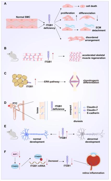 Fig. 1 ITGB1 and physiological function, benign disease.1
Key structural properties of ITGB1:
- Extracellular domain beta propeller structure formation, responsible for identifying ligands ecms
- Across the membrane area keep anchor protein on the cell membrane
- Intracellular tail region contains NPxY motif, such as mediated cytoskeleton connection
Functions of ITGB1
The main function of ITGB1 is to mediate the adhesion between cells and extracellular matrix and bidirectional signal transduction. In addition, it is also involved in regulating a variety of key cellular physiological processes.
| Function | Description |
| Cell adhesion | As a core component of integrin heterodimers, it recognizes and binds to extracellular matrices such as fibronectin and laminin, anchoring cells to their surrounding environment. |
| Mechanical signal transduction | Sensing the mechanical properties of the extracellular matrix (such as stiffness, tension), and converting mechanical signals into intracellular biochemical signals (such as FAK, Src kinase activation), affect cell behavior. |
| Cell migration and proliferation | By dynamically regulating the assembly and dissociation of adhesion spots, guide the directional migration of cells; At the same time, it transmits growth signals and promotes the progress of the cell cycle. |
| Tissue development and homeostasis | In embryonic development, angiogenesis, tissue repair process is crucial to its dysfunction can cause developmental defect or fibrosis and other diseases. |
| Immune regulation | Expression on the immune cells, lymphocytes homing, inflammation and the immune synapse formation. |
The affinity of ITGB1 for ligands is dynamically regulated by conformational changes (low, medium and high affinity states), a characteristic that enables it to precisely respond to microenvironmental changes and complete complex biological regulation.
Applications of ITGB1 and ITGB1 Antibody in Literature
1. Li, Yuexian, et al. "ITGB1 enhances the radioresistance of human non-small cell lung cancer cells by modulating the DNA damage response and YAP1-induced epithelial-mesenchymal transition." International journal of biological sciences 17.2 (2021): 635. https://doi.org/10.7150/ijbs.52319
Studies have shown that ITGB1 is highly expressed in non-small cell lung cancer cells and enhances radiation resistance by promoting DNA repair and YAP1-induced epithelial-mesenchymal transition, suggesting it as a potential therapeutic target for reversing radiotherapy tolerance.
2. Sun, Rui, et al. "ALKBH5 activates FAK signaling through m6A demethylation in ITGB1 mRNA and enhances tumor-associated lymphangiogenesis and lymph node metastasis in ovarian cancer." Theranostics 13.2 (2023): 833. https://doi.org/10.7150/thno.77441
Research has revealed that ALKBH5 promotes lymphangiogenesis and lymph node metastasis of ovarian cancer by reducing the m6A modification of ITGB1, enhancing its expression and activating the FAK/Src pathway. This signaling axis provides a new target for anti-metastasis therapy.
3. Yu-Jiao, Zhou, et al. "RNA-binding protein RPS7 promotes hepatocellular carcinoma progression via LOXL2-dependent activation of ITGB1/FAK/SRC signaling." Journal of Experimental and Clinical Cancer Research 43 (2024): 1. https://doi.org/10.1186/s13046-023-02929-1
Research reveals that RPS7 enhances the expression of LOXL2 mRNA by stabilizing it, thereby maintaining the stability of ITGB1 protein and activating the downstream FAK/SRC pathway, ultimately promoting the metastasis of hepatocellular carcinoma. This axis provides a new target for anti-metastasis treatment.
4. Gu, Wenchao, et al. "ITGB1 as a prognostic biomarker correlated with immune suppression in gastric cancer." Cancer Medicine 12.2 (2023): 1520-1531. https://doi.org/10.1002/cam4.5042
Studies have shown that the high expression of ITGB1 in gastric cancer is associated with tumor invasion, advanced stage and poor prognosis. It activates the Wnt/β-catenin pathway and mediates immunosuppression, suggesting that it can serve as a prognostic marker and a potential predictor of the efficacy of immunotherapy.
5. Zhu, Xingyu, et al. "ITGB1-mediated molecular landscape and cuproptosis phenotype induced the worse prognosis in diffuse gastric cancer." Frontiers in Oncology 13 (2023): 1115510. https://doi.org/10.3389/fonc.2023.1115510
Studies have shown that in diffuse gastric cancer, high expression of ITGB1 predicts a poor prognosis. It promotes tumor growth by regulating the ROCK1/PKA/AKT signaling pathway and inhibits the copper death metabolic pathway, suggesting that ITGB1 is a potential therapeutic target.
Creative Biolabs: ITGB1 Antibodies for Research
Creative Biolabs specializes in the production of high-quality ITGB1 antibodies for research and industrial applications. Our portfolio includes monoclonal antibodies tailored for ELISA, Flow Cytometry, Western blot, immunohistochemistry, and other diagnostic methodologies.
- Custom ITGB1 Antibody Development: Tailor-made solutions to meet specific research requirements.
- Bulk Production: Large-scale antibody manufacturing for industry partners.
- Technical Support: Expert consultation for protocol optimization and troubleshooting.
- Aliquoting Services: Conveniently sized aliquots for long-term storage and consistent experimental outcomes.
For more details on our ITGB1 antibodies, custom preparations, or technical support, contact us at email.
Reference
- Su, Chen, et al. "Integrinβ-1 in disorders and cancers: molecular mechanisms and therapeutic targets." Cell Communication and Signaling 22.1 (2024): 71.https://doi.org/10.1186/s12964-023-01338-3
Anti-ITGB1 antibodies
 Loading...


Hot products 
-
Mouse Anti-ALB Recombinant Antibody (V2-363290) (CBMAB-S0173-CQ)

-
Mouse Anti-F11R Recombinant Antibody (402) (CBMAB-0026-WJ)

-
Rabbit Anti-ENO2 Recombinant Antibody (BA0013) (CBMAB-0272CQ)

-
Rabbit Anti-ALDOA Recombinant Antibody (D73H4) (CBMAB-A2314-YC)

-
Mouse Anti-CD24 Recombinant Antibody (ALB9) (CBMAB-0176CQ)

-
Mouse Anti-FAS2 Monoclonal Antibody (1D4) (CBMAB-0071-CN)

-
Mouse Anti-ADAM29 Recombinant Antibody (V2-179787) (CBMAB-A1149-YC)

-
Mouse Anti-CCL18 Recombinant Antibody (64507) (CBMAB-C7910-LY)

-
Mouse Anti-DISP2 Monoclonal Antibody (F66A4B1) (CBMAB-1112CQ)

-
Mouse Anti-DLG1 Monolconal Antibody (4F3) (CBMAB-0225-CN)

-
Mouse Anti-AMOT Recombinant Antibody (CBYC-A564) (CBMAB-A2552-YC)

-
Mouse Anti-ENO1 Recombinant Antibody (CBYC-A950) (CBMAB-A4388-YC)

-
Mouse Anti-CAT Recombinant Antibody (724810) (CBMAB-C8431-LY)

-
Mouse Anti-ADV Recombinant Antibody (V2-503423) (CBMAB-V208-1364-FY)

-
Mouse Anti-AOC3 Recombinant Antibody (CBYY-0014) (CBMAB-0014-YY)

-
Mouse Anti-GFP Recombinant Antibody (28) (CBMAB-G3038-LY)

-
Mouse Anti-CD33 Recombinant Antibody (P67.6) (CBMAB-C10189-LY)

-
Mouse Anti-Acetyl SMC3 (K105/K106) Recombinant Antibody (V2-634053) (CBMAB-AP052LY)

-
Mouse Anti-ARG1 Recombinant Antibody (CBYCL-103) (CBMAB-L0004-YC)

-
Mouse Anti-ENPP1 Recombinant Antibody (CBFYE-0159) (CBMAB-E0375-FY)

- AActivation
- AGAgonist
- APApoptosis
- BBlocking
- BABioassay
- BIBioimaging
- CImmunohistochemistry-Frozen Sections
- CIChromatin Immunoprecipitation
- CTCytotoxicity
- CSCostimulation
- DDepletion
- DBDot Blot
- EELISA
- ECELISA(Cap)
- EDELISA(Det)
- ESELISpot
- EMElectron Microscopy
- FFlow Cytometry
- FNFunction Assay
- GSGel Supershift
- IInhibition
- IAEnzyme Immunoassay
- ICImmunocytochemistry
- IDImmunodiffusion
- IEImmunoelectrophoresis
- IFImmunofluorescence
- IGImmunochromatography
- IHImmunohistochemistry
- IMImmunomicroscopy
- IOImmunoassay
- IPImmunoprecipitation
- ISIntracellular Staining for Flow Cytometry
- LALuminex Assay
- LFLateral Flow Immunoassay
- MMicroarray
- MCMass Cytometry/CyTOF
- MDMeDIP
- MSElectrophoretic Mobility Shift Assay
- NNeutralization
- PImmunohistologyp-Paraffin Sections
- PAPeptide Array
- PEPeptide ELISA
- PLProximity Ligation Assay
- RRadioimmunoassay
- SStimulation
- SESandwich ELISA
- SHIn situ hybridization
- TCTissue Culture
- WBWestern Blot
