MFN2 Antibodies
Background
The MFN2 gene encodes a transmembrane GTPase called Mitofusin 2, which is mainly located on the outer mitochondrial membrane. This protein regulates the fusion process of mitochondria, maintaining the dynamic balance and normal function of the mitochondrial network, and plays a crucial role in cellular energy metabolism, calcium homeostasis, and apoptotic pathways. In mammals, mutations in the MFN2 gene have been proven to be directly related to the occurrence of CMT2A,which is a hereditary peripheral neuropathy caused by abnormal mitochondrial function in the central and peripheral nervous systems. Since its pathogenic mechanism was clearly reported in 2004, MFN2 has become one of the core molecules in studying the relationship between mitochondrial dynamics and neurodegenerative diseases. The continuous clarification of its structural mechanism and pathological significance has significantly advanced our understanding of organelle interactions, energy metabolism disorders, and treatment strategies for related diseases.
Structure of MFN2
The mitochondrial fusion protein 2 encoded by the MFN2 gene is a transmembrane GTPase with a molecular weight of approximately 86 kDa. Its molecular weight is highly conserved among different mammalian species, with the main differences arising from subtle amino acid sequence variations in the regulatory region. The basic amino acid sequence forms a complex spatial conformation consisting of a GTPase domain, multiple coiled-coil domains, and two transmembrane domains. The N-terminal and C-terminal ends of the protein face the cytoplasm, and it drives mitochondrial outer membrane fusion through GTP-dependent dimerization. The two heptapeptide repeat domains, HR1 and HR2, play a central role in mediating trans-oligomerization, while the transmembrane domains are responsible for anchoring the protein to the mitochondrial outer membrane. The function of this protein is highly dependent on the conformational change energy provided by the GTPase domain's GTP hydrolysis.
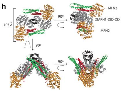 Fig. 1 Representative ribbon model of the docked MFN2.1
Key structural properties of MFN2:
- The core GTPase domain that provides energy for membrane fusion
- Two heptapeptide repeating coiled-coil domains, mediating trans-dimerization
- Two transmembrane area anchor protein in the mitochondrial outer membrane
Functions of MFN2
The core function of the MFN2 gene is to regulate the dynamics of the mitochondrial network, particularly by mediating the process of mitochondrial membrane fusion. However, it is also widely involved in a variety of crucial cellular physiological and pathological processes, including energy metabolism regulation, apoptosis signal transduction, and endoplasmic reticulum-mitochondria interaction.
| Function | Description |
| Mitochondrial Fusion | The MFN2 protein drives the tethering and fusion of the outer mitochondrial membrane through its GTPase domain, maintaining the continuity and integrity of the mitochondrial network. |
| Energy Metabolism Regulation | By maintaining a healthy mitochondrial network, it ensures the efficiency of oxidative phosphorylation in cells (especially nerve and muscle cells) and the stable generation of ATP. |
| Mitochondrial Quality Control | As one of the regulatory factors of mitochondrial autophagy, it participates in the identification and marking of dysfunctional mitochondria, ensuring the renewal of cellular organelles. |
| Endoplasmic Reticulum-Mitochondria Interaction | MFN2 is simultaneously located on the endoplasmic reticulum membrane, mediating and stabilizing the membrane contact sites between the endoplasmic reticulum and mitochondria, and regulating lipid transfer and calcium ion signaling. |
| Cell Apoptosis Involvement | In the mitochondrial-mediated apoptotic pathway, the degradation or dysfunction of MFN2 can promote mitochondrial division and the release of cytochrome c. |
Compared with the "single" function that only mediates mitochondrial fusion, the function of MFN2 exhibits a high degree of networked nature and context dependence. Its activity is finely regulated by post-translational modifications (such as ubiquitination and phosphorylation), and its functional imbalance is closely related to various neurodegenerative diseases such as fibrodysplasia ossificans progressiva type 2A.
Applications of MFN2 and MFN2 Antibody in Literature
1. Yepuri, Gautham, et al. "DIAPH1-MFN2 interaction regulates mitochondria-SR/ER contact and modulates ischemic/hypoxic stress." Nature Communications 14.1 (2023): 6900. https://doi.org/10.1038/s41467-023-42521-x
Research has confirmed that the actin regulatory factor DIAPH1 directly interacts with the mitochondrial fusion protein MFN2, which can shorten the distance between mitochondria and the endoplasmic reticulum, enhance the contact between organelles, thereby regulating mitochondrial metabolism and oxidative stress under ischemic conditions, providing a new target for the repair of ischemic tissues.
2. Zanfardino, Paola, et al. "The balance of MFN2 and OPA1 in mitochondrial dynamics, cellular homeostasis, and disease." Biomolecules 15.3 (2025): 433. https://doi.org/10.3390/biom15030433
Studies have confirmed that the mitochondrial fusion regulator MFN2 and OPA1 are not only structural proteins, but also regulate cellular energy metabolism and autophagy. Abnormalities in their functions can lead to neurodegenerative diseases, and related therapeutic strategies are becoming a new research direction.
3. Kirshenbaum, Lorrie A., et al. "DIAPH1-MFN2 interaction decreases the endoplasmic reticulum-mitochondrial distance and promotes cardiac injury following myocardial ischemia." nature communications 15.1 (2024): 1469. https://doi.org/10.1038/s41467-024-45560-0
Research has confirmed that the actin polymerization protein DIAPH1 forms an heteromeric complex with the mitochondrial fusion protein MFN2, which can shorten the distance between mitochondria and the endoplasmic reticulum and enhance their contact. This interaction plays a crucial regulatory role in myocardial ischemia/reperfusion injury.
4. Zhao, Fanpeng, et al. "Mfn2 overexpression attenuates MPTP neurotoxicity in vivo." International Journal of Molecular Sciences 22.2 (2021): 601. https://doi.org/10.3390/ijms22020601
The research found that in the Parkinson's disease model, overexpression of MFN2 could inhibit the mitochondrial fragmentation induced by MPTP, thereby significantly reducing the damage to dopaminergic neurons, oxidative stress, and motor disorders. This provides a new idea for treating the disease by regulating mitochondrial dynamics.
5. Zhang, Jingjing, et al. "MFN2 deficiency affects calcium homeostasis in lung adenocarcinoma cells via downregulation of UCP4." FEBS Open Bio 13.6 (2023): 1107-1124. https://doi.org/10.1002/2211-5463.13591
This study found that the mitochondrial fusion protein MFN2, in lung adenocarcinoma, jointly maintains cellular calcium homeostasis by regulating UCP4 and PINK1. Its low expression is associated with poor prognosis, suggesting that it could be a potential therapeutic target.
Creative Biolabs: MFN2 Antibodies for Research
Creative Biolabs specializes in the production of high-quality MFN2 antibodies for research and industrial applications. Our portfolio includes monoclonal and polyclonal antibodies tailored for ELISA, Flow Cytometry, Western blot, immunohistochemistry, and other diagnostic methodologies.
- Custom MFN2 Antibody Development: Tailor-made solutions to meet specific research requirements.
- Bulk Production: Large-scale antibody manufacturing for industry partners.
- Technical Support: Expert consultation for protocol optimization and troubleshooting.
- Aliquoting Services: Conveniently sized aliquots for long-term storage and consistent experimental outcomes.
For more details on our MFN2 antibodies, custom preparations, or technical support, contact us at info@creative-biolabs.com.
Reference
- Yeahuri, Gautham, et al. "DIAPH1-MFN2 interaction regulates mitochondria-SR/ER contact and modulates ischemic/hypoxic stress." Nature Communications 14.1 (2023): 6900. Distributed under Open Access license CC BY 4.0. Cropped from the original figure. https://doi.org/10.1038/s41467-023-42521-x
Anti-MFN2 antibodies
 Loading...


Hot products 
-
Mouse Anti-ASB9 Recombinant Antibody (1D8) (CBMAB-A0529-LY)

-
Mouse Anti-CRYAB Recombinant Antibody (A4345) (CBMAB-A4345-YC)

-
Mouse Anti-BCL6 Recombinant Antibody (CBYY-0442) (CBMAB-0445-YY)

-
Rabbit Anti-ABL1 (Phosphorylated Y245) Recombinant Antibody (V2-505716) (PTM-CBMAB-0465LY)

-
Mouse Anti-CECR2 Recombinant Antibody (CBWJC-2465) (CBMAB-C3533WJ)

-
Mouse Anti-GFP Recombinant Antibody (28) (CBMAB-G3038-LY)

-
Rabbit Anti-CCN1 Recombinant Antibody (CBWJC-3580) (CBMAB-C4816WJ)

-
Mouse Anti-CD33 Recombinant Antibody (P67.6) (CBMAB-C10189-LY)

-
Mouse Anti-FN1 Monoclonal Antibody (71) (CBMAB-1241CQ)

-
Mouse Anti-ENO1 Recombinant Antibody (CBYC-A950) (CBMAB-A4388-YC)

-
Mouse Anti-ALB Recombinant Antibody (V2-363290) (CBMAB-S0173-CQ)

-
Mouse Anti-C4B Recombinant Antibody (CBYY-C2996) (CBMAB-C4439-YY)

-
Rabbit Anti-BRCA2 Recombinant Antibody (D9S6V) (CBMAB-CP0017-LY)

-
Mouse Anti-ALDOA Recombinant Antibody (A2) (CBMAB-A2316-YC)

-
Mouse Anti-ESR1 Recombinant Antibody (Y31) (CBMAB-1208-YC)

-
Mouse Anti-GFAP Recombinant Antibody (24) (CBMAB-G2927-LY)

-
Mouse Anti-GFAP Recombinant Antibody (20) (CBMAB-G2914-LY)

-
Mouse Anti-CHRNA9 Recombinant Antibody (8E4) (CBMAB-C9161-LY)

-
Mouse Anti-CARTPT Recombinant Antibody (113612) (CBMAB-C2450-LY)

-
Mouse Anti-CD24 Recombinant Antibody (SN3) (CBMAB-C1037-CQ)

- AActivation
- AGAgonist
- APApoptosis
- BBlocking
- BABioassay
- BIBioimaging
- CImmunohistochemistry-Frozen Sections
- CIChromatin Immunoprecipitation
- CTCytotoxicity
- CSCostimulation
- DDepletion
- DBDot Blot
- EELISA
- ECELISA(Cap)
- EDELISA(Det)
- ESELISpot
- EMElectron Microscopy
- FFlow Cytometry
- FNFunction Assay
- GSGel Supershift
- IInhibition
- IAEnzyme Immunoassay
- ICImmunocytochemistry
- IDImmunodiffusion
- IEImmunoelectrophoresis
- IFImmunofluorescence
- IGImmunochromatography
- IHImmunohistochemistry
- IMImmunomicroscopy
- IOImmunoassay
- IPImmunoprecipitation
- ISIntracellular Staining for Flow Cytometry
- LALuminex Assay
- LFLateral Flow Immunoassay
- MMicroarray
- MCMass Cytometry/CyTOF
- MDMeDIP
- MSElectrophoretic Mobility Shift Assay
- NNeutralization
- PImmunohistologyp-Paraffin Sections
- PAPeptide Array
- PEPeptide ELISA
- PLProximity Ligation Assay
- RRadioimmunoassay
- SStimulation
- SESandwich ELISA
- SHIn situ hybridization
- TCTissue Culture
- WBWestern Blot
