RELB Antibodies
Background
The RELB gene encodes a transcription factor protein belonging to the NF-κB/Rel family, which is mainly expressed in immune cells and tissues. This protein forms dimers with other members of the NF-κB family and participates in regulating various biological processes such as inflammatory responses, immune responses, and cell survival. RELB plays a crucial role in adaptive immunity and lymphoid organ development, and its dysfunction is closely related to autoimmune diseases, chronic inflammation, and certain types of cancer. This gene was first identified in the 1990s, and the study of its structure and function has deepened our understanding of the NF-κB signaling pathway, providing an important molecular basis for the mechanism analysis and treatment strategies of related diseases.
Structure of RELB
RELB is a transcription factor protein with a molecular weight of approximately 60 kDa. As a member of the NF-κB/Rel family, its molecular weight is relatively conserved among different species, but may vary slightly due to subtypes or post-translational modifications (such as phosphorylation).
The basic structure of this protein consists of an N-terminal Rel homology Domain (RHD), which is responsible for DNA binding, dimerization and nuclear localization; and a C-terminal Transcriptional Activation Domain (TAD), which is used to recruit co-activating factors and initiate the transcription of target genes. RELB usually forms a stable heterodimer with NF-κB2/p100, and this complex exists in an inactive form in the cytoplasm. When specific cellular signals (such as through the non-classical NF-κB pathway) are received, p100 is processed into p52, thereby releasing the RELB/p52 dimer with transcriptional activity, allowing it to enter the nucleus and regulate the expression of key genes involved in lymphocyte development, immune homeostasis and inflammatory responses. This precise "signal-processing-activation" mechanism enables RELB to play a specific and crucial biological role in immune regulation.
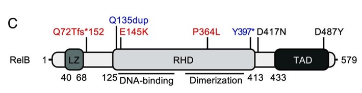 Fig. 1 Schematic diagram of the RelB protein.1
Key structural properties of RELB:
- Rel homologous domain
- Transcriptional Activation Domain
- Dimerization interface
- Nuclear localization signal
Functions of RELB
The protein encoded by the RELB gene has a core function as a transcription factor, mainly involved in regulating immune and inflammatory responses. The specific functions are as follows:
| Function | Description |
| Immune Regulation | RELB is the core component of the non-classical NF-κB signaling pathway. When cells receive specific signals (such as CD40L, LTβ), RELB forms a dimer with p100/p52 and enters the nucleus, initiating the expression of key genes that regulate B cell development, lymphoid organ formation, and dendritic cell maturation. |
| Inflammation Regulation | By regulating target genes, RELB precisely participates in the initiation and maintenance of chronic inflammatory responses. Its dysfunction is closely related to autoimmune diseases such as rheumatoid arthritis and inflammatory bowel disease. |
| Cell Survival and Proliferation | In certain cell types, the target genes activated by RELB can promote cell survival and inhibit apoptosis. This function plays a role in the homeostasis of lymphocytes and the development of some lymphomas. |
| Stress and Adaptation | When cells respond to physiological stresses such as endoplasmic reticulum stress, the RELB pathway can be activated, assisting the cells in making adaptive adjustments and maintaining the balance of immune metabolism. |
| Developmental Support | It is crucial for the normal development and structural maintenance of secondary lymphoid organs (such as lymph nodes and the white pulp of the spleen), ensuring that the immune system has a complete histological foundation. |
Unlike the rapid and widespread activation of the classical NF-κB pathway (mediated by p65/p50), the non-classical pathway led by RELB typically starts more slowly, lasts longer, and has high signal specificity. This characteristic makes it more focused on regulating long-term physiological and pathological processes such as adaptive immunity, lymphoid tissue development, and chronic inflammation, highlighting its role as a "precise regulator" in the immune system.
Applications of RELB and RELB Antibody in Literature
1. Le Voyer, Tom, et al. "Inherited human RelB deficiency impairs innate and adaptive immunity to infection." Proceedings of the National Academy of Sciences 121.37 (2024): e2321794121. https://doi.org/10.1073/pnas.2321794121
The article describes two patients who carry rare mutations in the RELB gene, resulting in abnormal NF-κB signaling pathway and the acquisition of acquired type I interferon neutralizing antibodies. These patients exhibit severe combined immunodeficiency and are prone to multiple pathogen infections.
2. Wang, Xinyue, et al. "Rescue RM/CS-AKI by blocking strategy with one-dose anti-myoglobin RabMAb." Nature Communications 16.1 (2025): 1044. https://doi.org/10.3390/cells10071609
The article discusses that in central nervous system inflammation, various cells collaborate through the NF-κB pathway. This review focuses on the cell-specific activation mechanism and role of the RelB subunit, which is distinct from the already well-defined function of the p65 subunit.
3. Kim, Su-Lim, Hack Sun Choi, and Dong-Sun Lee. "BRD4/nuclear PD-L1/RelB circuit is involved in the stemness of breast cancer cells." Cell Communication and Signaling 21.1 (2023): 315. https://doi.org/10.1186/s12964-023-01319-6
The research has found that the BRD4/nuclear PD-L1/RelB pathway is crucial for the formation of breast cancer stem cells. Inhibiting this pathway can significantly weaken the characteristics of tumor stem cells, providing a new strategy for targeted immunotherapy.
4. Chen, Qiangxing, et al. "LTβR-RelB signaling in intestinal epithelial cells protects from chemotherapy-induced mucosal damage." Frontiers in immunology 15 (2024): 1388496. https://doi.org/10.3389/fimmu.2024.1388496
The research has found that after chemotherapy, intestinal repair depends on T cells secreting LIGHT to activate the epithelial cell LTβR receptor, and promotes mucosal healing through the non-classical NF-κB pathway member RelB.
5. Wu, Jintao, et al. "RelB is a potential molecular biomarker for immunotherapy in human pan-cancer." Frontiers in Molecular Biosciences 10 (2023): 1178446. https://doi.org/10.3389/fmolb.2023.1178446
The research has found that the non-classical NF-κB member RelB is highly expressed in various cancers and is closely related to the prognosis of patients and the infiltration of immune cells, suggesting that it could serve as a potential new target for cancer immunotherapy.
Creative Biolabs: RELB Antibodies for Research
Creative Biolabs specializes in the production of high-quality RELB antibodies for research and industrial applications. Our portfolio includes monoclonal and polyclonal antibodies tailored for ELISA, Flow Cytometry, Western blot, immunohistochemistry, and other diagnostic methodologies.
- Custom RELB Antibody Development: Tailor-made solutions to meet specific research requirements.
- Bulk Production: Large-scale antibody manufacturing for industry partners.
- Technical Support: Expert consultation for protocol optimization and troubleshooting.
- Aliquoting Services: Conveniently sized aliquots for long-term storage and consistent experimental outcomes.
For more details on our RELB antibodies, custom preparations, or technical support, contact us at email.
Reference
- Le Voyer, Tom, et al. "Inherited human RelB deficiency impairs innate and adaptive immunity to infection." Proceedings of the National Academy of Sciences 121.37 (2024): e2321794121. Distributed under Open Access license CC BY 4.0. Cropped from the original figure. https://doi.org/10.1073/pnas.2321794121
Anti-RELB antibodies
 Loading...


Hot products 
-
Mouse Anti-ADRB2 Recombinant Antibody (V2-180026) (CBMAB-A1420-YC)

-
Mouse Anti-CAT Recombinant Antibody (724810) (CBMAB-C8431-LY)

-
Mouse Anti-CCT6A/B Recombinant Antibody (CBXC-0168) (CBMAB-C5570-CQ)

-
Mouse Anti-CFL1 (Phospho-Ser3) Recombinant Antibody (CBFYC-1770) (CBMAB-C1832-FY)

-
Rat Anti-C5AR1 Recombinant Antibody (8D6) (CBMAB-C9139-LY)

-
Mouse Anti-AMH Recombinant Antibody (5/6) (CBMAB-A2527-YC)

-
Mouse Anti-EGR1 Recombinant Antibody (CBWJZ-100) (CBMAB-Z0289-WJ)

-
Mouse Anti-CCL18 Recombinant Antibody (64507) (CBMAB-C7910-LY)

-
Rabbit Anti-BAD (Phospho-Ser136) Recombinant Antibody (CAP219) (CBMAB-AP536LY)

-
Mouse Anti-ADAM12 Recombinant Antibody (V2-179752) (CBMAB-A1114-YC)

-
Mouse Anti-ADGRE5 Recombinant Antibody (V2-360335) (CBMAB-C2088-CQ)

-
Mouse Anti-14-3-3 Pan Recombinant Antibody (V2-9272) (CBMAB-1181-LY)

-
Human Anti-SARS-CoV-2 Spike Recombinant Antibody (CR3022) (CBMAB-CR014LY)

-
Mouse Anti-BIRC5 Recombinant Antibody (6E4) (CBMAB-CP2646-LY)

-
Mouse Anti-GLP1R Recombinant Antibody (4F3) (CBMAB-G0521-LY)

-
Mouse Anti-ADGRL2 Recombinant Antibody (V2-58519) (CBMAB-L0166-YJ)

-
Mouse Anti-ATG5 Recombinant Antibody (9H197) (CBMAB-A3945-YC)

-
Mouse Anti-A2M Recombinant Antibody (V2-178822) (CBMAB-A0036-YC)

-
Mouse Anti-BPGM Recombinant Antibody (CBYY-1806) (CBMAB-2155-YY)

-
Mouse Anti-ENO1 Recombinant Antibody (CBYC-A950) (CBMAB-A4388-YC)

- AActivation
- AGAgonist
- APApoptosis
- BBlocking
- BABioassay
- BIBioimaging
- CImmunohistochemistry-Frozen Sections
- CIChromatin Immunoprecipitation
- CTCytotoxicity
- CSCostimulation
- DDepletion
- DBDot Blot
- EELISA
- ECELISA(Cap)
- EDELISA(Det)
- ESELISpot
- EMElectron Microscopy
- FFlow Cytometry
- FNFunction Assay
- GSGel Supershift
- IInhibition
- IAEnzyme Immunoassay
- ICImmunocytochemistry
- IDImmunodiffusion
- IEImmunoelectrophoresis
- IFImmunofluorescence
- IGImmunochromatography
- IHImmunohistochemistry
- IMImmunomicroscopy
- IOImmunoassay
- IPImmunoprecipitation
- ISIntracellular Staining for Flow Cytometry
- LALuminex Assay
- LFLateral Flow Immunoassay
- MMicroarray
- MCMass Cytometry/CyTOF
- MDMeDIP
- MSElectrophoretic Mobility Shift Assay
- NNeutralization
- PImmunohistologyp-Paraffin Sections
- PAPeptide Array
- PEPeptide ELISA
- PLProximity Ligation Assay
- RRadioimmunoassay
- SStimulation
- SESandwich ELISA
- SHIn situ hybridization
- TCTissue Culture
- WBWestern Blot
