RELT Antibodies
Background
The RELT gene encodes a cell surface protein belonging to the tumor necrosis factor receptor superfamily, which is mainly expressed in immune tissues and specific epithelial cells. This protein participates in immune regulation and cell survival processes by mediating intercellular signal transduction, especially playing a significant role in T cell activation and inflammatory responses. Its ligand binding characteristics and signaling pathways were gradually revealed in the early 21st century. Studies have shown that RELT can affect cell proliferation and differentiation through pathways such as activating NF-κB. Recent studies have found that the abnormal expression of this gene is associated with autoimmune diseases such as rheumatoid arthritis, and its unique extracellular domain provides a new molecular basis for the development of targeted immunotherapy.
Structure of RELT
RELT is a type I transmembrane protein with a molecular weight of approximately 45-50 kDa. The extracellular region of this protein contains a characteristic homologous domain of tumor necrosis factor receptor, and its amino acid sequence shows moderate conservation among different species.
| Species | Human | Mouse | Rat |
| Molecular Weight (kDa) | 430 | 427 | 428 |
| Primary Structural Differences | Extracellular region rich in cysteine, forming a typical structure of CRD | Extracellular area about 65% homology with humans | Intracellular section of the tail is shorter, the lack of obvious death domain structure |
The intracellular segment structure of RELT protein is relatively short, and it interacts with intracellular signal adaptation molecules through specific basic amino acid residues. The secondary structure of this protein is mainly β -folding, which together form a stable extracellular ligand binding interface. Conserved cysteine residues stabilize their spatial conformation by forming intramolecular disulfide bonds, which is crucial for their binding to ligands and triggering the downstream NF-κB signaling pathway.
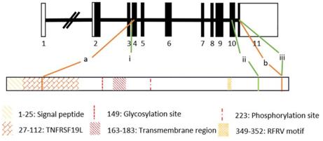 Fig. 1 Gene diagram and domain architecture of RELT.1
Key structural properties of RELT:
- The extracellular segment contains a conserved homologous domain of tumor necrosis factor receptor
- Across the membrane area consists of a single alpha helix stability anchor structure
- Intracellular lack of classic signal domain but contain characteristic basic amino acid cluster
- Cysteine residues form intramolecular disulfide bonds to maintain spatial conformation
Functions of RELT
The main function of RELT protein is to mediate signal transduction between immune cells and participate in various physiological and pathological processes. The specific functional system is as follows:
| Function | Description |
| Immune regulation | Specific expression in the surface of T cells, by activating the nf-kappa B signaling pathways regulating immune responses such as strength. |
| Regulation of cell survival | Transmit pro-survival signals and inhibit the programmed death of specific types of cells. |
| Involvement of inflammatory response | In diseases such as rheumatoid arthritis and high expression, and promote the release of proinflammatory factor. |
| Cell adhesion assistance | Mediate the interaction between immune cells and target cells through extracellular domains. |
| Influence of bone metabolism | Detect its expression in osteoclast, which may be involved in bone remodeling balance control. |
The binding curve of RELT to ligands shows typical receptor saturation characteristics, and its signal transduction efficiency is precisely regulated by the phosphorylation state of the intracellular basic amino acid cluster. This feature enables it to play a key role in maintaining immune homeostasis.
Applications of RELT and RELT Antibody in Literature
1. Cusick, John K., Jessa Alcaide, and Yihui Shi. "The RELT family of proteins: an increasing awareness of their importance for cancer, the immune system, and development." Biomedicines 11.10 (2023): 2695. https://doi.org/10.3390/biomedicines11102695
The article indicates that RELT and its homologous proteins RELL1/RELL2 (collectively referred to as RELTfms) are members of the tumor necrosis factor family and have attracted much attention in recent years due to their role in cancer. Studies have shown that RELT may suppress immune responses and promote tumor development, but its ligands and physiological functions have not been fully clarified. These proteins also play significant roles in processes such as inflammation and microbial infections.
2. Batiste, Maryann, et al. "RELT Is Upregulated in Breast Cancer and Induces Death in Breast Cancer Cells." Biomedicines 12.12 (2024): 2667. https://doi.org/10.3390/biomedicines12122667
Research has found that the expression of RELT is elevated in cancer cells such as breast cancer and lung cancer. It can induce apoptosis in breast cancer cells that is independent of OXSR1 phosphorylation. In addition, RELT has been found to be able to be located in the cell nucleus, which is a new function worth in-depth exploration.
3. Nikolopoulos, Georgios, et al. "New missense variants in RELT causing hypomineralised amelogenesis imperfecta." Clinical genetics 97.5 (2020): 688-695. https://doi.org/10.1111/cge.13721
Research has found that novel homozygous mutations in the RELT gene can lead to recessive enamel hypoplasia. Analysis of the patient's teeth indicated that RELT plays a key role in the coordination between ameloblasts and the enamel matrix. Furthermore, the previously reported syndrome symptoms were not found in all patients, which expands the understanding of the pathogenic mechanism of RELT.
4. Wang, Hongying, et al. "LILRB4 on multiple myeloma cells promotes bone lesion by p-SHP2/NF-κB/RELT signal pathway." Journal of Experimental & Clinical Cancer Research 43.1 (2024): 183. https://doi.org/10.1186/s13046-024-03110-y
This study reveals that LILRB4 is highly expressed in multiple myeloma and promotes osteoclast differentiation by secreting RELT, thereby exacerbating bone injury. Blocking the LILRB4 signal or using anti-LILRB4 antibodies can effectively inhibit this pathological process, providing a new target for the treatment of bone lesions in multiple myeloma.
5. Shahid, Shifa, et al. "ADAM10: Possible functions in enamel development." Frontiers in Physiology 13 (2022): 1032383. https://doi.org/10.3389/fphys.2022.1032383
Studies have shown that the metalloproteinase ADAM10 is a splicing protein of RELT and may play a role together in tooth development. Since RELT gene mutations can lead to enamel deformities, ADAM10 may play an important role in enamel formation by regulating proteins such as RELT.
Creative Biolabs: RELT Antibodies for Research
Creative Biolabs specializes in the production of high-quality RELT antibodies for research and industrial applications. Our portfolio includes monoclonal antibodies tailored for ELISA, Flow Cytometry, Western blot, immunohistochemistry, and other diagnostic methodologies.
- Custom RELT Antibody Development: Tailor-made solutions to meet specific research requirements.
- Bulk Production: Large-scale antibody manufacturing for industry partners.
- Technical Support: Expert consultation for protocol optimization and troubleshooting.
- Aliquoting Services: Conveniently sized aliquots for long-term storage and consistent experimental outcomes.
For more details on our RELT antibodies, custom preparations, or technical support, contact us at email.
Reference
- Nikolopoulos, Georgios, et al. "New missense variants in RELT causing hypomineralised amelogenesis imperfecta." Clinical genetics 97.5 (2020): 688-695. https://doi.org/10.1111/cge.13721
Anti-RELT antibodies
 Loading...


Hot products 
-
Mouse Anti-AMIGO2 Recombinant Antibody (CBYY-C0756) (CBMAB-C2192-YY)

-
Mouse Anti-APP Recombinant Antibody (5C2A1) (CBMAB-A3314-YC)

-
Mouse Anti-CD24 Recombinant Antibody (HIS50) (CBMAB-C10123-LY)

-
Mouse Anti-CARTPT Recombinant Antibody (113612) (CBMAB-C2450-LY)

-
Mouse Anti-ASH1L Monoclonal Antibody (ASH5H03) (CBMAB-1372-YC)

-
Mouse Anti-BMI1 Recombinant Antibody (CBYC-P026) (CBMAB-P0108-YC)

-
Mouse Anti-CD1C Recombinant Antibody (L161) (CBMAB-C2173-CQ)

-
Mouse Anti-C5b-9 Recombinant Antibody (aE11) (CBMAB-AO138LY)

-
Mouse Anti-CALR Recombinant Antibody (CBFYC-0763) (CBMAB-C0818-FY)

-
Mouse Anti-ARSA Recombinant Antibody (CBYC-A799) (CBMAB-A3679-YC)

-
Mouse Anti-ENO1 Recombinant Antibody (8G8) (CBMAB-E1329-FY)

-
Mouse Anti-AHCYL1 Recombinant Antibody (V2-180270) (CBMAB-A1703-YC)

-
Mouse Anti-DMPK Recombinant Antibody (CBYCD-324) (CBMAB-D1200-YC)

-
Mouse Anti-CIITA Recombinant Antibody (CBLC160-LY) (CBMAB-C10987-LY)

-
Mouse Anti-CEMIP Recombinant Antibody (3C12) (CBMAB-K0296-LY)

-
Mouse Anti-ALB Recombinant Antibody (V2-363290) (CBMAB-S0173-CQ)

-
Mouse Anti-CD247 Recombinant Antibody (6B10.2) (CBMAB-C1583-YY)

-
Mouse Anti-CORO1A Recombinant Antibody (4G10) (V2LY-1206-LY806)

-
Mouse Anti-APOE Recombinant Antibody (A1) (CBMAB-0078CQ)

-
Mouse Anti-CTCF Recombinant Antibody (CBFYC-2371) (CBMAB-C2443-FY)

- AActivation
- AGAgonist
- APApoptosis
- BBlocking
- BABioassay
- BIBioimaging
- CImmunohistochemistry-Frozen Sections
- CIChromatin Immunoprecipitation
- CTCytotoxicity
- CSCostimulation
- DDepletion
- DBDot Blot
- EELISA
- ECELISA(Cap)
- EDELISA(Det)
- ESELISpot
- EMElectron Microscopy
- FFlow Cytometry
- FNFunction Assay
- GSGel Supershift
- IInhibition
- IAEnzyme Immunoassay
- ICImmunocytochemistry
- IDImmunodiffusion
- IEImmunoelectrophoresis
- IFImmunofluorescence
- IGImmunochromatography
- IHImmunohistochemistry
- IMImmunomicroscopy
- IOImmunoassay
- IPImmunoprecipitation
- ISIntracellular Staining for Flow Cytometry
- LALuminex Assay
- LFLateral Flow Immunoassay
- MMicroarray
- MCMass Cytometry/CyTOF
- MDMeDIP
- MSElectrophoretic Mobility Shift Assay
- NNeutralization
- PImmunohistologyp-Paraffin Sections
- PAPeptide Array
- PEPeptide ELISA
- PLProximity Ligation Assay
- RRadioimmunoassay
- SStimulation
- SESandwich ELISA
- SHIn situ hybridization
- TCTissue Culture
- WBWestern Blot
