TFEB Antibodies
Background
TFEB is a transcription factor belonging to the MiT/TFE family, existing in a helical-ring-helical leucine zipper structure and mainly expressed in the cell nucleus. This protein regulates core life activities within cells such as lysosomal biogenesis, autophagy processes, and energy metabolism by binding to the promoter regions of lysosomal and autophagy-related genes. Under cellular stress conditions, TFEB can be transported into the nucleus to activate catabolic pathways, which is crucial for maintaining cellular homeostasis. This gene was first identified in 1990 by the team of Alessandro Ballabio from the University of Rome in their research. The core mechanism of it as a major regulatory factor was clarified in 2009, promoting a major breakthrough in the study of the association between cell metabolism and diseases. Its multi-level regulatory network remains a hot topic in molecular biology research to this day, providing a key theoretical framework for understanding cellular adaptive responses, metabolic diseases and cancer treatment.
Structure of TFEB
TFEB is a basic helical-ring-helical leucine zipper transcription factor with a molecular weight of approximately 53-65 kDa. Its exact molecular weight varies depending on the degree of post-translational modification and experimental conditions.
| Species | Human | Mouse | Rat | Zebrafish |
| Molecular Weight (kDa) | About 65 | About 64 | About 64 | About 58 |
| Primary Structural Differences | Contains multiple conserved phosphorylation sites (such as Ser142, Ser211) | Highly homologous to human TFEB, its function is conserved | Amino acid sequence and the rats are basically identical | Structure domain core conservative, N/C end sequence difference is significant |
This protein is composed of 476 amino acid residues, and its basic structure is made up of the transcriptional activation domain at the N-terminal, the central basic helical-ring-helical (bHLH) domain, and the leucine zipper (ZIP) motif at the C-terminal. The bHLH-ZIP domain is responsible for mediating protein dimerization and recognizing the E-box motifs on the target DNA sequence. The nuclear localization signal (NLS) and nuclear export signal (NES) of proteins are regulated through their spatial arrangement. The phosphorylation state of key serine residues (such as Ser211) directly determines the retention of TFEB in the cytoplasm or its transport to the nucleus, thereby controlling its transcriptional activity.
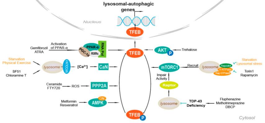 Fig. 1 Schematic of mechanisms of TFEB agonists.1
Key structural properties of TFEB:
- Alkaline helical-ring-helical-leucine zipper (bHLH-Zip) dimerization domain
- The N-terminal and C-terminal each contain a transcriptional activation domain (AD)
- Conserved nuclear localization signal (NLS) and nuclear export signal (NES)
- Multiple serine phosphorylation sites (such as Ser142, Ser211) regulation of nuclear mass
Functions of TFEB
The core function of TFEB is to serve as a global regulatory switch for the cell clearance process. In addition, it is also involved in a variety of physiological and pathological processes, including cell metabolic reprogramming, immune responses, and cell fate determination.
| Function | Description |
| Lysosomal biogenesis | Activate the gene network encoding lysosomal proteins, promote the generation of new lysosomes, and enhance the cell's degradation capacity. |
| Autophagy regulation | Up-regulate the expression of autophagy-related genes, drive the formation of autophagosomes, and mediate the clearance of damaged organelles and macromolecular substances. |
| Metabolic adaptation | When nutrients are lacking, coordinate catabolic pathways and recover nutrients to maintain cellular energy homeostasis. |
| Membrane transport regulation | Genes that regulate the endocytosomal pathway to ensure the connection between substance transport and degradation processes. |
| Stress defense | By enhancing the cell clearance ability, it helps cells cope with various internal and external stresses such as oxidative stress and protein toxic aggregation. |
Unlike most single-function transcription factors, TFEB functions by coordinating a vast gene network (CLEAR network), and its activity is precisely regulated by phosphorylation events of multiple kinases such as mTORC1 and ERK2. This multi-level regulatory mechanism makes it an integration center for cells to respond to environmental changes.
Applications of TFEB and TFEB Antibody in Literature
1. Cui, Zhicheng, et al. "Structure of the lysosomal mTORC1–TFEB–Rag–Ragulator megacomplex." Nature 614.7948 (2023): 572-579. https://doi.org/10.1038/s41586-022-05652-7
The article indicates that TFEB is a key factor regulating lysosomal production and autophagy. The phosphorylation process depends on mTORC1's recognition through a special mechanism: two Rag-Ragulator complexes work in synergy in a non-classical manner, where the GDP state of RagC is crucial for the binding of TFEB. This mechanism reveals the regulatory role of FLCN on the phosphorylation of TFEB.
2. Chen, Mingyue, et al. "TFEB biology and agonists at a glance." Cells 10.2 (2021): 333. https://doi.org/10.3390/cells10020333
The article indicates that the transcription factor TFEB is the core protein that regulates autophagy and lysosomes. It can eliminate pathogenic substances and thus has become an important target for treating related diseases. At present, the development of agonists for TFEB has made progress, and some have entered the preclinical or clinical trial stage, showing broad prospects.
3. Curnock, Rachel, et al. "TFEB‐dependent lysosome biogenesis is required for senescence." The EMBO journal 42.9 (2023): e111241. https://doi.org/10.15252/embj.2022111241
Research has found that in senescent cells, the transcription factors TFEB/TFE3 are continuously activated and enter the cell nucleus, driving lysosome production to maintain their survival. Although the lysosomal function of senescent cells is defective, the increase in quantity compensates for the degradation capacity. Inhibiting TFEB/TFE3 can affect the survival of senescent cells, revealing its key role.
4. Zhang, Weihuang, et al. "Regulation of TFEB activity and its potential as a therapeutic target against kidney diseases." Cell death discovery 6.1 (2020): 32. https://doi.org/10.1038/s41420-020-0265-4
Research has found that the transcription factor TFEB is a core protein that regulates autophagy and lysosomal function. It can promote the clearance of pathogenic substances within cells, thus becoming an important target for treating related diseases. At present, the development of agonists for TFEB has made progress, and some have entered the preclinical or clinical trial stage.
5. Corà, Davide, Federico Bussolino, and Gabriella Doronzo. "TFEB signalling-related MicroRNAs and autophagy." Biomolecules 11.7 (2021): 985. https://doi.org/10.3390/biom11070985
Research has found that the transcription factor TFEB is a core molecule that regulates autophagy and lysosomal production. Its activity is precisely regulated at multiple levels by microRNAs (miRNAs), including directly targeting its transcripts or indirectly influencing its modifying enzymes. Meanwhile, TFEB itself can also regulate the transcription of various miRNAs, and the two form a complex bidirectional regulatory network, jointly determining the fate and function of cells.
Creative Biolabs: TFEB Antibodies for Research
Creative Biolabs specializes in the production of high-quality TFEB antibodies for research and industrial applications. Our portfolio includes monoclonal antibodies tailored for ELISA, Flow Cytometry, Western blot, immunohistochemistry, and other diagnostic methodologies.
- Custom TFEB Antibody Development: Tailor-made solutions to meet specific research requirements.
- Bulk Production: Large-scale antibody manufacturing for industry partners.
- Technical Support: Expert consultation for protocol optimization and troubleshooting.
- Aliquoting Services: Conveniently sized aliquots for long-term storage and consistent experimental outcomes.
For more details on our TFEB antibodies, custom preparations, or technical support, contact us at email.
Reference
- Chen, Mingyue, et al. "TFEB biology and agonists at a glance." Cells 10.2 (2021): 333. https://doi.org/10.3390/cells10020333
Anti-TFEB antibodies
 Loading...


Hot products 
-
Mouse Anti-ENO1 Recombinant Antibody (8G8) (CBMAB-E1329-FY)

-
Mouse Anti-ENO1 Recombinant Antibody (CBYC-A950) (CBMAB-A4388-YC)

-
Mouse Anti-CD2AP Recombinant Antibody (BR083) (CBMAB-BR083LY)

-
Rabbit Anti-ABL1 (Phosphorylated Y185) Recombinant Antibody (V2-443434) (PTM-CBMAB-0001YC)

-
Mouse Anti-BrdU Recombinant Antibody (IIB5) (CBMAB-1038CQ)

-
Mouse Anti-BACE1 Recombinant Antibody (61-3E7) (CBMAB-1183-CN)

-
Mouse Anti-CCN2 Recombinant Antibody (CBFYC-2383) (CBMAB-C2456-FY)

-
Mouse Anti-CD19 Recombinant Antibody (CBXC-1224) (CBMAB-C1491-CQ)

-
Mouse Anti-FN1 Monoclonal Antibody (71) (CBMAB-1241CQ)

-
Mouse Anti-8-oxoguanine Recombinant Antibody (V2-7697) (CBMAB-1869CQ)

-
Mouse Anti-BLK Recombinant Antibody (CBYY-0618) (CBMAB-0621-YY)

-
Mouse Anti-CFL1 (Phospho-Ser3) Recombinant Antibody (CBFYC-1770) (CBMAB-C1832-FY)

-
Mouse Anti-FLT1 Recombinant Antibody (11) (CBMAB-V0154-LY)

-
Mouse Anti-C5B-9 Recombinant Antibody (CBFYA-0216) (CBMAB-X0304-FY)

-
Mouse Anti-CGAS Recombinant Antibody (CBFYM-0995) (CBMAB-M1146-FY)

-
Mouse Anti-BCL6 Recombinant Antibody (CBYY-0442) (CBMAB-0445-YY)

-
Mouse Anti-CCS Recombinant Antibody (CBFYC-1093) (CBMAB-C1150-FY)

-
Mouse Anti-ALPL Antibody (B4-78) (CBMAB-1009CQ)

-
Mouse Anti-CDKL5 Recombinant Antibody (CBFYC-1629) (CBMAB-C1689-FY)

-
Mouse Anti-EMP3 Recombinant Antibody (CBFYE-0100) (CBMAB-E0207-FY)

- AActivation
- AGAgonist
- APApoptosis
- BBlocking
- BABioassay
- BIBioimaging
- CImmunohistochemistry-Frozen Sections
- CIChromatin Immunoprecipitation
- CTCytotoxicity
- CSCostimulation
- DDepletion
- DBDot Blot
- EELISA
- ECELISA(Cap)
- EDELISA(Det)
- ESELISpot
- EMElectron Microscopy
- FFlow Cytometry
- FNFunction Assay
- GSGel Supershift
- IInhibition
- IAEnzyme Immunoassay
- ICImmunocytochemistry
- IDImmunodiffusion
- IEImmunoelectrophoresis
- IFImmunofluorescence
- IGImmunochromatography
- IHImmunohistochemistry
- IMImmunomicroscopy
- IOImmunoassay
- IPImmunoprecipitation
- ISIntracellular Staining for Flow Cytometry
- LALuminex Assay
- LFLateral Flow Immunoassay
- MMicroarray
- MCMass Cytometry/CyTOF
- MDMeDIP
- MSElectrophoretic Mobility Shift Assay
- NNeutralization
- PImmunohistologyp-Paraffin Sections
- PAPeptide Array
- PEPeptide ELISA
- PLProximity Ligation Assay
- RRadioimmunoassay
- SStimulation
- SESandwich ELISA
- SHIn situ hybridization
- TCTissue Culture
- WBWestern Blot
