TMPO Antibodies
Background
The TMPO gene encodes the nuclear membrane protein LAP2β, which is mainly present in the nuclear membrane and nucleoplasm of vertebrate cells. This protein interacts with chromatin and the nuclear scaffold, participating in maintaining the structural stability of the cell nucleus and regulating gene expression and the cell cycle process. Studies have found that TMPO has a special function in cardiomyocytes, and abnormal expression of this protein may affect cardiac development and functional homeostasis. This gene was first identified in the 1990s, and the protein encoded by it, as a member of the nuclear lamina-related polypeptide family, provides a key model for understanding the structure of the nuclear membrane and the regulatory mechanism of cell division. Its multi-level functional characteristics continuously drive in-depth research on the dynamic assembly of the cell nucleus, the spatial organization of chromatin, and the mechanisms of disease-related mutations.
Structure of TMPO
The molecular weight of the thymopentin protein encoded by the TMPO gene is approximately 75 kDa. Due to differences in splicing variants and post-translational modifications among different species, there is a certain fluctuation range in the molecular weight.
| Species | Human | Mouse | Rat |
| Molecular Weight (kDa) | ~75 | ~74 | ~73 |
| Primary Structural Differences | Mainly located in the nuclear membrane, it participates in cell cycle regulation and chromatin organization. | Consistently highly conserved in function with its human homologues | There are differences in expression patterns among specific cell types such as neurons. |
This protein is composed of multiple domains. Among them, the chromatin-binding domain at the N-terminus and the nuclear membrane localization signal at the C-terminus are particularly crucial, enabling it to anchor chromatin to the nuclear lamina in a "bridge" form. Its secondary structure contains typical α-helical bundles, forming the core interface for interaction with DNA and nuclear lamina proteins Lamin A/C. These features jointly maintain the structural integrity of the cell nucleus and the regulation of gene expression.
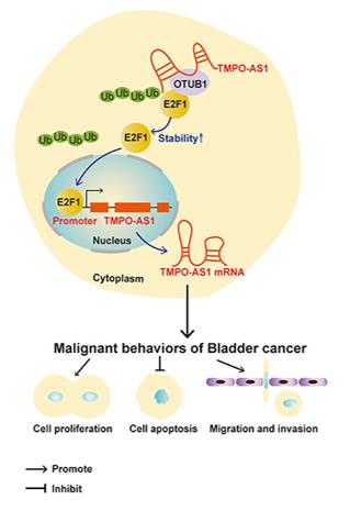 Fig. 1 TMPO-AS1 Drives Bladder Cancer: A Schematic Mechanism.1
Key structural properties of TMPO:
- Multi-domain modular architecture
- Conservative nuclear lamina interaction interface
- Variable phosphorylation regulatory sites
Functions of TMPO
The thymoprotein encoded by the TMPO gene mainly functions to maintain the structure of the nuclear membrane and regulate the cell cycle process. Additionally, it is also involved in key cellular activities such as chromatin spatial organization and gene expression regulation.
| Function | Description |
| Maintaining Nuclear Membrane Structure | As a nuclear lamina-related protein, it anchors chromatin to the inner layer of the nuclear membrane, maintaining the stability of the cell nucleus shape. |
| Cell Cycle Regulation | During the interphase and mitotic phase of cell division, dynamic phosphorylation occurs, regulating the subcellular localization and function of the molecules, and influencing the cell cycle progression. |
| Chromatin Organization | By interacting with chromatin, it participates in the formation and maintenance of heterochromatin structure, influencing gene silencing and expression. |
| Transcriptional Regulation | As a scaffold protein, it participates in the formation of transcription complexes and regulates the expression levels of specific genes (such as those related to the cell cycle). |
| Organ-specific functions | In specific cell types such as cardiac muscle cells and neurons, they are involved in maintaining cellular homeostasis and responding to stress. |
The functional state of this protein is precisely regulated by its phosphorylation modification. Its binding affinity to nucleolar proteins and chromatin changes dynamically along with the cell cycle stage, thereby coordinating the structural integrity and functional requirements of the cell nucleus.
Applications of TMPO and TMPO Antibody in Literature
1. Zhang, Yeyu, et al. "The long non-coding RNA TMPO-AS1 promotes bladder cancer growth and progression via OTUB1-induced E2F1 deubiquitination." Frontiers in oncology 11 (2021): 643163. https://doi.org/10.3389/fonc.2021.643163
The article indicates that TMPO-AS1 is upregulated in bladder cancer and promotes tumor proliferation and metastasis by forming a positive feedback loop with E2F1/OTUB1. This study reveals its crucial role in bladder cancer for the first time, providing a new target for treatment.
2. Jin, Yingmin, et al. "Long noncoding RNA TMPO-AS1 accelerates glycolysis by regulating the miR-1270/PKM2 axis in colorectal cancer." BMC cancer 24.1 (2024): 238. https://doi.org/10.1186/s12885-024-11964-w
The article indicates that long non-coding RNA TMPO-AS1 is highly expressed in colorectal cancer and has a poor prognosis. It upregulates PKM2 by adsorbing miR-1270, thereby promoting tumor proliferation, invasion and glycolysis, and it is a potential therapeutic target.
3. Wang, Min, et al. "The long transcript of lncRNA TMPO-AS1 promotes bone metastases of prostate cancer by regulating the CSNK2A1/DDX3X complex in Wnt/β-catenin signaling." Cell Death Discovery 9.1 (2023): 287. https://doi.org/10.1038/s41420-023-01585-w
The article indicates that in metastatic prostate cancer, the expression of long transcript TMPO-AS1L is upregulated. It activates the Wnt/β-catenin pathway by promoting the interaction between CSNK2A1 and DDX3X, thereby driving bone metastasis. This can serve as a potential prognostic marker and therapeutic target.
4. Guo, Xiaobo, and Yun Wang. "LncRNA TMPO‐AS1 promotes hepatocellular carcinoma cell proliferation, migration and invasion through sponging miR‐329‐3p to stimulate FOXK1‐mediated AKT/mTOR signaling pathway." Cancer medicine 9.14 (2020): 5235-5246. https://doi.org/10.1002/cam4.3046
The article indicates that in hepatocellular carcinoma, TMPO-AS1 is highly expressed and upregulates FOXK1 by adsorbing miR-329-3p, activating the AKT/mTOR pathway, and promoting tumor proliferation, invasion and migration. It is a potential therapeutic target.
5. Vadrot, Nathalie, et al. "Abnormal cellular phenotypes induced by three TMPO/LAP2 variants identified in men with cardiomyopathies." Cells 12.2 (2023): 337. https://doi.org/10.3390/cells12020337
The article indicates that three new heterozygous mutations of TMPO were identified in patients with cardiomyopathy, which can lead to abnormal function of LAP2 protein, affecting cell proliferation, chromatin structure and gene regulatory pathways, and suggesting that they have gender-specific effects in cardiomyopathy.
Creative Biolabs: TMPO Antibodies for Research
Creative Biolabs specializes in the production of high-quality TMPO antibodies for research and industrial applications. Our portfolio includes monoclonal antibodies tailored for ELISA, Flow Cytometry, Western blot, immunohistochemistry, and other diagnostic methodologies.
- Custom TMPO Antibody Development: Tailor-made solutions to meet specific research requirements.
- Bulk Production: Large-scale antibody manufacturing for industry partners.
- Technical Support: Expert consultation for protocol optimization and troubleshooting.
- Aliquoting Services: Conveniently sized aliquots for long-term storage and consistent experimental outcomes.
For more details on our TMPO antibodies, custom preparations, or technical support, contact us at email.
Reference
- Zhang, Yeyu, et al. "The long non-coding RNA TMPO-AS1 promotes bladder cancer growth and progression via OTUB1-induced E2F1 deubiquitination." Frontiers in oncology 11 (2021): 643163. https://doi.org/10.3389/fonc.2021.643163
Anti-TMPO antibodies
Products List
 Loading...


Hot products 
-
Mouse Anti-ATP5F1A Recombinant Antibody (51) (CBMAB-A4043-YC)

-
Mouse Anti-AAV8 Recombinant Antibody (V2-634028) (CBMAB-AP022LY)

-
Mouse Anti-DDC Recombinant Antibody (8E8) (CBMAB-0992-YC)

-
Mouse Anti-AGO2 Recombinant Antibody (V2-634169) (CBMAB-AP203LY)

-
Rabbit Anti-DLK1 Recombinant Antibody (9D8) (CBMAB-D1061-YC)

-
Mouse Anti-DLL4 Recombinant Antibody (D1090) (CBMAB-D1090-YC)

-
Mouse Anti-DLG1 Monolconal Antibody (4F3) (CBMAB-0225-CN)

-
Mouse Anti-CECR2 Recombinant Antibody (CBWJC-2465) (CBMAB-C3533WJ)

-
Mouse Anti-CTCF Recombinant Antibody (CBFYC-2371) (CBMAB-C2443-FY)

-
Mouse Anti-BANF1 Recombinant Antibody (3F10-4G12) (CBMAB-A0707-LY)

-
Mouse Anti-GFAP Recombinant Antibody (20) (CBMAB-G2914-LY)

-
Mouse Anti-DMPK Recombinant Antibody (CBYCD-324) (CBMAB-D1200-YC)

-
Rabbit Anti-ADRA1A Recombinant Antibody (V2-12532) (CBMAB-1022-CN)

-
Mouse Anti-BRCA2 Recombinant Antibody (CBYY-1728) (CBMAB-2077-YY)

-
Mouse Anti-ADRB2 Recombinant Antibody (V2-180026) (CBMAB-A1420-YC)

-
Mouse Anti-CD24 Recombinant Antibody (ALB9) (CBMAB-0176CQ)

-
Mouse Anti-CFL1 Recombinant Antibody (CBFYC-1771) (CBMAB-C1833-FY)

-
Mouse Anti-BZLF1 Recombinant Antibody (BZ.1) (CBMAB-AP705LY)

-
Mouse Anti-ARSA Recombinant Antibody (CBYC-A799) (CBMAB-A3679-YC)

-
Mouse Anti-ADGRE2 Recombinant Antibody (V2-261270) (CBMAB-C0813-LY)

- AActivation
- AGAgonist
- APApoptosis
- BBlocking
- BABioassay
- BIBioimaging
- CImmunohistochemistry-Frozen Sections
- CIChromatin Immunoprecipitation
- CTCytotoxicity
- CSCostimulation
- DDepletion
- DBDot Blot
- EELISA
- ECELISA(Cap)
- EDELISA(Det)
- ESELISpot
- EMElectron Microscopy
- FFlow Cytometry
- FNFunction Assay
- GSGel Supershift
- IInhibition
- IAEnzyme Immunoassay
- ICImmunocytochemistry
- IDImmunodiffusion
- IEImmunoelectrophoresis
- IFImmunofluorescence
- IGImmunochromatography
- IHImmunohistochemistry
- IMImmunomicroscopy
- IOImmunoassay
- IPImmunoprecipitation
- ISIntracellular Staining for Flow Cytometry
- LALuminex Assay
- LFLateral Flow Immunoassay
- MMicroarray
- MCMass Cytometry/CyTOF
- MDMeDIP
- MSElectrophoretic Mobility Shift Assay
- NNeutralization
- PImmunohistologyp-Paraffin Sections
- PAPeptide Array
- PEPeptide ELISA
- PLProximity Ligation Assay
- RRadioimmunoassay
- SStimulation
- SESandwich ELISA
- SHIn situ hybridization
- TCTissue Culture
- WBWestern Blot
