TNFSF10 Antibodies
Background
The protein encoded by the TNFSF10 gene, also known as TRAIL, is a transmembrane signaling molecule belonging to the tumor necrosis factor family. This protein mainly plays a key role in inducing cell apoptosis in the immune system, and can specifically recognize and bind to death receptors on the surface of tumor cells, thereby initiating the programmed death pathway to eliminate abnormal cells. Since its discovery in 1995, TNFSF10 has become an important target in cancer treatment research due to its role in tumor-selective apoptosis. The precise receptor recognition mechanism and apoptotic signaling pathway have been deeply analyzed, providing a theoretical basis for the development of targeted anti-cancer drugs and promoting the development of tumor immunotherapy.
Structure of TNFSF10
The protein encoded by the TNFSF10 (tumor necrosis factor superfamily member 10) gene, also known as TRAIL/Apo-2L, has a molecular weight of approximately 32.5 kDa. The molecular weight may vary within a certain range due to different subtypes and post-translational modifications (such as glycosylation), and its functional form usually exists in the form of homotrimer.
| Species | Human | Mouse | Rat |
| Molecular Weight (kDa) | ~32.5 | ~33.0 | ~32.8 |
| Primary Structural Differences | Standard type, containing death receptor binding domain | Highly similar to humans and with conserved functions | Similar core structure domain, receptor specificity is slightly different |
The TNFSF10 protein belongs to the type II transmembrane protein. Its extracellular region contains a typical tumor necrosis factor homologous domain (THD), which forms a stable trimeric structure through reverse parallel β-sheet folds. This trimerization is the necessary structural basis for its binding to death receptors (such as DR4/DR5) and triggering downstream apoptotic signals. The receptor binding interface is mainly composed of conserved amino acid residues, which determines the specificity of its induction of apoptosis.
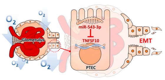 Fig. 1 The Role of the miR-545-3p–TNFSF10 Axis in Hypoxia-driven EMT of PTECs.1
Key structural properties of TNFSF10:
- Conservative tumor necrosis factor homologous domain
- Reverse-parallel β -folded sheets form homologous trimers
- The C-terminal region is responsible for specific binding to death receptors (DR4/DR5)
Functions of TNFSF10
The core function of the TNFSF10 (TRAIL) protein is to selectively induce apoptosis in various tumor cells and virus-infected cells. Additionally, it is also involved in regulating various physiological and pathological processes such as immune homeostasis and inflammatory responses.
| Function | Description |
| Induction of apoptosis | By binding to the death receptors (such as DR4/DR5) on the surface of target cells, it initiates the Caspase cascade reaction, leading to programmed cell death. |
| Immune Surveillance | It plays a role in innate immunity and is expressed by immune cells (such as NK cells, T cells) to eliminate abnormal or infected cells. |
| Immune Regulation | It participates in regulating the function of T cells and maintaining self-immune tolerance. The dysregulation of its signaling pathways may be associated with autoimmune diseases. |
| Antiviral Defense | It can induce the apoptosis of cells infected by the virus, thereby limiting the replication and spread of the virus. |
| Maintenance of Organ Homeostasis | Under normal physiological conditions, it participates in the removal of aged or damaged cells, maintaining tissue renewal and stability. |
Unlike most cytokines, TNFSF10 can distinguish between normal and abnormal cells through its unique receptor system (decepting receptors DcR1/DcR2), thereby achieving selective killing of tumor cells while having a relatively minor impact on most normal cells. This characteristic makes it an important target for tumor treatment research.
Applications of TNFSF10 and TNFSF10 Antibody in Literature
1. Kuo, Mei-Chuan, et al. "Hypoxia-Induced epithelial-to-Mesenchymal transition in proximal tubular epithelial cells through miR-545-3p–TNFSF10." Biomolecules 11.7 (2021): 1032. https://doi.org/10.3390/biom11071032
This study reveals that hypoxia inhibits the expression of TNFSF10 by upregulating miR-545-3p, thereby inducing epithelial-mesenchymal transition (EMT) in renal tubular epithelial cells and promoting renal fibrosis. This mechanism provides a new therapeutic target for hypoxic renal injury.
2. Han, Yoo Jane, et al. "An enhancer variant associated with breast cancer susceptibility in Black women regulates TNFSF10 expression and antitumor immunity in triple-negative breast cancer." Human Molecular Genetics 32.1 (2023): 139-150. https://doi.org/10.1093/hmg/ddac168
This study reveals that the genetic variant rs13074711, which is associated with the high risk of breast cancer in African-American women, can regulate the expression of TNFSF10. TNFSF10 is involved in the anti-viral immune response in TNBC. Its absence leads to a reduction in interferon-induced apoptosis of cancer cells and a decrease in tumor-infiltrating T cells, suggesting that the genetic variant participates in cancer occurrence by influencing the immune mechanism.
3. Zeng, Ziliang, et al. "Circulating monocytes act as a common trigger for the calcification paradox of osteoporosis and carotid atherosclerosis via TGFB1-SP1 and TNFSF10-NFKB1 Axis." Frontiers in Endocrinology 13 (2022): 944751. https://doi.org/10.3389/fendo.2022.944751
This study reveals that CD14+ monocytes, by delivering TGFB1 and TNFSF10, activate SP1 and NFKB1, delaying osteoblast differentiation in osteoporosis and promoting osteogenic transformation of vascular smooth muscle in atherosclerosis, thereby mediating the "calcification paradox". Their senescent state further exacerbates the progression of both diseases.
4. Baijer, Jan, et al. "TNFSF10/TRAIL regulates human T4 effector memory lymphocyte radiosensitivity and predicts radiation-induced acute and subacute dermatitis." Oncotarget 7.16 (2016): 21416. https://doi.org/10.18632/oncotarget.7893
The study found that the radiation sensitivity of T4 effector memory lymphocytes is regulated by the level of membrane-bound TRAIL (TNFSF10) and exhibits Mendelian inheritance characteristics. SNPs in the TRAIL gene are significantly associated with cell radiation sensitivity and dermatitis in breast cancer patients after radiotherapy, suggesting that they may serve as potential predictive markers for individual radiation sensitivity.
5. Ho, Ming-Fen, et al. "Plasma TNFSF10 levels associated with acamprosate treatment response in patients with alcohol use disorder." Frontiers in Pharmacology 13 (2022): 986238. https://doi.org/10.3389/fphar.2022.986238
In this study, through plasma proteomics analysis of patients with alcohol use disorder, it was found that the baseline concentration of TNFSF10 was significantly correlated with the intensity of craving and the therapeutic outcome of acamprosate treatment. This indicates that TNFSF10 may serve as a potential biomarker for predicting the efficacy of acamprosate.
Creative Biolabs: TNFSF10 Antibodies for Research
Creative Biolabs specializes in the production of high-quality TNFSF10 antibodies for research and industrial applications. Our portfolio includes monoclonal and polyclonal antibodies tailored for ELISA, Flow Cytometry, Western blot, immunohistochemistry, and other diagnostic methodologies.
- Custom TNFSF10 Antibody Development: Tailor-made solutions to meet specific research requirements.
- Bulk Production: Large-scale antibody manufacturing for industry partners.
- Technical Support: Expert consultation for protocol optimization and troubleshooting.
- Aliquoting Services: Conveniently sized aliquots for long-term storage and consistent experimental outcomes.
For more details on our TNFSF10 antibodies, custom preparations, or technical support, contact us at info@creative-biolabs.com.
Reference
- Kuo, Mei-Chuan, et al. "Hypoxia-Induced epithelial-to-Mesenchymal transition in proximal tubular epithelial cells through miR-545-3p–TNFSF10." Biomolecules 11.7 (2021): 1032. Distributed under Open Access license CC BY 4.0, without modification. https://doi.org/10.3390/biom11071032
Anti-TNFSF10 antibodies
 Loading...


Hot products 
-
Mouse Anti-ATP1B3 Recombinant Antibody (1E9) (CBMAB-A4021-YC)

-
Mouse Anti-BIRC3 Recombinant Antibody (16E63) (CBMAB-C3367-LY)

-
Mouse Anti-CD19 Recombinant Antibody (CBXC-1224) (CBMAB-C1491-CQ)

-
Mouse Anti-BrdU Recombinant Antibody (IIB5) (CBMAB-1038CQ)

-
Mouse Anti-CCL18 Recombinant Antibody (64507) (CBMAB-C7910-LY)

-
Mouse Anti-AMIGO2 Recombinant Antibody (CBYY-C0756) (CBMAB-C2192-YY)

-
Mouse Anti-AKR1C3 Recombinant Antibody (V2-12560) (CBMAB-1050-CN)

-
Mouse Anti-AKT1/AKT2/AKT3 (Phosphorylated T308, T309, T305) Recombinant Antibody (V2-443454) (PTM-CBMAB-0030YC)

-
Mouse Anti-FN1 Monoclonal Antibody (D6) (CBMAB-1240CQ)

-
Rabbit Anti-BRCA2 Recombinant Antibody (D9S6V) (CBMAB-CP0017-LY)

-
Mouse Anti-ENO2 Recombinant Antibody (H14) (CBMAB-E1341-FY)

-
Mouse Anti-ARIH1 Recombinant Antibody (C-7) (CBMAB-A3563-YC)

-
Mouse Anti-ADV Recombinant Antibody (V2-503423) (CBMAB-V208-1364-FY)

-
Mouse Anti-CD63 Recombinant Antibody (CBXC-1200) (CBMAB-C1467-CQ)

-
Mouse Anti-GDF5 Recombinant Antibody (1F4) (CBMAB-G2740-LY)

-
Mouse Anti-ALDOA Recombinant Antibody (A2) (CBMAB-A2316-YC)

-
Mouse Anti-BAX Recombinant Antibody (CBYY-0216) (CBMAB-0217-YY)

-
Mouse Anti-CRYAB Recombinant Antibody (A4345) (CBMAB-A4345-YC)

-
Rabbit Anti-CCN1 Recombinant Antibody (CBWJC-3580) (CBMAB-C4816WJ)

-
Mouse Anti-BACE1 Recombinant Antibody (61-3E7) (CBMAB-1183-CN)

- AActivation
- AGAgonist
- APApoptosis
- BBlocking
- BABioassay
- BIBioimaging
- CImmunohistochemistry-Frozen Sections
- CIChromatin Immunoprecipitation
- CTCytotoxicity
- CSCostimulation
- DDepletion
- DBDot Blot
- EELISA
- ECELISA(Cap)
- EDELISA(Det)
- ESELISpot
- EMElectron Microscopy
- FFlow Cytometry
- FNFunction Assay
- GSGel Supershift
- IInhibition
- IAEnzyme Immunoassay
- ICImmunocytochemistry
- IDImmunodiffusion
- IEImmunoelectrophoresis
- IFImmunofluorescence
- IGImmunochromatography
- IHImmunohistochemistry
- IMImmunomicroscopy
- IOImmunoassay
- IPImmunoprecipitation
- ISIntracellular Staining for Flow Cytometry
- LALuminex Assay
- LFLateral Flow Immunoassay
- MMicroarray
- MCMass Cytometry/CyTOF
- MDMeDIP
- MSElectrophoretic Mobility Shift Assay
- NNeutralization
- PImmunohistologyp-Paraffin Sections
- PAPeptide Array
- PEPeptide ELISA
- PLProximity Ligation Assay
- RRadioimmunoassay
- SStimulation
- SESandwich ELISA
- SHIn situ hybridization
- TCTissue Culture
- WBWestern Blot
