TPM1 Antibodies
Background
The TPM1 gene encodes myosin 1, which is an α-helical coiled-coil protein highly expressed in cardiac and skeletal muscles. This protein binds to actin filaments and precisely controls the calcium ion-mediated sarcomere contraction process by regulating the conformational changes of the troponin complex. Mutations in the TPM1 gene have been found in patients with congenital heart defects. These variations, by disrupting the normal response of myofilaments to calcium signals, ultimately lead to hypertrophic cardiomyopathy. The TPM1 gene was first localized to chromosome 15 through linkage analysis in 1990. TPM1 is the first disease-causing gene confirmed to be associated with familial hypertrophic cardiomyopathy. The mechanism of phosphorylation modification of its encoded product remains a hot topic in muscle physiology research, and the related achievements have laid a molecular foundation for developing targeted treatments for abnormal myocardial contractility.
Structure of TPM1
The protein encoded by the TPM1 gene has a molecular weight of approximately 32.8 kDa. There are sequence differences among different species, but the α-helical coiling structure is highly conserved.
| Species | Human | Mouse | Rat | Pig | Bovine |
| Molecular Weight (kDa) | 32.8 | 32.7 | 32.8 | 32.9 | 32.8 |
| Primary Structural Differences | Highly conserved sequence | Two amino acid substitutions | Similar to mice | There is a slight variation at the C end | Has the highest homology with humans |
This protein is composed of 284 amino acids and forms a coiled-coil dimer consisting of two α-helices intertwined with each other. Each peptide chain is arranged in a heptapeptide repeating pattern, and the hydrophobic residues are located at the contact surface of the helices to stabilize the dimer structure. The actinomyosin molecule is connected end-to-end along the actin filament and maintains the polymer stability through the periodically distributed cysteine residues. Its C-terminal region specifically binds to the troponin T subunit, while the N-terminal region is involved in the regulation of head-to-tail polymerization.
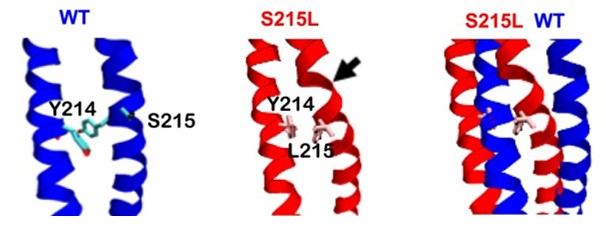 Fig. 1 A detailed view of the isolated TPM1 structure: WT (left), S215L (center), and overlay (right).1
Key structural properties of TPM1:
- Coiled-coil dimers formed by the winding of two α-helices
- Hydrophobic residues are enriched in the helical contact surface to stabilize the structure
- Seven peptide repeat mode decision periodic charge distribution
- Cysteine residues mediate the polymerization of the dimer
- N-terminal acetylation modification regulates the efficiency of head-tail connection
- The central region combines actin filaments
- The C-terminal domain anchors the troponin complex
Functions of TPM1
The core function of the TPM1 gene is to regulate the contraction activities of cardiac muscle and skeletal muscle. Additionally, it also plays multiple roles in cytoskeleton stability, signal transduction, and development processes.
| Function | Description |
| Muscle contraction regulation | Binding to actin filaments, through conformational changes, regulates the response of myosin protein complex to calcium ions, initiating striated muscle contraction. |
| Cellular skeleton stability | Arranged along actin microfilaments, maintains the integrity of myofibrils and affects cell morphology maintenance. |
| Cardiomyopathy association | Genetic mutations lead to hypertrophic or dilated cardiomyopathy, the mechanism involving changes in calcium sensitivity or abnormal myofilament assembly. |
| Developmental process participation | Expressed upregulated during embryonic myofiber formation, regulates the process of myocardial maturation through isoform conversion. |
| Oxidative stress protection | Some isoforms can respond to reactive oxygen levels, regulate contraction efficiency through phosphorylation modification to adapt to metabolic stress. |
The regulatory network of TPM1 is tissue-specific. Different splicing isoforms show differential expression in fast muscle, slow muscle and cardiac muscle, and their allosteric effects directly affect the efficiency of excitation-contraction coupling.
Applications of TPM1 and TPM1 Antibody in Literature
1. Halder, Saiti S., et al. "Mechanisms of pathogenicity in the hypertrophic cardiomyopathy-associated TPM1 variant S215L." PNAS nexus 2.3 (2023): pgad011. https://doi.org/10.1093/pnasnexus/pgad011
This study, through calculation and experimentation, confirmed that the TPM1 S215L mutation disrupts the stability of actin-myosin, increases calcium sensitivity and contractility, leading to diastolic dysfunction and myocardial hypertrophy, supporting its pathogenic role in causing hypertrophic cardiomyopathy.
2. Li, Rong, et al. "TPM1 mediates inflammation downstream of TREM2 via the PKA/CREB signaling pathway." Journal of neuroinflammation 19.1 (2022): 257.https://doi.org/10.1186/s12974-022-02619-3
The study found that TPM1 mediates the pro-inflammatory response of retinal microglia through the PKA/CREB signaling pathway, and its effect is negatively regulated by TREM2. The absence of TPM1 can alleviate inflammatory damage, suggesting that it may potentially serve as a therapeutic target for neuroinflammatory diseases.
3. Tian, Zhihui, Jian Zhao, and Yusheng Wang. "The prognostic value of TPM1–4 in hepatocellular carcinoma." Cancer Medicine 11.2 (2022): 433-446. https://doi.org/10.1002/cam4.4453
The study found that the expressions of TPM1-4 were elevated in hepatocellular carcinoma. Among them, the high expression of TPM3 was associated with poor prognosis and could serve as an independent prognostic factor, as well as participate in the regulation of the tumor immune microenvironment.
4. Halder, Saiti S., et al. "Distinct mechanisms drive divergent phenotypes in hypertrophic and dilated cardiomyopathy–associated TPM1 variants." The Journal of Clinical Investigation 134.24 (2024). https://doi.org/10.1172/JCI179135
The study found that the E62Q (hypertrophic cardiomyopathy) and E54K (dilated cardiomyopathy) mutations of the TPM1 gene alter myofilament function through different mechanisms. The former enhances calcium sensitivity, while the latter alters the binding of troponin, resulting in opposite cardiac phenotypes.
5. Man, Yilong, et al. "Identification of a novel missense mutation in the TPM1 gene via exome sequencing in a Chinese family with dilated cardiomyopathy: A case report and literature review." Medicine 101.2 (2022): e28551.https://doi.org/10.1097/MD.0000000000028551
The study found a new mutation c.340G>C (p.E114Q) in the TPM1 gene in a Chinese Han DCM family, confirming it as the DCM1Y type. This expands the mutation spectrum of the pathogenic gene and is helpful for clinical diagnosis.
Creative Biolabs: TPM1 Antibodies for Research
Creative Biolabs specializes in the production of high-quality TPM1 antibodies for research and industrial applications. Our portfolio includes monoclonal and polyclonal antibodies tailored for ELISA, Flow Cytometry, Western blot, immunohistochemistry, and other diagnostic methodologies.
- Custom TPM1 Antibody Development: Tailor-made solutions to meet specific research requirements.
- Bulk Production: Large-scale antibody manufacturing for industry partners.
- Technical Support: Expert consultation for protocol optimization and troubleshooting.
- Aliquoting Services: Conveniently sized aliquots for long-term storage and consistent experimental outcomes.
For more details on our TPM1 antibodies, custom preparations, or technical support, contact us at info@creative-biolabs.com.
Reference
- Halder, Saiti S., et al. "Mechanisms of pathogenicity in the hypertrophic cardiomyopathy-associated TPM1 variant S215L." PNAS nexus 2.3 (2023): pgad011. Distributed under Open Access license CC BY 4.0. Cropped from the original figure. https://doi.org/10.1093/pnasnexus/pgad011
Anti-TPM1 antibodies
 Loading...


Hot products 
-
Mouse Anti-ASB9 Recombinant Antibody (1D8) (CBMAB-A0529-LY)

-
Rabbit Anti-ALOX5AP Recombinant Antibody (CBXF-1219) (CBMAB-F0750-CQ)

-
Rabbit Anti-ADRA1A Recombinant Antibody (V2-12532) (CBMAB-1022-CN)

-
Mouse Anti-GFAP Recombinant Antibody (5) (CBMAB-G0346-LY)

-
Mouse Anti-CRYAB Recombinant Antibody (A4345) (CBMAB-A4345-YC)

-
Mouse Anti-BIRC5 Recombinant Antibody (6E4) (CBMAB-CP2646-LY)

-
Rat Anti-CCR2 Recombinant Antibody (475301) (CBMAB-C1338-LY)

-
Rabbit Anti-CAMK2A Recombinant Antibody (BA0032) (CBMAB-0137CQ)

-
Mouse Anti-CTNND1 Recombinant Antibody (CBFYC-2414) (CBMAB-C2487-FY)

-
Mouse Anti-ARHGDIA Recombinant Antibody (CBCNA-009) (CBMAB-R0415-CN)

-
Mouse Anti-CDKL5 Recombinant Antibody (CBFYC-1629) (CBMAB-C1689-FY)

-
Mouse Anti-COL1A2 Recombinant Antibody (CF108) (V2LY-1206-LY626)

-
Mouse Anti-BrdU Recombinant Antibody (IIB5) (CBMAB-1038CQ)

-
Rat Anti-CD34 Recombinant Antibody (MEC 14.7) (CBMAB-C10196-LY)

-
Mouse Anti-CD33 Recombinant Antibody (6C5/2) (CBMAB-C8126-LY)

-
Mouse Anti-ANXA7 Recombinant Antibody (A-1) (CBMAB-A2941-YC)

-
Mouse Anti-BMI1 Recombinant Antibody (CBYC-P026) (CBMAB-P0108-YC)

-
Rabbit Anti-ABL1 (Phosphorylated Y245) Recombinant Antibody (V2-505716) (PTM-CBMAB-0465LY)

-
Mouse Anti-AMOT Recombinant Antibody (CBYC-A564) (CBMAB-A2552-YC)

-
Mouse Anti-GFAP Recombinant Antibody (24) (CBMAB-G2927-LY)

- AActivation
- AGAgonist
- APApoptosis
- BBlocking
- BABioassay
- BIBioimaging
- CImmunohistochemistry-Frozen Sections
- CIChromatin Immunoprecipitation
- CTCytotoxicity
- CSCostimulation
- DDepletion
- DBDot Blot
- EELISA
- ECELISA(Cap)
- EDELISA(Det)
- ESELISpot
- EMElectron Microscopy
- FFlow Cytometry
- FNFunction Assay
- GSGel Supershift
- IInhibition
- IAEnzyme Immunoassay
- ICImmunocytochemistry
- IDImmunodiffusion
- IEImmunoelectrophoresis
- IFImmunofluorescence
- IGImmunochromatography
- IHImmunohistochemistry
- IMImmunomicroscopy
- IOImmunoassay
- IPImmunoprecipitation
- ISIntracellular Staining for Flow Cytometry
- LALuminex Assay
- LFLateral Flow Immunoassay
- MMicroarray
- MCMass Cytometry/CyTOF
- MDMeDIP
- MSElectrophoretic Mobility Shift Assay
- NNeutralization
- PImmunohistologyp-Paraffin Sections
- PAPeptide Array
- PEPeptide ELISA
- PLProximity Ligation Assay
- RRadioimmunoassay
- SStimulation
- SESandwich ELISA
- SHIn situ hybridization
- TCTissue Culture
- WBWestern Blot
